What Is An Echocardiogram

Key Takeaways
- An echocardiogram uses ultrasound to show the heart in motion.
- It can help assess pumping strength, valves, chambers, and blood flow.
- The test is generally painless and does not use radiation.
- Different types of echocardiograms are used for different clinical questions.
- Results should always be interpreted together with symptoms and other medical findings.
An echocardiogram is a noninvasive ultrasound test that uses sound waves to create moving images of the heart. It helps doctors assess heart structure and function, often guiding diagnosis and treatment in a wide range of cardiac conditions.
Overview
An echocardiogram, often called an echo or heart ultrasound, is a test that lets doctors look at the heart while it is beating. Instead of using radiation, it uses sound waves to build live images of the heart’s chambers, valves, muscle, and blood flow.
For many people, the test becomes part of a first clear picture of what may be happening inside the chest: shortness of breath that appears only when climbing stairs, a new heart murmur, swelling in the legs, or a follow-up after a known heart condition. Because it is noninvasive and widely available, echocardiography is one of the most common tools in modern cardiology.
In practical terms, the value of the test is not just in showing anatomy. It also helps clinicians understand how well the heart squeezes, whether valves open and close properly, and whether the heart is handling blood flow as expected. That information often shapes next steps, from reassurance and monitoring to medication changes or more advanced testing.
Symptoms and Reasons Doctors Order the Test
An echocardiogram is usually ordered when symptoms or examination findings suggest the heart may need a closer look. A doctor may recommend it for chest discomfort, unexplained fatigue, breathlessness, palpitations, fainting, swelling, or a changed heartbeat heard on examination.
It is also commonly used after a heart attack, to monitor known valve disease, to evaluate heart failure, or to check for congenital heart conditions. In some people, the test is done as part of a wider assessment before surgery or before starting certain treatments that may affect the heart.
Sometimes there are no symptoms at all. A person may have an echocardiogram because a routine physical exam, an ECG, or another scan raised questions that can only be answered by seeing the heart in motion.
Causes and Risk Factors

An echocardiogram does not identify a single disease on its own; rather, it helps reveal the effects of different heart problems. Conditions such as valve narrowing or leakage, weakened heart muscle, high blood pressure effects, fluid around the heart, or structural abnormalities can all be assessed with this test.
Risk factors that make clinicians more likely to consider echocardiography include a history of hypertension, coronary artery disease, diabetes, smoking, pregnancy-related heart symptoms, family history of inherited heart disease, and past infections that may affect the heart. Age can also play a role, since some structural changes become more common over time.
For international patients traveling for evaluation, the test is often part of a broader cardiovascular workup. That broader context matters: the echo is most useful when the doctor understands the person’s symptoms, medical history, medications, and any prior imaging or lab results brought from home.
How Diagnosis Is Made
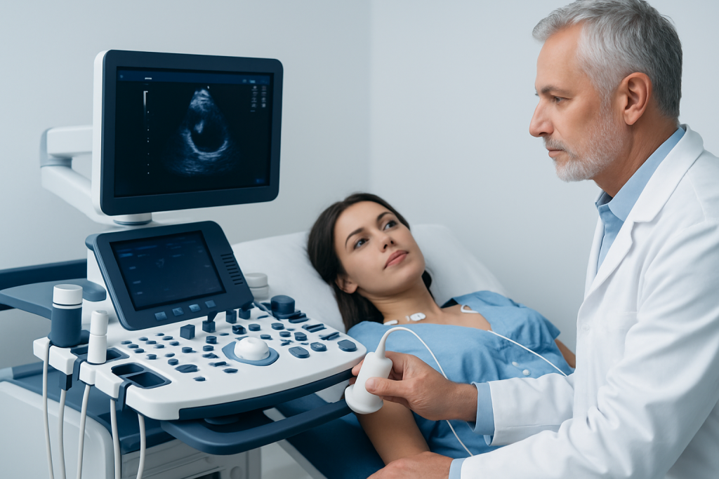
The test itself is straightforward, but the way it is interpreted is careful and detailed. During a transthoracic echocardiogram, a sonographer places a handheld probe on the chest and moves it to capture different views of the heart. The images are then reviewed by a cardiologist who looks at measurements and the overall pattern of heart movement.
Depending on the question being asked, other forms may be used. A transesophageal echocardiogram, or TEE, places a special probe in the esophagus for a closer look at structures that are harder to see through the chest wall. A stress echocardiogram examines how the heart performs during exercise or after medication that simulates exertion.
Results are usually reported in the context of the person’s symptoms and other tests. Terms such as ejection fraction, valve regurgitation, chamber size, wall motion, or pericardial fluid may appear in the report, but they should be explained by the care team in plain language.
Treatment Options
An echocardiogram is not a treatment itself; it is a guide that helps choose treatment. If the scan shows high blood pressure effects, valve disease, reduced pumping function, or another issue, the doctor may recommend medicines, lifestyle changes, monitoring, or a procedure depending on the cause and severity.
In some cases, the result simply confirms that no major structural problem is present, which can be just as helpful. In others, it provides the detail needed to decide whether the person should see a cardiologist, repeat the scan later, or move ahead with more specialized care.
For patients traveling from another country, treatment planning should include time for discussion, record review, and a clear follow-up plan after return home. A good cardiology team will explain which findings require immediate attention, which can be observed, and how continuity of care should be arranged with the local physician.
What to Expect During the Test
Most people have a standard transthoracic echocardiogram while lying on an examination table. Gel is applied to the chest to help the probe make good contact, and the room may be dimmed so the images are easier to view. The exam usually lasts from a short visit to a longer appointment depending on the type of study and the complexity of the case.
The test is generally comfortable, although the probe may be pressed at different angles to capture the best pictures. The person may be asked to hold their breath briefly, turn onto one side, or change position during the scan. Afterward, there is no recovery period for the standard chest ultrasound, and normal activities can usually resume right away.
For a transesophageal study, preparation is more involved because the throat needs to be numbed and sedation may be used. That type of exam may require fasting beforehand and a short observation period afterward, which is why patients should follow the clinic’s instructions carefully.
Prevention and Self-care
People cannot always prevent the need for an echocardiogram, but they can help protect heart health in general. Managing blood pressure, cholesterol, diabetes, and smoking exposure lowers the chance of many conditions that lead to abnormal heart findings.
Good self-care also includes taking prescribed medicines as directed, staying physically active within personal limits, eating a balanced diet, and seeking medical review if symptoms change. For those with a known heart condition, keeping prior test reports and medication lists organized can make follow-up much smoother, especially when care is shared between countries.
If a doctor recommends repeat echocardiography, it is usually because the heart needs to be tracked over time. Follow-up is not a sign that something is wrong by itself; it is often the safest way to confirm stability or catch a change early.
When to See a Doctor
A doctor should be consulted if symptoms such as shortness of breath, chest pain, fainting, leg swelling, or palpitations appear, especially if they are new or worsening. These symptoms do not always mean a serious problem, but they do deserve proper assessment.
Medical advice is also important after a concerning ECG, a heart murmur, unexplained reduced exercise tolerance, or if there is a family history of inherited heart disease. When symptoms begin during travel or while seeking care abroad, it helps to bring previous records, test results, and a list of medicines so the evaluation can start efficiently.
If a cardiologist recommends an echocardiogram, it is usually because the test can answer an important question about the heart. In centers experienced with international patients, including Acibadem Health Point, multidisciplinary specialists and JCI-accredited hospitals can diagnose and treat these conditions with coordinated planning and follow-up.
Frequently asked questions
Is an echocardiogram the same as an ECG?
No. An ECG records the heart’s electrical activity, while an echocardiogram shows the heart’s structure and how it moves. The two tests provide different information and are often used together.
Does an echocardiogram hurt?
A standard transthoracic echocardiogram is usually painless. Some pressure from the probe may be felt, but most people tolerate it well.
Do I need to prepare for the test?
For a regular chest echo, preparation is often minimal. If a transesophageal or stress echocardiogram is planned, the clinic may give specific instructions such as fasting or adjusting certain medicines.
Can an echocardiogram show blocked arteries?
It may suggest reduced blood flow or areas of the heart muscle that are not moving normally, but it does not directly visualize all coronary artery blockages. If blockage is suspected, a doctor may recommend additional tests.
How long does it take to get results?
That depends on the clinic and the complexity of the study. In many settings, the images are reviewed the same day or shortly afterward, and the doctor explains what the findings mean in context.
Is an echocardiogram safe in pregnancy?
Yes, it is generally considered safe because it uses ultrasound and does not involve radiation. Doctors may use it to evaluate symptoms or known heart conditions during pregnancy.
References
- American Heart Association
- Mayo Clinic
- National Heart, Lung, and Blood Institute
- European Society of Cardiology
This article is for general information only and is not a substitute for professional medical advice. Please consult a qualified doctor about your individual situation.