Transthoracic Echocardiogram

Key Takeaways
- A transthoracic echocardiogram uses sound waves to create moving images of the heart.
- It helps assess heart chambers, valves, pumping strength, and blood flow patterns.
- The test is painless for most people and usually takes place in an outpatient setting.
- Results can support the diagnosis of valve disease, heart failure, cardiomyopathy, and other cardiac conditions.
- Preparation is often simple, and most people can return to normal activities right away.
A transthoracic echocardiogram is a noninvasive heart ultrasound that helps doctors look at the heart’s structure and pumping function in real time. It is commonly used to assess symptoms such as chest discomfort, shortness of breath, palpitations, or a known heart condition.
Overview
A transthoracic echocardiogram, often shortened to TTE, is one of the most common ways doctors evaluate the heart without using radiation or surgery. It works like an ultrasound exam of the chest: a probe sends sound waves through the chest wall, and a moving picture of the heart appears on a monitor.
For many patients, this test is part of answering a practical question: is the heart pumping as expected, are the valves opening and closing well, and is there any sign of strain or fluid around the heart? Because the scan is noninvasive, it is often chosen early in the workup of symptoms or as follow-up for an existing heart diagnosis.
International patients often appreciate that the test is straightforward to schedule and usually does not require a long recovery period. In many care pathways, it becomes one useful piece of a larger cardiology assessment, helping the doctor decide whether further testing is needed and how urgently treatment should begin.
Symptoms and Reasons It May Be Ordered
A transthoracic echocardiogram is not performed for one single symptom. Instead, it is commonly ordered when a doctor wants to understand the source of signs that may involve the heart. Shortness of breath, swelling in the legs, chest pain, fainting episodes, or a new heart murmur are frequent reasons for the exam.
The test may also be used when a person notices palpitations, unusual fatigue, reduced exercise tolerance, or symptoms that appear after an infection, pregnancy, chemotherapy, or a known heart event. In other situations, it is used to monitor a previously diagnosed condition such as valve disease, heart failure, congenital heart disease, or cardiomyopathy.
Sometimes there are no symptoms at all. A doctor may order the exam after an abnormal electrocardiogram, a concerning physical examination finding, or to establish a baseline before treatment. In this way, the echocardiogram becomes less about treating a symptom and more about mapping the heart’s condition with clarity.
Causes and Risk Factors

A TTE itself does not identify a single cause; it helps reveal what may be affecting the heart. Findings can point toward valve narrowing or leakage, weakened pumping function, enlarged chambers, thickened heart muscle, or fluid around the heart. Each of these patterns can have different underlying reasons.
Risk factors that often lead doctors to request this test include high blood pressure, diabetes, high cholesterol, smoking, family history of heart disease, previous heart attack, known congenital heart abnormalities, and certain infections or inflammatory conditions. Age can also matter, since some valve problems and changes in heart function become more common over time.
In a travel-based care journey, the history is especially important. A cardiologist may want details about prior scans, medication use, prior hospitalizations, exercise limits, and whether symptoms change with altitude, long flights, or stress. These details help the imaging findings fit into the patient’s wider story rather than stand alone.
Diagnosis: What the Test Can Show
The transthoracic echocardiogram gives the doctor a live look at the heart in motion. It can show how strongly the left and right ventricles contract, whether the chambers are enlarged, and whether the valves are opening and closing in the expected way. The exam can also estimate pressure patterns and detect some structural abnormalities.
Depending on the reason for the test, the report may comment on ejection fraction, wall motion, valve function, chamber size, and whether there is evidence of fluid around the heart. For some people, the findings are immediately reassuring. For others, the result helps explain symptoms and guides the next step, which may be medication changes, more detailed imaging, or a follow-up test such as a transesophageal echocardiogram or stress test.
It is helpful to remember that TTE is one part of diagnosis, not the entire diagnosis itself. Doctors interpret it together with the physical exam, ECG, lab work, and the patient’s symptom history. That broader approach reduces the chance of overreading a single image and keeps care grounded in the full clinical picture.
Treatment Options
The echocardiogram does not treat heart disease, but it plays an important role in choosing treatment. If the scan shows valve disease, the care plan may include regular monitoring, medicines, or evaluation for a procedure. If reduced pumping function is found, doctors may consider therapies used for heart failure or additional testing to understand the cause.
When the scan suggests a problem related to rhythm, pressure, or previous heart damage, treatment is tailored to the underlying issue rather than the image alone. That may involve blood pressure control, cholesterol management, anticoagulation in selected cases, lifestyle changes, or specialist referral. In some patients, no treatment is needed beyond observation and periodic reassessment.
For international patients, treatment planning may also include timing follow-up before they return home, sharing electronic copies of the report, and arranging care coordination with a local physician abroad. Clear documentation is especially useful when the next appointment will happen in another country.
Preparation, Procedure, and What to Expect
Most people need very little preparation for a standard transthoracic echocardiogram. They may be asked to wear a two-piece outfit or change into a gown so the chest can be accessed easily. Jewelry around the chest area may need to be removed, and the patient may be asked about previous heart procedures or current symptoms before the scan begins.
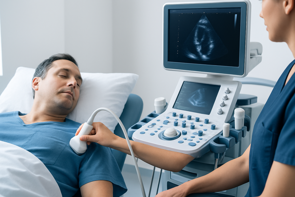
During the test, the patient lies on an examination table while a technician or clinician places gel on the chest and moves the probe across several positions. The room is usually dim so the screen images are easier to see. Patients may be asked to briefly hold their breath, lie on the side, or shift position so the heart can be viewed from different angles.
The procedure is generally comfortable. Some people feel slight pressure from the probe or notice the coolness of the gel, but there is no incision and no radiation exposure. Afterward, most people can return to normal activities immediately, including travel plans in many cases, unless their doctor gives different instructions based on the reason for the exam.
Prevention and Self-care
A transthoracic echocardiogram is not something people typically need to prevent. What they can help prevent is avoidable strain on the heart that may later require repeated imaging or treatment. Practical steps include following advice for blood pressure, diabetes, and cholesterol management, staying physically active within personal limits, and avoiding smoking.
If a person already has a known heart condition, keeping follow-up appointments matters. Echo results can change over time, and regular reassessment helps the doctor notice early shifts before symptoms become severe. Patients who are traveling should carry a medication list, prior test results, and the name of their treating cardiologist when possible.
- Take prescribed heart medicines consistently unless a doctor advises otherwise.
- Track symptoms such as swelling, breathlessness, chest pain, or palpitations.
- Ask whether repeat imaging is needed and how soon follow-up should occur.
- Share prior echocardiogram reports if care is transferred to another country or clinic.
When to See a Doctor
Medical review is appropriate when chest discomfort, shortness of breath, fainting, new swelling, or persistent palpitations occur, even if the symptoms seem mild at first. A doctor can determine whether a transthoracic echocardiogram or another heart test is the right next step. The exam is especially important when symptoms are new, worsening, or occurring with exertion.
After the scan, the doctor should explain what the findings mean in plain language and how they fit with the rest of the evaluation. If a result suggests a more complex heart issue, timely referral to a cardiologist is the safest path. If the result is normal, the patient may still need a plan for symptom monitoring or a different explanation for the complaints.
Acibadem Health Point’s multidisciplinary specialists and JCI-accredited hospitals diagnose and treat heart conditions for international patients, helping coordinate imaging, specialist review, and follow-up care across borders.
Frequently asked questions
What is a transthoracic echocardiogram used for?
It is used to look at the heart’s structure and function with ultrasound. Doctors use it to assess heart valves, pumping strength, chamber size, and signs of fluid or other abnormalities.
Is a transthoracic echocardiogram painful?
It is usually not painful. Some people feel mild pressure from the probe or discomfort from lying in one position, but the test is generally well tolerated.
Do I need to fast before the test?
Most people do not need to fast for a standard transthoracic echocardiogram. The care team will let the patient know if any special preparation is needed based on the reason for the exam.
How long does the exam take?
The scan often takes a short outpatient visit, though the exact time can vary depending on how many views are needed. Additional explanation from the doctor may take longer than the imaging itself.
Can a transthoracic echocardiogram show a heart attack?
It can sometimes show areas of the heart muscle that are not moving normally, which may suggest prior heart damage. However, doctors usually combine it with other tests and the symptom history before reaching a conclusion.
What happens if the result is abnormal?
An abnormal result does not always mean something urgent or severe. The doctor will explain the finding, discuss whether more testing is needed, and decide on follow-up or treatment based on the overall picture.
References
- American Heart Association
- Mayo Clinic
- National Heart, Lung, and Blood Institute
- British Society of Echocardiography
This article is for general information only and is not a substitute for professional medical advice. Please consult a qualified doctor about your individual situation.