Echocardiogram vs. Cardiac MRI vs. CT: Which Scan Answers Which Question?

Key Takeaways
- An echocardiogram is usually the first-line test for heart structure and pumping function.
- Cardiac MRI provides detailed information about heart muscle, scarring, inflammation, and complex anatomy.
- Cardiac CT is especially useful for looking at the coronary arteries and some structural problems.
- No single scan is best for every situation; the right choice depends on the question being asked.
- Your doctor will also consider kidney function, heart rhythm, implanted devices, and how urgent the answer is.
Medically reviewed by the Acıbadem clinical team — June 13, 2026
Echocardiogram, cardiac MRI, and cardiac CT all look at the heart, but each answers a different clinical question. The best test depends on what needs to be seen: structure, function, blood flow, tissue detail, or the coronary arteries.
Overview
When someone needs heart imaging, the real question is usually not “Which scan is best?” but “What exactly needs to be answered?” An echocardiogram, cardiac MRI, and cardiac CT can all help doctors evaluate the heart, yet they do so in very different ways.
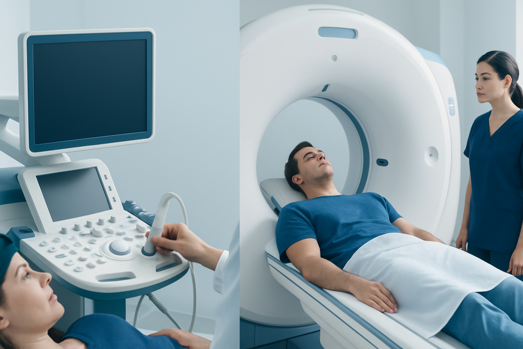
An echocardiogram uses ultrasound to create moving images of the heart. Cardiac MRI uses a strong magnetic field and radio waves to show the heart in remarkable detail. Cardiac CT uses X-rays to produce fast, cross-sectional pictures, often with a strong focus on the coronary arteries. Each test has a different strength, and that is why a cardiologist may choose one, two, or sometimes a sequence of scans.
For international patients, the practical question can also include timing, travel, and follow-up. A person may need a quick test before a procedure, a more detailed scan after an unclear result, or a study that helps plan care from afar. The most efficient pathway is the one that matches the medical question while fitting the patient’s overall situation.
Symptoms and Clinical Clues That Lead to Imaging
Heart scans are not usually ordered simply because a person feels unwell in a general way. They are recommended when symptoms, examination findings, blood tests, or prior tests suggest that the heart needs closer evaluation. Shortness of breath, chest discomfort, palpitations, fainting, leg swelling, or reduced exercise tolerance may all prompt imaging, depending on the context.
Sometimes imaging is requested even when symptoms are subtle. A doctor may hear a murmur, notice a change in blood pressure, or see signs of heart enlargement on an electrocardiogram or chest X-ray. In other cases, the scan is part of follow-up for a known condition such as valve disease, cardiomyopathy, coronary artery disease, congenital heart disease, or heart failure.
The pattern of symptoms can hint at which scan will be most useful. For example, a valve problem often begins with an echocardiogram. Concerns about scar tissue, inflammation, or inherited heart muscle disease may point toward cardiac MRI. Questions about the coronary arteries, especially when calcium or narrowing is suspected, may lead to cardiac CT.
Causes and Risk Factors: Why Doctors Choose One Scan Over Another

Each imaging test answers a different kind of medical question. An echocardiogram is excellent for seeing how the heart valves open and close, how strongly the heart pumps, and whether fluid surrounds the heart. It is often used first because it is widely available, does not use radiation, and can be done at the bedside or in an outpatient setting.
Cardiac MRI becomes especially valuable when the doctor needs tissue-level detail. It can help distinguish old scar from active inflammation, assess heart muscle thickness, evaluate congenital abnormalities, and clarify complex findings that ultrasound cannot fully define. It is often chosen when a more complete map of the heart is needed before deciding on treatment.
Cardiac CT is usually selected when speed and precision matter, particularly for the coronary arteries. It can show plaque, narrowing, calcium, and some structural abnormalities with high clarity. A doctor may favor CT when symptoms suggest coronary disease, when other tests were inconclusive, or when a detailed road map is needed before a procedure. Choice of scan can also depend on kidney function, contrast allergy history, implanted devices, body size, heart rate, and the need to avoid radiation or time-consuming appointments.
Diagnosis: What Each Scan Actually Shows
An echocardiogram shows the heart in motion. It evaluates chamber size, valve function, blood flow patterns, pumping strength, and pressure estimates. Because it is dynamic, it is especially useful for seeing how the heart behaves in real time, not just how it looks as a still image.
Cardiac MRI is often the most detailed test for heart muscle and tissue characterization. It can show whether a muscle is thickened, weak, scarred, inflamed, or infiltrated by an abnormal process. Doctors may use it to assess myocarditis, cardiomyopathies, viability after a heart attack, congenital anatomy, and some valve or pericardial conditions.
Cardiac CT offers a different kind of clarity. It is particularly strong for the coronary arteries and the anatomy around the heart. It can identify calcium buildup, narrowing, bypass graft patency, and some aortic or structural problems. In practical terms, the scan is chosen not by prestige or complexity, but by whether the doctor needs movement, tissue detail, or a fast arterial map.
- Echocardiogram: best for pumping function, valves, and bedside assessment.
- Cardiac MRI: best for tissue detail, scarring, inflammation, and complex anatomy.
- Cardiac CT: best for coronary arteries, calcium, and rapid anatomic mapping.
Treatment Options: How Imaging Shapes Care
Imaging itself is not treatment, but it often determines the next step in treatment. An echocardiogram may reveal a leaky valve, weakened pumping, or fluid around the heart, which then guides medication, monitoring, or a procedure. In that sense, the scan helps doctors decide whether the heart needs support, repair, or closer follow-up.
Cardiac MRI can change treatment by showing whether symptoms are caused by scar, inflammation, or a specific cardiomyopathy. This distinction matters because the management of active inflammation, chronic damage, and inherited muscle disease may be very different. MRI can also help doctors judge whether part of the heart muscle may recover after an injury or whether a procedure is likely to help.
Cardiac CT can influence decisions about medication, stenting, surgery, or reassurance when coronary disease is less likely than expected. In some cases, it helps doctors avoid unnecessary invasive testing. For patients traveling for care, this can be particularly useful because a clear anatomic answer may streamline the rest of the evaluation and reduce repeated appointments.
Prevention and Self-care: Preparing for the Right Scan
Good preparation starts with sharing a complete medical history. Patients should tell the care team about kidney problems, allergies to contrast material, asthma, pregnancy, implanted devices, pacemakers, metal fragments, prior heart surgery, and any difficulty lying flat or holding still. These details help the team choose the safest and most informative scan.
It also helps to ask what the scan is meant to answer. A patient who understands the purpose is better able to prepare for the experience, whether that means fasting, arriving early, removing metal objects, or arranging support after the appointment. International patients may also want to confirm how results will be delivered, whether follow-up can be virtual, and what documents to bring from prior care.
Self-care after the scan is usually straightforward. Most people can return to normal activity quickly, but they should follow the team’s instructions about hydration, resuming medicines, and watching for delayed contrast reactions. If the scan involves travel, it is wise to leave enough time for review of the images before departing so that any next step can be planned without delay.
When to See a Doctor
Medical attention is appropriate when symptoms suggest a heart problem, especially chest pain, unexplained shortness of breath, fainting, new swelling, or palpitations that do not settle. A doctor should also be consulted if a previous test was unclear or if a known heart condition is changing over time.
People should seek prompt care if a clinician recommends urgent imaging after an abnormal exam, blood test, or emergency evaluation. The right scan may need to be arranged quickly, and the order in which tests are done can affect treatment decisions. In many cases, the fastest path is not the most advanced test, but the one that answers the immediate question cleanly.
For patients coming from another country, it is especially helpful to coordinate imaging with a cardiologist who can interpret the findings in the context of symptoms, medicines, and previous reports. Acibadem Health Point’s multidisciplinary specialists and JCI-accredited hospitals diagnose and treat heart conditions for international patients, which can help make the imaging and follow-up process more coordinated.
Choosing Between Echocardiogram, Cardiac MRI, and CT
The best scan depends on the question. If the concern is how well the heart pumps or whether a valve is working properly, echocardiography is often the starting point. If the doctor needs fine detail about the heart muscle, scars, inflammation, or congenital anatomy, cardiac MRI may be more informative. If the key question is what is happening in the coronary arteries or whether there is calcified plaque, cardiac CT often provides the clearest answer.
It is also common for the tests to complement one another. A patient may start with an echocardiogram, then move to MRI for deeper tissue assessment, or to CT for coronary imaging. This stepwise approach reduces unnecessary testing and helps build a complete picture of the heart without forcing one scan to do every job.
Ultimately, the most useful scan is the one that answers the clinician’s question safely and efficiently. Patients are usually best served by asking, “What are we trying to learn?” rather than “Which technology sounds most advanced?”
Frequently asked questions
Is an echocardiogram the same as a cardiac MRI?
No. An echocardiogram uses ultrasound, while cardiac MRI uses a magnetic field and radio waves. They provide different kinds of information, so a doctor may choose one over the other depending on the question.
Why would a doctor order cardiac CT instead of MRI?
Cardiac CT is often preferred when the coronary arteries need to be seen clearly or when a fast anatomic picture is needed. It can be especially helpful when the goal is to assess plaque, narrowing, or bypass grafts.
Which scan is best for heart valves?
An echocardiogram is usually the first test for valve problems because it shows movement in real time. In some cases, MRI or CT may add detail if the anatomy is complex or the ultrasound images are limited.
Does cardiac MRI use radiation?
No, cardiac MRI does not use ionizing radiation. That is one reason it is often chosen when detailed tissue information is needed and radiation is best avoided.
Can these scans replace a cardiac catheterization?
Sometimes they can reduce the need for an invasive procedure, but not always. If a doctor needs direct pressure measurements or treatment during the same procedure, catheterization may still be necessary.
How should a patient prepare for these scans?
Preparation depends on the test and the reason for it. Patients should follow the imaging team’s instructions, share any kidney issues, allergies, or implanted devices, and ask when they can resume normal activities afterward.
References
- American College of Cardiology
- American Heart Association
- Society for Cardiovascular Magnetic Resonance
- Radiological Society of North America
- National Heart, Lung, and Blood Institute
This article is for general information only and is not a substitute for professional medical advice. Please consult a qualified doctor about your individual situation.