

Navicular Bone

Key Takeaways
- The navicular bone sits in the midfoot and helps connect the foot’s inner arch to nearby bones.
- Pain in the top or inner midfoot, especially with walking or running, can be a sign of navicular injury.
- Stress fractures may develop gradually, while acute fractures usually follow a specific injury.
- Imaging is often needed because navicular problems can be difficult to diagnose on symptoms alone.
- Treatment depends on the type of injury and may include rest, immobilization, or surgery in some cases.
The navicular bone is a small but important bone in the midfoot that helps support movement and weight-bearing. When it is injured, irritated, or fractured, it can cause persistent pain that often deserves careful orthopedic evaluation.
Overview
The navicular bone is a small, boat-shaped bone on the inner side of the midfoot. Even though it is not large, it plays an important role in maintaining the shape of the arch and in transferring force through the foot during walking, running, and jumping.
Because of the way it sits in the foot, the navicular can be involved in several different problems. Some people develop inflammation or irritation around the bone, while others experience an acute fracture after a twist, fall, or direct impact. Athletes and active travelers who suddenly increase their walking or training loads may also develop a stress fracture, which can be easy to overlook at first.
When someone is deciding whether to seek care at home or while traveling, persistent midfoot pain should not be dismissed as a minor sprain if it is not improving. Navicular injuries are often managed best when diagnosed early, before pain changes the way a person walks and places extra strain on nearby structures.
Symptoms
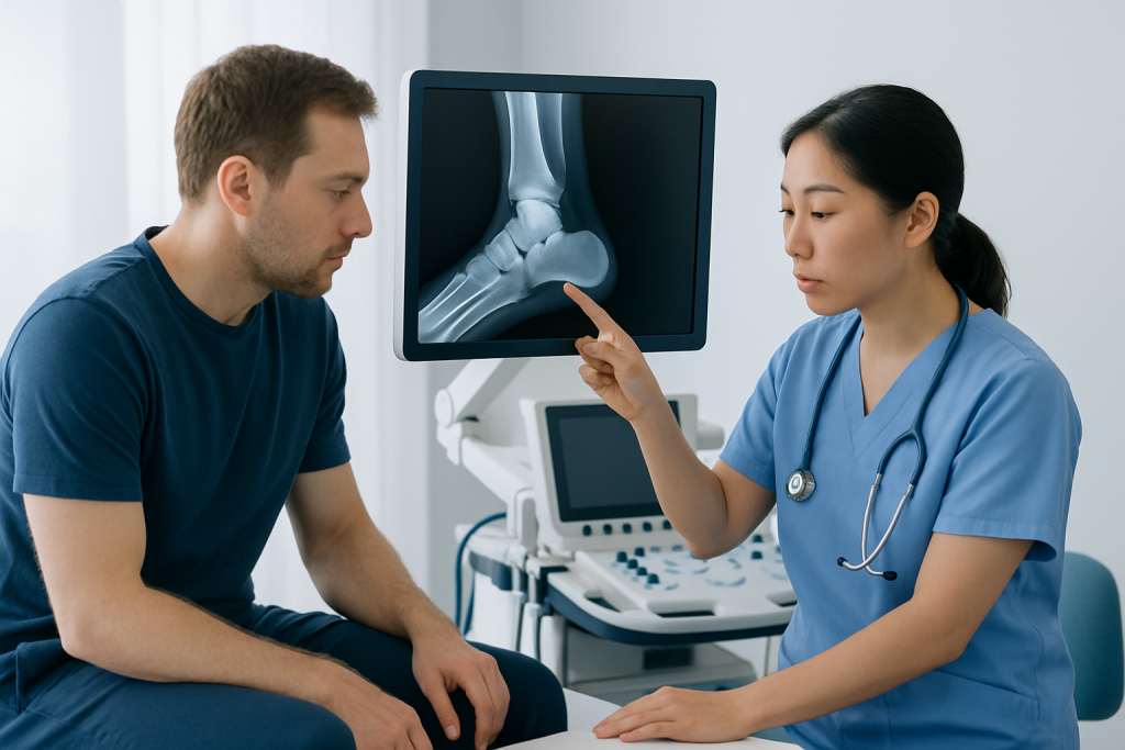
Navicular bone problems usually cause pain in the middle of the foot, often on the inner or top side. The discomfort may be sharp when weight is placed on the foot, or it may feel like a deep ache that becomes more noticeable during activity.
Some people notice tenderness when pressing on a specific point in the midfoot. Swelling can occur, although it is not always dramatic. With stress fractures, symptoms may begin gradually and become worse with repeated walking, running, or standing, then ease somewhat with rest before returning again.
Possible signs include:
- Pain in the midfoot, especially during walking or exercise
- Tenderness over the inner arch or top of the foot
- Swelling around the midfoot
- Pain that worsens with pushing off the foot
- Limping or avoiding full weight-bearing
Because midfoot pain can come from several different structures, the pattern of symptoms matters. A person who has recently increased activity, changed footwear, or had a minor twist injury may be experiencing a navicular issue even if the foot still looks normal from the outside.
Causes & Risk Factors

Navicular injuries can happen in more than one way. An acute fracture may result from a direct blow, a fall, or a twisting injury to the foot. Stress fractures, by contrast, develop over time when repeated loading gradually exceeds the bone’s ability to repair itself.
Several factors can increase the chance of a navicular problem. High-impact sports such as running, dancing, and court sports are common settings for stress injuries. Sudden changes in training, poor footwear, tight calves, foot alignment differences, and a history of previous foot injuries may also contribute.
Risk can be higher when the foot is exposed to repeated strain during long periods of travel, especially if a person walks more than usual while sightseeing or carrying luggage. In international patients, this sometimes becomes noticeable only after a trip has already started, which is why early attention to worsening midfoot pain matters.
Common contributors include:
- Repetitive impact from sports or prolonged walking
- Sudden increase in physical activity
- Direct trauma or twisting injuries
- Foot mechanics that place extra stress on the midfoot
- Inadequate recovery after prior foot pain
Diagnosis
Diagnosis starts with a careful history and physical examination. A doctor will ask where the pain is located, how it began, whether it worsens with impact, and whether the person can bear weight. They will also examine the foot for tenderness, swelling, and movement patterns that suggest a navicular injury.
Imaging is often important because the navicular can be difficult to assess by examination alone. X-rays may identify some fractures, but stress fractures are not always visible early. If symptoms continue or the diagnosis is uncertain, a doctor may recommend MRI, CT, or other imaging to look more closely at the bone and surrounding tissues.
For people who are abroad when symptoms begin, it is reasonable to seek a local orthopedic assessment rather than wait for a return flight if pain is significant or walking is becoming difficult. Clear communication about when the pain started, how much walking has been involved, and whether there was an injury can help the clinician choose the right tests.
Treatment Options
Treatment depends on whether the problem is a stress fracture, acute fracture, or another type of navicular injury. Some mild overuse injuries can improve with rest and activity modification, while more serious fractures may require immobilization in a boot or cast to protect healing.
Doctors often recommend limiting weight-bearing when the navicular is injured, especially if walking causes pain. This may be paired with crutches, a walking boot, or a cast. Knee Pain Relief: Causes and Treatment" class="ahp-ilk">Pain relief measures can help, but the larger goal is to reduce stress on the bone long enough for healing to begin.
In certain fractures, especially when the bone is displaced, unstable, or not healing as expected, surgery may be considered. The exact approach depends on the fracture pattern, the person’s activity level, and how much support the foot needs to regain normal function. After treatment, rehabilitation is often used to restore strength, balance, and walking mechanics.
Typical treatment pathways may include:
- Relative rest and activity modification
- Immobilization with a boot or cast
- Temporary reduction in weight-bearing
- Physical therapy during recovery
- Surgery for selected fractures or non-healing cases
For international patients, treatment planning may also include practical follow-up questions: how long the foot should stay protected, when it is safe to fly, and how to coordinate rehab once back home. A clear discharge plan can make recovery much easier to manage across borders.
Prevention & Self-care
Not every navicular injury can be prevented, but many overuse problems can be reduced by paying attention to training load and foot support. A gradual return to exercise is usually safer than making sudden jumps in distance, speed, or intensity.
Supportive shoes that fit well can help distribute pressure more evenly through the foot. Athletes often benefit from reviewing technique, warm-up habits, and recovery time. People with flat feet, high arches, or recurring midfoot pain may need individualized guidance from an orthopedic specialist or physical therapist.
Self-care is most helpful when it complements, rather than replaces, medical evaluation. If pain is present, reducing impact and avoiding “pushing through” symptoms can keep a small injury from becoming a longer recovery.
- Increase activity gradually
- Choose supportive footwear for walking and exercise
- Allow time for recovery between high-impact sessions
- Address persistent foot pain early
- Follow rehabilitation exercises as advised
When to See a Doctor
Medical evaluation is appropriate when midfoot pain lasts more than a few days, keeps returning, or interferes with walking. Pain that is focused over the navicular area, especially after sports or a twist injury, deserves prompt attention because delayed diagnosis can lengthen recovery.
A doctor should be seen sooner if there is swelling, marked tenderness, limping, or inability to bear weight. Even if the injury seems minor, persistent pain in the midfoot is not something to ignore, particularly in active people or those who have recently increased walking or training.
Emergency assessment is less commonly needed than for severe trauma, but urgent review is sensible if the foot looks deformed, pain is severe, or numbness develops. With the right diagnosis and protection early on, many navicular injuries heal more predictably and a person can return to normal activity more safely.
Acibadem Health Point’s multidisciplinary specialists and JCI-accredited hospitals diagnose and treat navicular bone problems for international patients, with care plans that can be coordinated around travel and follow-up needs.
Recovery and Follow-up
Recovery time varies widely depending on the type of injury and how quickly treatment begins. Stress fractures often need a more cautious return to activity than minor irritation, and some fractures require a period of protection before the foot can be loaded normally again.
Follow-up visits are important because healing may need to be checked with repeat examination or imaging. A doctor may adjust weight-bearing, recommend a transition from a boot to regular shoes, or guide the person through physical therapy once the bone is stable enough.
When recovery is managed well, the goal is not only pain relief but also a return to steady walking, sport, and daily movement without compensation. That often means rebuilding calf flexibility, foot strength, and balance so the midfoot is not overloaded again.
Living With Navicular Pain While Waiting for Care
If a person is awaiting an appointment or traveling to a clinic, it helps to keep the foot as calm as possible. Reducing long walks, avoiding jumping or running, and using supportive footwear can make symptoms more manageable until a clinician evaluates the problem.
It is also useful to note when the pain occurs, what makes it worse, and whether swelling or limping changes over time. These details help the doctor distinguish a bone injury from tendon, joint, or soft tissue causes of midfoot pain.
Most importantly, the navicular should not be self-treated for weeks if pain remains focused and activity-related. A timely assessment can lead to clearer answers and a more confident recovery plan.
Frequently asked questions
What is the navicular bone?
The navicular is a small bone in the midfoot on the inner side of the foot. It helps support the arch and contributes to normal movement when walking and running.
Can a navicular stress fracture heal without surgery?
Many navicular stress fractures can be treated without surgery if they are recognized early and protected properly. Treatment often involves rest, immobilization, and limited weight-bearing, depending on the fracture pattern.
How is navicular pain different from a regular sprain?
Navicular pain often feels centered deep in the midfoot and may be tender in one specific spot. A sprain can cause more diffuse pain around the joints and ligaments, but only an examination and imaging can reliably tell the difference.
Why can navicular injuries be missed at first?
Symptoms may start gradually and the foot may still look fairly normal. Early X-rays can also miss some stress fractures, which is why persistent pain may need more advanced imaging.
Should someone keep walking if the pain is mild?
It is usually safer to reduce impact and avoid pushing through pain until the cause is clear. Continuing to load an injured navicular can make healing slower and symptoms harder to control.
When can normal activity resume?
Return to activity depends on the type of injury, the treatment used, and how healing progresses. A doctor usually guides this step-by-step so the foot is not overloaded too early.
References
- American Academy of Orthopaedic Surgeons
- Mayo Clinic
- National Health Service
- Merck Manual Professional Edition
- RadiologyInfo.org
This article is for general information only and is not a substitute for professional medical advice. Please consult a qualified doctor about your individual situation.
More from the Health Library



Related Specialists

Prof. Dr. Ahmet Alanay
Orthopedic Surgery & Traumatology
Dr. Sinan Aksu
Orthopedic Surgery & Traumatology
Dr. Sirri Baştürk
Orthopedic Surgery & Traumatology
Prof. Dr. Korhan Özkan
Orthopedic Surgery & Traumatology