Fluoroscopy

Key Takeaways
- Fluoroscopy creates moving X-ray images, allowing doctors to watch body structures in real time.
- It is commonly used to guide procedures and assess how organs, joints, or contrast material move.
- Preparation depends on the exam and may include fasting, clothing changes, or stopping certain medicines only if instructed.
- Radiation exposure is generally kept as low as reasonably possible, but the benefits and risks should always be discussed.
- Most people return to normal activities quickly, although recovery instructions may vary after a procedure.
Fluoroscopy is a type of real-time X-ray imaging that helps clinicians see movement inside the body and guide certain procedures with precision. It is used across many specialties, from digestive and joint evaluations to catheter placement and treatment planning.
Overview
Fluoroscopy is an imaging method that shows continuous, moving X-ray images on a screen. Instead of a single still picture, it gives clinicians a live view of what is happening inside the body, which can be especially useful when motion matters: swallowing, blood flow, joint movement, contrast passage, or the path of a needle or catheter.
For many patients, the word “X-ray” suggests a quick snapshot. Fluoroscopy is different because it helps the care team see function as well as anatomy. That makes it valuable for both diagnosis and guidance during procedures. In international-patient care, it can also be a practical tool because it often helps doctors make decisions efficiently, sometimes in a single visit or during a planned treatment journey.
Depending on the reason for the exam, fluoroscopy may be performed in radiology, gastroenterology, orthopedics, urology, cardiology, or interventional suites. Some studies are purely diagnostic, while others are used to guide treatment in real time. The exact experience depends on the body area being examined and whether a contrast agent is used.
Symptoms and Common Reasons for the Exam
Fluoroscopy is not a symptom in itself; it is a tool used when a doctor needs to see movement or guidance that ordinary imaging may not provide. A person may be referred for this test when symptoms suggest a problem with swallowing, bowel function, joint mechanics, urinary flow, or the need for a procedure that benefits from live imaging.
Examples of situations where fluoroscopy may be helpful include trouble swallowing, chest discomfort linked to reflux or esophageal movement, unexplained pain with motion, suspected blockages, or the placement of tubes, stents, catheters, or needles. It can also support certain orthopedic assessments, spine-related injections, and vascular procedures.
- Difficulty swallowing or the feeling that food “sticks”
- Unclear abdominal or pelvic symptoms needing contrast evaluation
- Joint pain or instability that requires movement-based imaging
- Need for image guidance during a minimally invasive procedure
- Follow-up after surgery or treatment when function needs to be checked
Because the test is matched to a particular clinical question, patients often arrive with a very specific purpose rather than a broad screening appointment. Understanding that purpose can make the visit feel more predictable and less intimidating.
Causes and Risk Factors
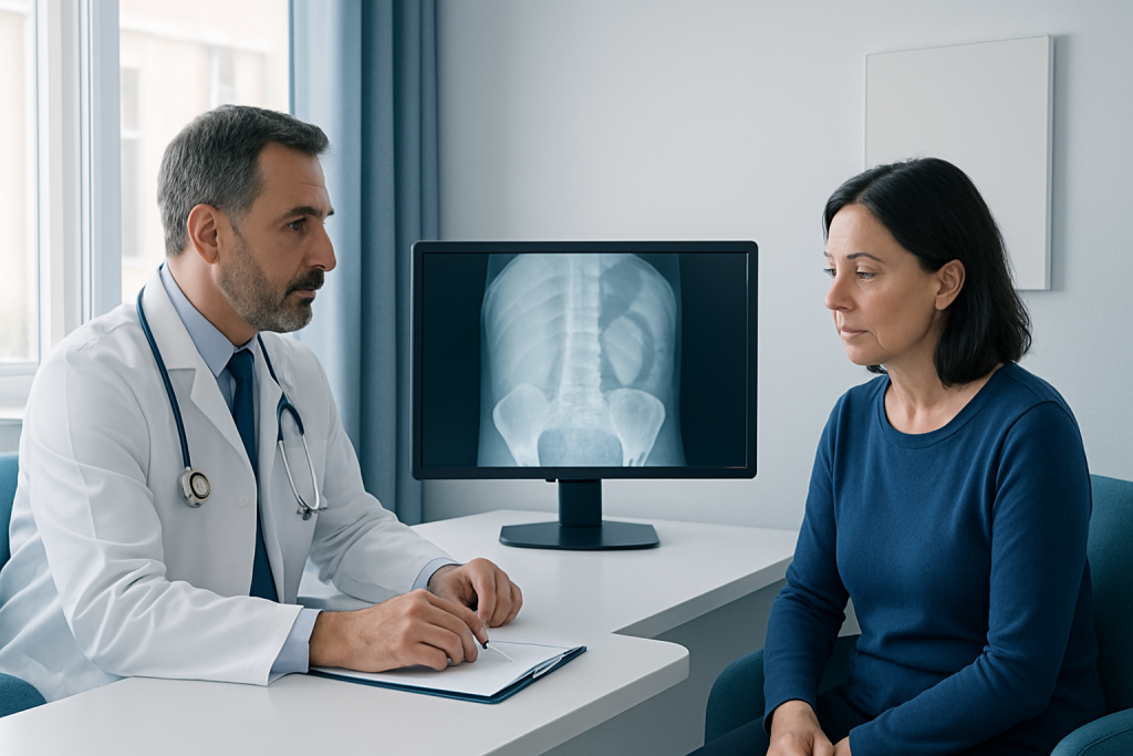
Since fluoroscopy is an imaging technique, it does not have causes in the usual sense. The reasons it is chosen are tied to the clinical problem being investigated. Doctors may prefer it when they need to observe motion, deliver contrast material, or guide instruments with high precision.
Risk factors for needing fluoroscopy vary by specialty. People with swallowing complaints, digestive symptoms, chronic joint problems, urinary tract concerns, vascular disease, or a need for minimally invasive procedures may be more likely to be referred. In some cases, prior surgery or an implanted device also makes live imaging more useful.
There are also practical considerations before the exam. Pregnancy, a history of contrast reaction, kidney disease, asthma, or certain medications may require special planning. Patients should always tell the care team about allergies, recent procedures, and the possibility of pregnancy so the study can be tailored safely.
How Fluoroscopy Is Performed
The details vary, but the process usually starts with checking the patient’s history and confirming the reason for the study. Depending on the exam, the patient may be asked to change into a gown and remove metal objects. If contrast material is needed, it may be swallowed, injected into a vein, placed in the rectum, or introduced through another route depending on the body area under study.
During the test, the fluoroscopy machine sends a controlled X-ray beam through the body and displays a moving image on a monitor. The room may have several staff members if the procedure is being used to guide treatment. The patient may be asked to hold still, change position, breathe in a certain way, or follow simple instructions to help produce clear images.
Some fluoroscopy exams are brief. Others take longer when they are part of a procedure or when the clinician needs to observe movement over time. The care team usually explains what will happen before starting, which is especially helpful for patients traveling from another country who may need to coordinate timing, interpreter support, and follow-up plans in advance.
Diagnosis and What the Images Can Show
Fluoroscopy can reveal both structure and motion. In the digestive tract, it may show how contrast moves through the esophagus, stomach, or intestines. In the joints or spine, it can show alignment, movement, and the precise placement of needles or instruments. In other settings, it can help confirm that a tube or catheter is in the correct location.
Because the images are real time, the clinician can adjust the view during the exam and sometimes reach a diagnosis immediately. In other cases, fluoroscopy is one part of a broader workup that includes ultrasound, CT, MRI, laboratory tests, or endoscopy. The results are interpreted in the context of symptoms, physical examination, and previous studies.
Patients should keep in mind that fluoroscopy does not answer every question. It is chosen because it can answer a specific one well: How does this move? Is the device positioned correctly? Does contrast pass as expected? Those focused answers often help guide the next step in care.
Treatment Options and Procedure-Guided Care
Fluoroscopy is often used to support treatment rather than act as the treatment itself. It may guide injections for pain management, placement of drains or feeding tubes, stenting of narrowed passageways, catheter-based heart or vascular procedures, and certain orthopedic or spine interventions. The value lies in precision: the doctor can see the instrument as it moves and make adjustments immediately.
When fluoroscopy is used during treatment, the care plan usually includes preparation, the procedure itself, and a short observation period afterward. Depending on the intervention, patients may receive local anesthesia, sedation, or more involved anesthesia. Recovery instructions are specific to the procedure and may include activity limits, wound care, hydration, or follow-up imaging.
For international patients, a coordinated approach is especially important. Reviewing prior scans, bringing medication lists, sharing allergy information, and organizing postoperative follow-up before travel can make the overall experience smoother and reduce unnecessary delays. Acibadem Health Point’s multidisciplinary specialists and JCI-accredited hospitals diagnose and treat this condition for international patients as part of a coordinated care pathway.
Prevention, Radiation Safety, and Self-Care
Fluoroscopy itself cannot always be avoided, because it is sometimes the best or safest way to answer a clinical question. What can be prevented is unnecessary exposure. Modern imaging teams follow the principle of using radiation only when needed and keeping it as low as reasonably possible while still obtaining clear images.
Patients can support safe care by sharing accurate medical information, especially pregnancy status, prior contrast reactions, kidney problems, and all current medicines and supplements. They should also ask whether fasting is required, whether they may drive afterward, and whether someone should accompany them home after sedation or a procedure.
- Follow all preparation instructions exactly as given
- Tell the team if there is any chance of pregnancy
- Report allergies, especially to contrast material or iodine-related products
- Drink fluids if advised after a contrast study
- Rest as instructed after procedures and watch for new symptoms
Self-care after a fluoroscopy exam is usually straightforward. If contrast was used, the care team may recommend hydration unless there is a reason not to. If a procedure was performed, patients should follow the discharge plan closely and avoid guessing about medication changes or wound care.
When to See a Doctor
A doctor’s advice is appropriate whenever symptoms suggest a problem that may benefit from live imaging, such as unexplained swallowing difficulty, persistent joint pain, digestive blockage symptoms, or concerns about a medical device. Fluoroscopy is not something patients order on their own; it is best used when a clinician has a clear question that this test can answer.
Patients should seek medical guidance promptly if they notice worsening pain, fever, breathing difficulty, blood in vomit or stool, new weakness, or a reaction after contrast exposure such as rash, swelling, or trouble breathing. These symptoms do not always mean something serious, but they do deserve timely assessment.
For people arranging care from abroad, it helps to request a written explanation of the proposed study, what it is expected to show, and what might happen next depending on the result. That kind of planning can make the journey more manageable and helps ensure the right follow-up once the patient returns home.
Living Well With an Imaging-Based Care Plan
Many patients feel relieved once they understand that fluoroscopy is a practical tool, not a diagnosis by itself. It is often used because doctors want answers that are dynamic and immediate, especially when the next step in care depends on seeing how something moves or where something is placed.
Good outcomes often come from good coordination. Bringing prior imaging, knowing the reason for the test, asking about preparation, and confirming follow-up can make the process smoother. This is true whether the patient is local or traveling internationally for evaluation or treatment.
When questions remain, a conversation with the radiologist or treating specialist can clarify what the images mean and whether additional tests are needed. Clear explanations are an important part of safe, patient-centered imaging care.
Frequently asked questions
What is fluoroscopy used for?
Fluoroscopy is used to see moving structures inside the body in real time. It helps doctors evaluate function and guide procedures such as injections, catheter placement, and contrast studies of the digestive tract.
Is fluoroscopy the same as a regular X-ray?
No. A regular X-ray provides a still image, while fluoroscopy shows a continuous moving picture. That live view is useful when motion or precise guidance matters.
Does fluoroscopy use contrast material?
It often does, but not always. Contrast may be swallowed, injected, or placed in another way to make certain structures easier to see and to show how material moves through the body.
Is fluoroscopy safe?
Fluoroscopy uses radiation, so it is ordered only when the expected medical benefit is worth it. Imaging teams work to keep exposure as low as possible and will consider special precautions when needed.
How should a patient prepare for fluoroscopy?
Preparation depends on the type of exam. A patient may need to fast, wear comfortable clothing, or avoid certain medicines only if the doctor specifically says so.
What happens after the test?
Many people can return to normal activities quickly after a simple exam. If fluoroscopy was part of a procedure or sedation was used, the care team will give specific recovery instructions and follow-up advice.
References
- RadiologyInfo.org
- American College of Radiology
- National Cancer Institute
- U.S. Food and Drug Administration
- World Health Organization
This article is for general information only and is not a substitute for professional medical advice. Please consult a qualified doctor about your individual situation.