Knee MRI or X-Ray: Which Scan Helps Most Before Orthopedic Treatment?

Key Takeaways
- Knee X-rays are best for bones, joint alignment, and arthritis-related changes.
- Knee MRI gives a detailed view of ligaments, menisci, cartilage, and other soft tissues.
- Doctors often start with an X-ray when bone injury, wear-and-tear, or alignment problems are suspected.
- MRI is usually more helpful when the knee looks stable on X-ray but pain, swelling, locking, or instability continues.
- The right scan depends on the medical question, not on which test is “better” overall.
- A doctor should guide imaging decisions, especially if symptoms are severe, persistent, or follow an injury.
Medically reviewed by the Acıbadem clinical team — June 13, 2026
A knee X-ray and a knee MRI answer different questions, so the better scan depends on the symptoms and the suspected problem. This guide explains how each test works, what it can reveal, and how doctors choose the most useful image before orthopedic treatment.
Overview
When a knee begins to hurt, swell, catch, or feel unstable, imaging can help the orthopedic team understand what is happening inside the joint. The most useful scan is not always the most detailed one. Instead, the right test is the one that best answers the doctor’s specific question.
A knee X-ray and a knee MRI are often compared, but they are designed for different jobs. X-rays are usually the first step when doctors want to look at bones, joint space, arthritis, fractures, or alignment. MRI is more detailed for soft tissues, including the meniscus, ligaments, cartilage, tendons, and bone marrow.
For patients planning treatment from another country, it helps to think of imaging as part of the treatment roadmap. A clear scan can support a more focused conversation with the orthopedic surgeon, reduce unnecessary tests, and help the care team decide whether rehabilitation, injections, arthroscopy, or another treatment is most appropriate.
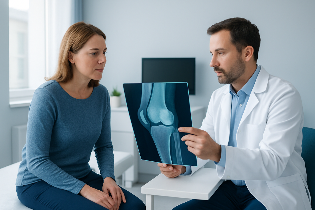
What a Knee X-Ray Can Show
A knee X-ray uses a small amount of radiation to create images of the hard structures in and around the joint. It is quick, widely available, and often the first imaging test ordered when a person has knee pain after walking, sports, a fall, or long-term strain.
X-rays are especially useful for showing fractures, dislocation, bone spurs, narrowing of the joint space, and signs of osteoarthritis. They can also help doctors assess whether the knee is aligned normally or whether one side of the joint is carrying more load than the other.
Because X-rays show bones much better than soft tissue, they are less useful for injuries involving the meniscus, ligaments, or cartilage. Still, they remain an important starting point because many knee problems are related to bone structure, arthritis, or trauma that can be seen clearly on this test.
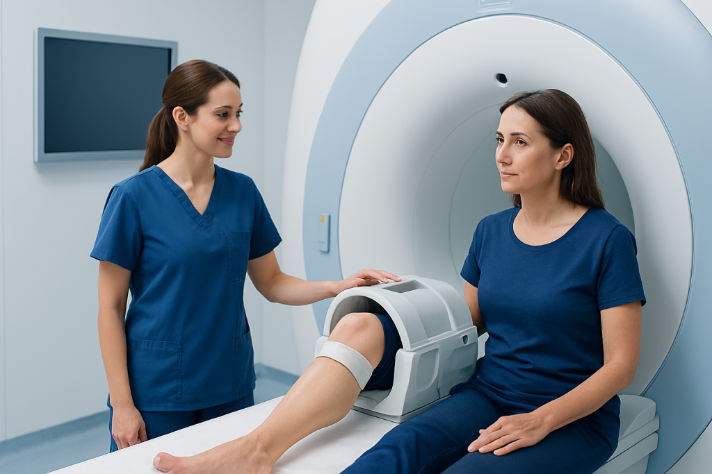
What a Knee MRI Can Show
An MRI uses magnetic fields and radio waves instead of radiation. It produces detailed cross-sectional images of the knee, making it especially valuable for structures that do not show well on X-rays.
MRI is often the best test when doctors suspect a meniscus tear, ligament injury such as an ACL problem, cartilage damage, tendon irritation, or internal swelling that is not explained by the X-ray. It can also show bone marrow edema, which may appear after an impact or stress injury even when the X-ray looks normal.
For patients with ongoing pain, locking, catching, or the feeling that the knee gives way, MRI can help define the underlying issue more precisely. That detail can be important when deciding whether physical therapy is enough or whether a surgical opinion is needed.
How Doctors Decide Between MRI and X-Ray
The choice usually starts with the story of the symptoms. A recent twist, fall, or sports injury may raise concern for ligament or meniscus damage, while long-standing stiffness and aching with movement may point more toward arthritis or alignment changes. The physical examination then helps narrow the possibilities.
If the main question involves bone injury, joint space, or degenerative change, an X-ray is often the first and most practical test. If the knee exam suggests soft-tissue injury, or if pain continues despite a normal X-ray, MRI may be the more useful next step.
Doctors also consider age, previous surgery, how the knee feels during motion, and whether the person has swelling, instability, locking, or reduced range of motion. The scan is chosen to support treatment decisions, not simply to gather more images.
- Suspected fracture or arthritis: X-ray first
- Suspected ligament, meniscus, or cartilage injury: MRI often more helpful
- Persistent symptoms with a normal X-ray: MRI may be considered
- Pre-treatment planning: imaging depends on what the surgeon needs to know
When One Scan Is Enough — and When Both Help
In many cases, one scan is enough to move forward. A clear X-ray may already show arthritis or a fracture that explains the symptoms and helps guide treatment. In other cases, MRI provides the detail needed to decide whether surgery is necessary or whether conservative care remains the better option.
Sometimes both tests are useful. A patient may first have an X-ray to evaluate the bones and then an MRI to study the soft tissues more closely. This stepwise approach is common because it balances clarity, speed, cost, and medical usefulness.
Doctors may avoid ordering MRI too early if the result is unlikely to change treatment. Likewise, they may not rely on X-ray alone when symptoms strongly suggest a soft-tissue injury. The best pathway is the one that answers the clinical question with the fewest unnecessary procedures.
Preparing for the Scan and What to Expect
Knee X-rays usually require little preparation. The person may be asked to remove metal objects from the area and position the leg in one or more ways while the images are taken. The test is typically fast and does not require recovery time.
MRI takes longer and requires the person to remain still in the scanner. Some centers may ask about implants, pacemakers, or metal fragments before scheduling the test. If the patient feels anxious in enclosed spaces, the care team can often discuss comfort measures in advance.
For international patients, it is helpful to bring previous imaging, operation notes, medication lists, and a summary of symptoms. Comparing older and current scans can give the orthopedic specialist a better sense of how the knee has changed over time and whether a new injury is involved.
Prevention and Self-Care While Waiting for Imaging
While waiting for an appointment or reviewing results, gentle self-care can help protect the knee. Resting from the activity that triggers pain, using support if advised, and following the care team’s guidance on movement can reduce irritation without causing stiffness from complete inactivity.
Ice, elevation, and controlled exercise are sometimes recommended depending on the problem. If the knee feels unstable or swollen, it is wise to avoid forceful twisting, jumping, or heavy loading until a doctor has reviewed the case.
Keeping track of symptoms can also be useful. Notes about when pain started, whether the knee locks or gives way, and what movements worsen symptoms can help the doctor decide whether a knee MRI or X-ray will be more informative.
When to See a Doctor
Medical review is important if knee pain does not improve, keeps returning, or starts limiting daily activity. A doctor should also assess the knee after a significant injury, especially if the person cannot bear weight, has major swelling, or feels instability.
Urgent assessment is appropriate if the knee is visibly deformed, very hot and swollen, or associated with fever, severe pain, or sudden inability to move the joint. These signs do not always mean something serious, but they deserve prompt attention so the right imaging and treatment can be arranged.
For patients organizing care from abroad, an orthopedic consultation can help decide whether the next best step is X-ray, MRI, or both. Acibadem Health Point’s multidisciplinary specialists and JCI-accredited hospitals diagnose and treat knee conditions for international patients, with imaging plans tailored to the treatment question at hand.
Frequently asked questions
Is a knee MRI better than an X-ray for knee pain?
Not always. An X-ray is better for bones, alignment, and arthritis, while MRI is better for soft tissues such as ligaments, menisci, and cartilage. The best scan depends on what the doctor suspects is causing the symptoms.
Why do doctors often start with an X-ray?
X-rays are quick, widely available, and very useful for seeing fractures and joint changes. They also help doctors decide whether an MRI is truly needed next.
Can an MRI show things that an X-ray cannot?
Yes. MRI can show soft-tissue injuries, cartilage damage, and internal swelling that X-rays usually miss. It can also reveal some bone problems that are not visible on an X-ray.
Do I need both scans before orthopedic treatment?
Sometimes, but not always. Many patients need only one imaging test, while others benefit from both if the doctor needs a broader view of the knee problem. The decision is usually based on symptoms and examination findings.
Is a knee MRI safe?
MRI does not use radiation, which is one reason it is often preferred for soft-tissue evaluation. However, it is not suitable for everyone, so the imaging team will review implants and other safety details first.
What should I bring to my orthopedic imaging appointment?
If possible, bring previous scans, surgery reports, and a list of current symptoms or medications. This is especially helpful for patients traveling from another country, because older records can make the new scan easier to interpret.
References
- American College of Radiology
- Radiological Society of North America
- American Academy of Orthopaedic Surgeons
- National Institute of Arthritis and Musculoskeletal and Skin Diseases
- Mayo Clinic
This article is for general information only and is not a substitute for professional medical advice. Please consult a qualified doctor about your individual situation.
More from the Health Library


Related Specialists

Assoc. Prof. Dr. Şule Göncü Ayhan
Gynecology & Obstetrics
Dr. Canan Savaş İyigün
Dermatology
Dt. Türkan Subaşıoğlu
Oral & Dental Health
Prof. Dr. Faruk Buyru
Gynecology & Obstetrics