Spine Imaging for Back Pain: When MRI Changes the Treatment Plan

Key Takeaways
- MRI is most helpful when the cause of back pain is unclear or when symptoms suggest nerve or spinal cord involvement.
- Many people with back pain improve without imaging, so an MRI is not always the first step.
- MRI can reveal disc problems, spinal stenosis, inflammation, infection, fractures, or tumors that may need different care.
- A doctor usually orders MRI when red flags, persistent symptoms, or treatment failure make a more detailed look worthwhile.
- Imaging results should be interpreted alongside symptoms and physical examination, not in isolation.
Medically reviewed by the Acıbadem clinical team — June 13, 2026
Back pain is common, but not every case needs an MRI. The scan becomes most useful when symptoms, exam findings, or recovery do not match a simple strain and the result could change the treatment plan.
Overview
Back pain can be straightforward or surprisingly stubborn. In many people, it comes from muscles, ligaments, or joints and improves with time, movement, and conservative care. In those situations, an MRI often adds little at the beginning because the treatment approach is usually the same whether the scan shows a minor disc bulge or another common age-related change.
Spine imaging becomes more valuable when the pattern changes. If pain spreads into a leg, numbness or weakness appears, walking becomes difficult, or recovery stalls despite appropriate care, MRI can help clarify whether a nerve, disc, spinal canal, or other structure is involved. That is the point where the scan may truly change the plan, not just the label.
For international patients arranging care from another country, this distinction matters. A well-timed MRI can prevent unnecessary travel for procedures that are not needed, or it can help a specialist team prepare a focused treatment plan before the patient arrives. In that sense, imaging is not simply about “seeing more”; it is about making the next step more precise.
Symptoms That Make MRI More Useful
Not every backache deserves advanced imaging, but certain symptoms make MRI more helpful. A clinician may consider it when pain travels below the knee, when there is persistent tingling or numbness, or when weakness affects lifting the foot, climbing stairs, or standing from a chair. These features can suggest irritation or compression of a spinal nerve.
Other patterns also raise the value of MRI. Pain that is severe at night, worsens steadily rather than improving, follows a significant fall or accident, or appears together with fever or unexplained weight loss deserves careful assessment. In these settings, imaging can help look for causes that are less common but more urgent to identify.
Bladder or bowel changes, numbness in the groin or inner thighs, or rapidly worsening leg weakness are especially important. These symptoms need prompt medical review, and MRI may be used quickly because the result can guide time-sensitive decisions.
- Pain radiating into the buttock, thigh, or calf
- Persistent numbness or tingling
- Noticeable weakness in a leg or foot
- Pain after trauma or a fall
- Fever, cancer history, or unexplained weight loss
- Problems with bladder, bowel, or saddle sensation
Causes & Risk Factors

Back pain has many possible causes, and MRI is most useful when the clinician needs to separate one from another. A disc herniation, spinal stenosis, vertebral fracture, infection, inflammatory disease, or tumor may all create different imaging patterns and lead to different treatment decisions. MRI is particularly good at showing soft tissues, nerves, spinal discs, and inflammation.
Risk factors help determine whether imaging is likely to be useful sooner. Older age, osteoporosis, a history of cancer, immune suppression, recent infection, significant trauma, or chronic inflammatory conditions can shift the threshold toward MRI. So can persistent symptoms that do not behave like a typical strain, especially when physical examination shows nerve-related changes.
It is also important to know what MRI is not for. A scan may show disc degeneration or small bulges in people who have no pain at all. For that reason, a clinician looks at the whole picture: the story of the pain, the examination, prior treatments, and whether the MRI findings would actually change the next step.
Diagnosis: How MRI Fits Into the Workup
The diagnostic process usually begins with a careful history and physical examination. A doctor asks when the pain started, where it is located, whether it shoots down a leg, what makes it better or worse, and whether there are numbness, weakness, fever, injury, or weight loss. Examination may include testing strength, sensation, reflexes, posture, and walking pattern.
In many cases, this first assessment is enough to start treatment without imaging. If symptoms are mild and improving, or if the problem looks like an uncomplicated mechanical back strain, immediate MRI is often unnecessary. This is because many imaging findings are common and may not explain the pain.
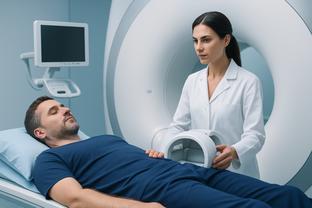
When MRI is ordered, it is usually used to answer a specific question: Is a nerve compressed? Is there narrowing in the spinal canal? Is there a fracture, infection, inflammation, or mass? The scan itself is painless, but it may feel long and noisy, so patients who are anxious or claustrophobic should mention this beforehand. For international patients, a team may also review prior scans or records before travel so the most appropriate study is performed once, rather than repeated.
Treatment Options When MRI Changes the Plan
An MRI can meaningfully change treatment when it reveals a problem that needs a different approach than simple rest or pain control. For example, a large disc herniation with nerve compression may lead to targeted physical therapy, medication changes, an injection, or, in some cases, a surgical consultation. Spinal stenosis may prompt a more specific rehabilitation plan or discussion of decompression if symptoms are limiting daily life.
Sometimes MRI helps avoid unnecessary procedures. If the scan does not show a structural reason for severe symptoms, the care team may focus more on nonsurgical management, pain education, posture, movement, and follow-up. That can spare patients from treatments that are unlikely to help.
When more serious conditions are found, the scan allows the team to act sooner. Infection, fracture, tumor, or inflammatory disease each requires its own pathway, often involving multiple specialists. For patients traveling internationally, this can be especially useful because a coordinated MRI-based assessment helps the receiving team plan the right consultations, timing, and recovery support before the visit begins.
- Physical therapy and guided exercise
- Anti-inflammatory or other pain-relief strategies, as advised by a doctor
- Targeted spinal injections in selected cases
- Activity modification and posture education
- Surgical evaluation when there is nerve compression or structural instability
Prevention & Self-care
While MRI is a diagnostic tool, many back pain episodes can be reduced or managed through everyday habits. Regular movement is usually better than prolonged bed rest. Gentle walking, stretching, and core-strengthening exercises may support spinal health when they are appropriate for the person’s condition.
Good ergonomics also matter. A supportive chair, frequent position changes, sensible lifting technique, and attention to sleep posture can reduce strain. People with osteoporosis, a prior spine injury, or inflammatory disease should follow individualized advice because their prevention needs may be different from those of someone with a simple muscle strain.
If pain has already been evaluated and imaging was not needed, self-care still has value. Keeping track of symptom changes, knowing which movements aggravate pain, and attending follow-up appointments help doctors decide whether MRI becomes useful later. For patients receiving care away from home, maintaining a written summary of symptoms, medications, and prior tests can make follow-up smoother across borders and time zones.
When to See a Doctor
Medical evaluation is appropriate when back pain lasts more than a short period, keeps returning, or interferes with work, sleep, or walking. It is also wise to seek care when pain spreads into a leg, numbness or weakness develops, or the symptoms are different from a usual strain. A clinician can decide whether an MRI is needed or whether a physical exam and conservative care are enough.
Urgent assessment is important if back pain comes with fever, significant trauma, fainting, new bladder or bowel problems, rapidly progressing weakness, or numbness in the groin area. These features do not automatically mean a serious diagnosis, but they do deserve prompt attention because they may change the timing and type of treatment.
Patients considering travel for spine care may benefit from an initial specialist review before making plans. Acibadem Health Point’s multidisciplinary specialists and JCI-accredited hospitals diagnose and treat spine conditions for international patients, helping align imaging, consultation, and follow-up in a coordinated way.
Why Imaging Results Need Context
One of the most important lessons about spine MRI is that an image alone does not equal a diagnosis. Many adults have disc degeneration, small protrusions, or age-related changes that are visible on MRI yet cause no symptoms at all. If those findings are mistaken for the true cause of pain, treatment may drift in the wrong direction.
For that reason, experienced clinicians interpret MRI together with the symptom pattern and examination findings. A scan becomes valuable when it confirms what the body is already suggesting, or when it reveals a hidden cause that explains why the pain is not behaving like a routine strain.
In practical terms, that is when MRI changes the treatment plan: it does not merely describe the spine, it helps decide what should happen next. Used well, it can support safer care, avoid unnecessary interventions, and give patients a clearer path forward.
Frequently asked questions
Do all people with back pain need an MRI?
No. Many cases of back pain improve with time and conservative care, so MRI is not always helpful at the start. A doctor usually considers it when symptoms suggest nerve involvement, when there are red flags, or when treatment is not working as expected.
What can a spine MRI show that an X-ray cannot?
MRI shows soft tissues in much greater detail, including discs, nerves, spinal cord, inflammation, and some infections or tumors. X-rays are better for bones and alignment, but they do not show nerve compression or many soft tissue problems as clearly.
Can MRI findings be present even if the pain is mild?
Yes. Some MRI findings, such as disc degeneration or small bulges, are common and may not cause symptoms. That is why the scan must be interpreted alongside the person’s history and examination.
Is MRI always the next step after physical therapy or medication?
Not necessarily. If symptoms are improving, a doctor may continue conservative treatment without imaging. MRI becomes more useful when the pain persists, worsens, or changes in a way that suggests a different cause.
What should a patient tell the doctor before the scan?
It helps to mention any metal implants, pacemakers, pregnancy, kidney problems if contrast might be used, and any history of claustrophobia. Sharing prior test results and a clear description of symptoms can also help the team choose the right scan and interpret it correctly.
How should someone prepare if they are traveling abroad for spine care?
Bringing prior reports, imaging disks or files, medication lists, and a summary of symptoms can make the consultation more efficient. If an MRI is likely to affect treatment, reviewing records before travel may help the receiving team plan the right appointment and avoid repeat testing.
References
- American College of Radiology
- National Institute of Neurological Disorders and Stroke
- American Academy of Orthopaedic Surgeons
- NHS
- Mayo Clinic
This article is for general information only and is not a substitute for professional medical advice. Please consult a qualified doctor about your individual situation.