Mri Advantages Over Ct

Key Takeaways
- MRI uses magnetic fields and radio waves, while CT uses X-rays.
- MRI is often better for soft tissues such as the brain, spinal cord, ligaments, and pelvic organs.
- CT is usually faster and may be preferred in emergencies, trauma, or when looking for bleeding or fractures.
- MRI does not use ionizing radiation, which can be an important advantage for some patients.
- The best scan depends on the body part, the clinical question, and the patient’s medical history.
MRI and CT are both valuable imaging tests, but they do not answer the same questions in the same way. MRI often offers superior soft-tissue detail and avoids ionizing radiation, which can make it the preferred choice for many brain, spine, joint, and pelvic evaluations.
Overview
When patients hear that a doctor has ordered imaging, the next question is often simple: why this scan and not another one? For many situations, the choice comes down to what information the doctor needs most. MRI and CT can both produce detailed pictures inside the body, but they are built on very different technologies and each excels in different settings.
MRI, or magnetic resonance imaging, uses a strong magnetic field and radio waves to create images. CT, or computed tomography, uses X-rays to build cross-sectional pictures quickly. The main advantage of MRI over CT is its ability to show soft tissues with exceptional contrast, often making it more informative for nerves, muscles, ligaments, organs, and many structures inside the brain and spine.
For international patients deciding where to have a scan, the practical side also matters. A good imaging plan should be efficient, clearly explained, and matched to the body area being studied so that travel, scheduling, and follow-up are as smooth as possible. The “best” test is the one that answers the clinical question with the least burden to the patient.
Why MRI Often Shows More Detail Than CT
MRI is especially strong at distinguishing between different soft tissues. This makes it useful when doctors need to examine subtle changes in the brain, spinal cord, discs, ligaments, tendons, or internal organs. In many cases, MRI can reveal inflammation, edema, scar tissue, or small lesions that may be harder to appreciate on CT.
Another advantage is that MRI can image in multiple planes without moving the patient much, which helps doctors evaluate complex anatomy. In neurologic and musculoskeletal care, that clarity may improve diagnostic confidence and help guide treatment decisions.
CT remains excellent for other questions, especially bone detail, acute bleeding, or chest and abdominal emergencies. Still, when the issue is soft tissue definition rather than speed, MRI is often the more informative test.
- Brain and spinal cord evaluation
- Joint, tendon, and ligament assessment
- Pelvic organ imaging
- Liver and some abdominal questions
- Certain tumors and inflammatory conditions
Advantages of MRI Over CT

The most widely recognized advantage of MRI over CT is the absence of ionizing radiation. This can be important for people who may need repeated imaging over time, and it is one reason MRI is often preferred when appropriate for younger patients or for conditions requiring follow-up scans.
MRI also tends to provide better soft-tissue contrast. In practical terms, that means doctors may be able to tell the difference between tissues that look similar on CT. This matters in problems involving the brain, spine, joints, pelvis, and many cancers, where small differences in tissue characteristics can change diagnosis or treatment planning.
There are also situations where MRI can add functional information, not just anatomy. Specialized MRI techniques may help doctors evaluate blood flow, tissue activity, or the extent of disease in ways that a standard CT cannot fully match. That said, the best test is still the one that fits the patient’s specific question, not the one with the longest feature list.
When CT May Still Be the Better Choice
CT is often faster and easier to perform in urgent situations. In emergency care, speed matters, especially after trauma, when doctors may need to look for bleeding, fractures, or organ injury right away. CT is also widely used for lungs, many abdominal concerns, and situations where MRI would take too long or be harder to tolerate.
Some patients cannot have MRI because of certain implanted devices, severe claustrophobia, or other safety concerns. CT may be the more practical option in those cases. It can also be less sensitive to motion, which may help when patients have pain, difficulty staying still, or trouble holding their breath.
The important point is not that one scan is universally better. Rather, MRI and CT are complementary tools. A good clinical team chooses the one that is most likely to answer the question safely and efficiently.
How Doctors Decide Which Scan to Order
Doctors usually choose between MRI and CT based on the suspected condition, the body part involved, the urgency of the problem, and whether the patient has any factors that affect scan safety. For example, a neurologic symptom, a sports injury, or a pelvic mass may point toward MRI, while a sudden chest pain workup or suspected fracture may lead to CT.
Before imaging is scheduled, the care team may ask about past surgeries, implants, pregnancy, kidney function, allergies to contrast agents, and whether the patient has had previous scans. These details help avoid delays and prevent unnecessary repeat imaging, which is especially helpful for patients traveling from another country and trying to complete care within a limited window.
For many people, the decision is made quickly, but it should still feel thoughtful. Patients are entitled to ask why a particular scan was chosen and what result the doctor expects to learn from it.
What to Expect During an MRI
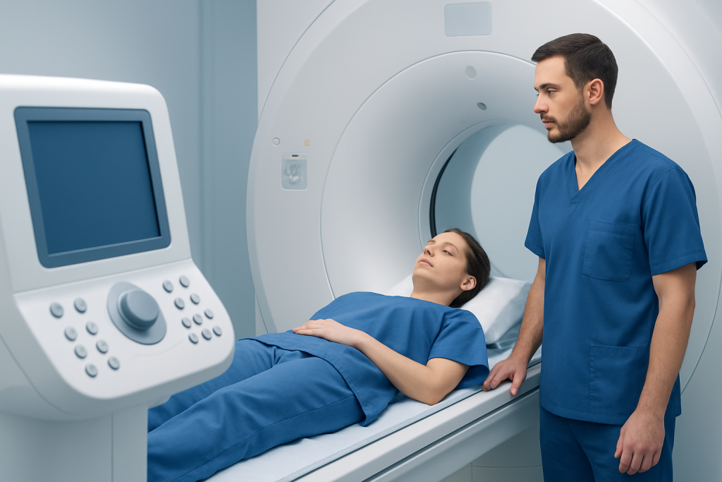
An MRI appointment is usually more involved than a CT scan, but it is still straightforward. The patient removes metal objects, changes into a gown if needed, and lies on a table that moves into the scanner. The machine makes loud tapping or humming sounds, and ear protection is commonly provided.
Staying still is important because movement can blur the images. Depending on the exam, the scan may take longer than CT, and in some cases contrast material may be used to improve the visibility of certain tissues or blood vessels. Patients should always tell the team if they have a pacemaker, metal fragments, or any device that could affect MRI safety.
Most patients complete the scan without difficulty. If anxiety is a concern, the imaging team may discuss comfort measures in advance so the experience feels manageable and predictable.
Prevention, Preparation, and Self-care
Imaging itself is not something patients “prevent,” but thoughtful preparation can reduce stress and help the exam go smoothly. Before the appointment, patients should follow the facility’s instructions about eating, drinking, medications, and contrast use. Bringing previous reports or images can also help the radiology team compare findings accurately.
Patients who are nervous about enclosed spaces can mention this early, rather than waiting until the day of the scan. Simple planning—such as arranging transportation, confirming the schedule, and allowing extra time for registration—can make a big difference, especially when coordinating care while abroad.
After the scan, most people can return to normal activities right away unless their doctor gives different instructions. If contrast was used, the team may advise additional hydration or specific follow-up depending on the situation. The most helpful self-care is often practical: keeping copies of reports, asking when results will be available, and knowing who will review them next.
- Bring all prior imaging and medical records if available.
- Tell the staff about implants, allergies, pregnancy, or kidney problems.
- Follow instructions about food, drink, and medications.
- Ask how results will be shared and who will explain them.
When to See a Doctor
Patients should speak with a doctor if they have symptoms that may need imaging, such as persistent headaches, neurologic changes, joint swelling, back pain with nerve symptoms, unexplained pelvic pain, or an abnormal finding on another test. Imaging should be interpreted in the context of the full clinical picture, not as a stand-alone answer.
If a patient has questions about whether MRI or CT is more appropriate, that discussion belongs with the referring doctor or radiologist. This is especially important for people with implanted devices, reduced kidney function, a history of contrast reactions, or a need for repeated follow-up scans.
For international patients, a coordinated second opinion or imaging plan can save time and reduce uncertainty. Acibadem Health Point’s multidisciplinary specialists and JCI-accredited hospitals diagnose and treat a wide range of conditions for international patients, helping ensure that the scan choice fits both the medical need and the travel plan.
Frequently asked questions
Is MRI always better than CT?
No. MRI is often better for soft tissues, but CT can be faster and more useful in emergencies, trauma, or when looking closely at bone and bleeding. The right test depends on the clinical question.
Why does MRI avoid radiation while CT does not?
MRI uses magnetic fields and radio waves, not X-rays, so it does not involve ionizing radiation. CT uses X-rays to create images, which is why it does involve radiation exposure.
Can MRI replace CT for all body parts?
Not usually. Each test has strengths, and some conditions are evaluated better with CT, some with MRI, and some with both. Doctors choose based on what needs to be seen most clearly.
Why might a doctor order MRI after a CT?
A CT may identify a problem but not fully define it. MRI can then provide more detail about soft tissue, inflammation, nerve involvement, or the extent of a lesion.
Is MRI safe for everyone?
MRI is safe for many people, but it is not appropriate for every patient. Certain implants, devices, or metal fragments may require special screening or a different imaging test.
How long does an MRI take compared with a CT scan?
MRI usually takes longer than CT because it gathers more detailed data. CT is often completed more quickly, which is one reason it is commonly used in urgent settings.
References
- Radiological Society of North America
- American College of Radiology
- National Institute of Biomedical Imaging and Bioengineering
- Mayo Clinic
- World Health Organization
This article is for general information only and is not a substitute for professional medical advice. Please consult a qualified doctor about your individual situation.