Circle Of Willis

Key Takeaways
- The Circle of Willis is a vital arterial network that supports blood supply to the brain.
- Some people have natural variations in this structure, and many never notice them.
- Problems such as aneurysms or blocked arteries may affect brain circulation and increase stroke risk.
- Doctors may evaluate it with MRI, CT angiography, or catheter-based angiography.
- Sudden neurological symptoms always deserve urgent medical attention.
The Circle of Willis is a ring of arteries at the base of the brain that helps maintain blood flow when one vessel narrows or is blocked. Understanding its role can make stroke symptoms, aneurysm concerns, and imaging findings easier to discuss with a doctor.
Overview
The Circle of Willis is a ring-shaped connection of arteries located at the base of the brain. Its main role is not to create blood, but to help redistribute it. When one vessel has reduced flow, the circle can sometimes provide a detour through other arteries so the brain still receives oxygen-rich blood.
This arrangement is one reason brain circulation is often described as “collateral.” In practical terms, it acts like a traffic junction with alternate routes. That does not mean it can always fully compensate for a blockage, but it can reduce the impact of some vascular changes.
For international patients who receive a brain scan far from home, the Circle of Willis may appear in the report even when no problem is found. Learning what it is can make the conversation with a neurologist or neurosurgeon less intimidating and more useful.
What the Circle of Willis includes
The Circle of Willis is formed by several arteries that meet and connect near the underside of the brain. These include the internal carotid arteries, the anterior cerebral arteries, the anterior communicating artery, the posterior cerebral arteries, and the posterior communicating arteries. Together, they create a loop that can share blood flow between the front and back parts of the brain.
The circle is often shown as perfectly symmetrical in diagrams, but real anatomy is rarely that neat. Many people have small differences in vessel size or shape. These variations are common and do not automatically mean disease.
Because the circle sits deep inside the skull, it is usually seen on imaging rather than on physical examination. It is discussed when doctors are assessing stroke risk, aneurysm risk, headaches with concerning features, or the blood supply before certain brain procedures.
Symptoms and why the Circle of Willis matters

The Circle of Willis itself does not usually cause symptoms. Instead, symptoms arise when blood flow through the brain is interrupted or when a vessel in this region becomes weakened or enlarged. In those situations, people may notice sudden weakness on one side of the body, facial drooping, speech difficulty, vision changes, severe dizziness, or a sudden, intense headache.
Some people learn about the circle after imaging for a stroke, transient ischemic attack, aneurysm, or unexplained headaches. Others discover an anatomical variation incidentally during tests done for a different reason. An incidental finding can still be important, because it may influence how a doctor interprets future symptoms or plans treatment.
A useful way to think about this structure is that it matters most when something else goes wrong. If a vessel is narrowed, blocked, or abnormal, the Circle of Willis may either help preserve flow or reveal that the brain has less backup circulation than expected.
Causes and risk factors
There is no single “cause” of the Circle of Willis, because it is a normal anatomical structure present from birth. What doctors do pay attention to are conditions that affect the vessels within it. Atherosclerosis, high blood pressure, diabetes, smoking, high cholesterol, and certain inherited conditions can all damage blood vessels over time.
Some people are born with incomplete or smaller connections in the circle. These variations are usually harmless on their own, but they may reduce the brain’s ability to reroute blood during a blockage. In a person with other vascular risks, that can become more relevant.
Risk factors for aneurysm formation in this region can include family history, smoking, uncontrolled hypertension, and some connective tissue disorders. Aneurysms may remain stable for years, yet doctors monitor them carefully because changes in size or shape can affect treatment decisions.
Diagnosis and imaging
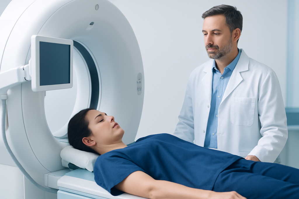
Doctors do not diagnose the Circle of Willis with a physical exam alone. They typically use imaging to look at the brain’s arteries. Common tests include MRI and MRA, CT angiography, and in some cases catheter angiography, which gives a very detailed view of the vessels.
The choice of test depends on the question being asked. For example, a clinician may want to assess blood flow after stroke-like symptoms, map an aneurysm before treatment, or understand whether a patient has enough collateral circulation before a procedure. Each test has its own strengths, limitations, and preparation steps.
For patients traveling for care, it helps to bring previous scans and reports on a CD, USB drive, or secure digital link if available. Comparing older and newer images can show whether a finding is stable, newly changed, or more important than it first appeared.
Treatment options
Treatment depends on what, if anything, is wrong with the arteries connected to the Circle of Willis. If the issue is a blocked vessel, doctors may focus on preventing another stroke with medications and risk-factor control, and sometimes procedures are considered in selected cases. If an aneurysm is found, the team may recommend observation, endovascular treatment, or surgery depending on its size, shape, location, and the person’s overall health.
When a patient has had a stroke or transient ischemic attack, the treatment plan often combines medication, rehabilitation, and vascular risk reduction. That may include managing blood pressure, controlling diabetes, stopping smoking, improving cholesterol, and addressing sleep or heart rhythm problems that can influence circulation.
People are sometimes relieved to hear that not every Circle of Willis variation needs treatment. In many cases, the finding simply informs medical decision-making and helps doctors plan safely. The right approach is usually individualized rather than automatic.
Prevention and self-care
Healthy brain circulation starts with everyday vascular care. Keeping blood pressure under control, avoiding tobacco, staying physically active as advised, and following a balanced eating pattern can all support the arteries that feed the brain. These steps are especially valuable for people with diabetes, high cholesterol, or a family history of stroke.
Self-care also includes paying attention to warning signs. Sudden weakness, trouble speaking, abrupt vision changes, or the “worst headache” of a person’s life should not be monitored at home. Prompt medical assessment is the safer choice, even if symptoms improve quickly.
For patients recovering away from home, follow-up planning matters. That may mean arranging local lab work, confirming medication names in advance, and knowing which symptoms should trigger an emergency visit. Clear discharge instructions and a copy of the imaging report can make the transition much smoother.
When to see a doctor
A person should seek medical care if they have symptoms that suggest a stroke, aneurysm, or other brain circulation problem. Emergency evaluation is important for sudden numbness or weakness, confusion, facial droop, speech difficulty, loss of balance, sudden severe headache, fainting, or a new seizure.
Non-urgent but important evaluation is reasonable if a scan shows an abnormal Circle of Willis, an aneurysm, or reduced blood flow and the person is unsure what it means. A neurologist or neurosurgeon can explain whether the finding is incidental, whether follow-up imaging is needed, and how it fits with the person’s overall risk profile.
Acibadem Health Point’s multidisciplinary specialists and JCI-accredited hospitals diagnose and treat conditions related to the Circle of Willis for international patients, helping coordinate imaging, consultation, and follow-up in one care pathway.
Living with the finding and planning follow-up
Many people feel more at ease once they understand that a Circle of Willis finding is often a map of blood flow rather than a diagnosis by itself. The key question is usually not “Does it exist?” but “Is it functioning well, and is any part of it at risk?” That perspective helps patients and families interpret scan results more calmly.
Follow-up may involve repeat imaging, especially if an aneurysm or other vascular concern is being watched over time. If treatment is not needed, the focus may shift to reducing vascular risk and returning to normal life with clear guidance on what symptoms should prompt reassessment.
For international patients, continuity is especially important. A well-organized plan should include the imaging type used, the reason for the finding, any medication changes, and the date for the next review. That makes it easier to continue care confidently after returning home.
Frequently asked questions
What is the main purpose of the Circle of Willis?
Its main purpose is to help maintain blood flow to the brain by connecting major arteries. If one route becomes narrowed or blocked, blood may sometimes be redirected through another pathway. It is a backup system, not a guarantee of full protection.
Can a person have a normal life with a Circle of Willis variation?
Yes. Many people have natural variations in this arterial ring and never have symptoms. Doctors usually pay attention to the finding only if it affects stroke risk, aneurysm risk, or treatment planning.
Does the Circle of Willis cause headaches?
Not by itself. Headaches are more often related to migraine, tension, infection, blood pressure issues, or other causes. A sudden, severe, or unusual headache should be assessed promptly because it can sometimes signal a vascular problem.
How do doctors check the Circle of Willis?
They usually use imaging such as MRI/MRA, CT angiography, or catheter angiography. The best test depends on what the doctor needs to see and how urgently the information is needed. A specialist can explain the result in the context of the whole brain circulation.
Is an aneurysm in the Circle of Willis dangerous?
It can be important, but the level of concern depends on its size, shape, location, and whether it has changed over time. Some aneurysms are monitored, while others are treated. A specialist should review the imaging and discuss the safest option.
When should emergency help be sought?
Emergency care is needed for sudden weakness, face drooping, trouble speaking, sudden vision loss, severe imbalance, seizure, fainting, or a very sudden severe headache. These symptoms can indicate a stroke or another urgent brain blood vessel problem. Quick assessment is the safest approach.
References
- National Institute of Neurological Disorders and Stroke
- American Heart Association
- Mayo Clinic
- Johns Hopkins Medicine
- World Health Organization
This article is for general information only and is not a substitute for professional medical advice. Please consult a qualified doctor about your individual situation.