

Osteochondroma

Key Takeaways
- Osteochondroma is a noncancerous bone growth that often appears near the knee, shoulder, or other long bones.
- It may be found by chance or noticed as a painless lump that becomes bothersome with growth or pressure.
- Imaging studies such as X-rays usually help confirm the diagnosis, and other tests may be used if the appearance is unclear.
- Treatment is often observation, while surgery may be recommended if the growth causes pain, nerve pressure, limited motion, or cosmetic concerns.
- Changes after skeletal maturity deserve prompt medical review because they can signal a need for closer assessment.
Osteochondroma is a benign bone growth that usually develops near the ends of long bones during childhood or adolescence. Many people never need treatment, but evaluation is important if the lump grows, causes pain, or affects movement.
Overview
Osteochondroma is the most common benign bone tumor. It grows from the surface of a bone and is made of both bone and cartilage, which is why doctors sometimes describe it as a cartilage-capped bony outgrowth. It typically develops during periods of bone growth, often in childhood or the teen years, and may stop enlarging once growth is complete.
These growths are often found near the ends of long bones, especially around the knee, shoulder, or hip region. Some people discover an osteochondroma because they feel a hard, painless bump. Others learn about it only after an X-ray is taken for an unrelated reason. For international patients weighing where to seek care, the main question is usually not urgency but whether the mass is truly benign and whether it is affecting nearby structures.
Most osteochondromas do not lead to serious problems. Still, they deserve proper evaluation because their location can matter. A growth near a tendon, nerve, blood vessel, or joint can cause discomfort, stiffness, or mechanical irritation even when the tumor itself is noncancerous.
Symptoms
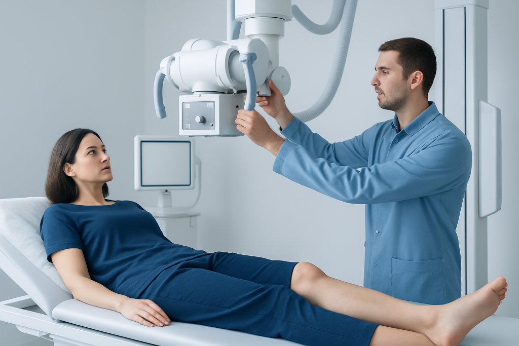
An osteochondroma may produce no symptoms at all. When symptoms do appear, they often begin gradually and relate to pressure on nearby tissue rather than the tumor being painful on its own. A firm lump under the skin is one of the most common findings, and it may be noticed incidentally by the person, a parent, or a clinician during a routine exam.
Possible symptoms can include aching after activity, a sense of tightness around a joint, reduced range of motion, or irritation from clothing or movement. If the growth presses on a nerve or blood vessel, a person may notice numbness, tingling, weakness, swelling, or changes in circulation in the nearby limb. In children and adolescents, a growth that changes the way a limb moves may be more noticeable during sports or physical play.
- Painless, hard lump near a joint or bone
- Local tenderness or soreness with activity
- Stiffness or restricted movement
- Numbness, tingling, or weakness if nearby nerves are affected
- Swelling or pressure symptoms if surrounding structures are compressed
Because many osteochondromas are slow-growing and harmless, the pattern of symptoms matters more than the presence of a lump alone. A change in pain, size, or function is usually what prompts further assessment.
Causes & Risk Factors
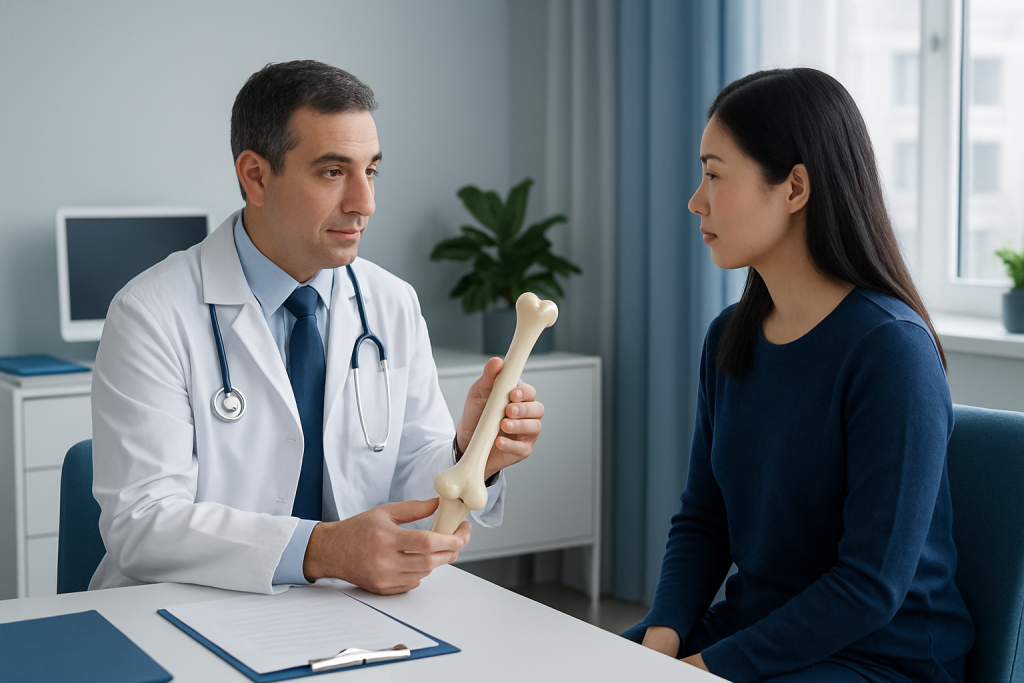
Osteochondroma develops when a portion of the growth plate grows outward instead of forming normal bone in the expected direction. In most people, the exact reason this happens is not fully understood. The lesion may occur as a single growth, or less commonly as part of a condition called multiple hereditary exostoses, which is an inherited disorder that causes several osteochondromas.
Age is one of the main factors, since these growths usually begin while bones are still developing. They are commonly identified in children, teenagers, and young adults, then become stable once skeletal growth is finished. A family history of multiple osteochondromas can be important, especially when several bumps are present or when more than one family member has similar findings.
Risk is also influenced by location. Bones that grow rapidly and have active growth plates are more likely to be involved. Although the condition is benign, doctors pay attention to whether an osteochondroma changes after growth has stopped, because a new increase in size later in life needs careful review.
Diagnosis
Diagnosis usually begins with a medical history and physical examination. A clinician will ask when the lump was first noticed, whether it has changed, and whether it is causing pain, stiffness, or nerve-related symptoms. The location, firmness, and mobility of the mass help guide the next steps.
X-rays are often the first and most useful test. They can show the characteristic bony outgrowth and whether it appears connected to the underlying bone. If the growth is close to joints or soft tissues, or if the appearance is not typical, additional imaging such as MRI or CT may be recommended to evaluate the cartilage cap and the relationship to nearby muscles, vessels, or nerves. These details matter when planning treatment, particularly for patients traveling from abroad who may need a clear answer before deciding whether to remain for surgery or return home for follow-up.
Doctors may consider other conditions when the image is not classic, including other bone tumors or surface lesions. In selected cases, especially when there are concerning changes, tissue sampling or removal with laboratory analysis may be needed. The goal is not to alarm the patient, but to confirm exactly what the growth is and whether observation is enough.
Treatment Options
Treatment depends on symptoms, size, location, and whether the lesion is stable. Many osteochondromas are simply observed over time, especially when they are not painful and are not affecting movement. Follow-up visits and repeat imaging may be used to make sure the growth remains unchanged after skeletal maturity.
When symptoms are present, surgery may be recommended to remove the osteochondroma and, when appropriate, its cartilage cap and attachment to the bone. This is more likely when the growth causes persistent pain, limits motion, compresses a nerve or blood vessel, creates repeated irritation, or is cosmetically bothersome. Surgery may also be considered if the diagnosis is uncertain or if the lesion shows changes that need closer evaluation.
For international patients, treatment planning often includes practical decisions as well as medical ones: whether imaging can be completed in one center, whether surgery can be done with coordinated rehabilitation, and how follow-up will be arranged after returning home. A multidisciplinary orthopedic team can help match the plan to the person’s symptoms, age, and travel needs.
After surgery, recovery usually involves wound care, gradual return to activity, and monitoring for improvement in pain or function. The exact recovery course depends on where the lesion was removed and how close it was to nearby structures.
Prevention & Self-care
There is no proven way to prevent osteochondroma, especially when it develops because of normal bone growth patterns or inherited factors. What can help is early recognition and sensible follow-up, particularly in children and adolescents who have a known bony lump or a family history of multiple lesions.
Self-care is mostly about protecting the area and noticing changes. Avoid repeated pressure or friction over the lump if it is already irritated. Keep track of whether pain, size, or movement changes over time, since those details can be helpful at a medical appointment. If a doctor recommends monitoring, keeping the suggested follow-up schedule is a practical way to catch any change early.
- Watch for new pain or faster growth after bone maturity
- Note numbness, tingling, weakness, or swelling
- Use activity adjustments if a joint feels irritated
- Attend planned follow-up imaging or orthopedic visits
- Seek care before long travel if symptoms are changing
People returning home after evaluation or surgery should ask for a clear summary of findings, imaging reports, and recovery instructions. That documentation makes it easier for a local clinician to continue care smoothly.
When to See a Doctor
A medical evaluation is wise whenever a new lump is noticed near a bone, even if it is painless. Osteochondromas are usually benign, but only a clinician can confirm the diagnosis and decide whether observation is appropriate. This is especially important if the person is still growing, since the lesion may be changing along with the skeleton.
Prompt assessment is recommended if the lump becomes painful, grows after skeletal maturity, limits movement, or causes numbness, tingling, weakness, or swelling. These signs do not automatically mean something serious, but they do mean the growth is affecting nearby tissue and should be checked. Sudden or unexplained changes are worth attention even if the lesion has been present for years.
Anyone considering care abroad should try to obtain an orthopedic review before or soon after travel if the lump is new, enlarging, or symptomatic. At Acibadem Health Point, multidisciplinary specialists and JCI-accredited hospitals diagnose and treat osteochondroma for international patients, helping coordinate imaging, treatment, and follow-up in a structured way.
Living With an Osteochondroma
For many people, living with an osteochondroma means little more than periodic observation. The experience becomes more important when the growth is in a spot that rubs against shoes, restricts a shoulder, or sits near a nerve. In those situations, the focus is usually on maintaining comfort and preserving normal function rather than chasing every visible change on the skin or X-ray.
Children and young adults may need reassurance that a benign bone growth is not the same as cancer. Clear communication helps families understand why doctors may suggest monitoring instead of immediate removal. If surgery is chosen, the decision is usually based on symptoms or anatomy, not because the condition is dangerous by default.
Patients who split care between countries often do best when one clinician leads the plan and shares records with the next team. That continuity makes it easier to interpret follow-up imaging, compare measurements accurately, and avoid unnecessary repeat testing.
With the right assessment and a reasonable follow-up plan, most people with osteochondroma can continue daily activities with confidence.
Frequently asked questions
Is osteochondroma cancerous?
Osteochondroma is usually benign, meaning it is not cancer. Doctors still evaluate it carefully because a change in pain, growth, or appearance may need further testing. A small number of cases can require closer review, especially if symptoms develop after growth has finished.
Does every osteochondroma need surgery?
No, many do not. If the growth is painless and not interfering with nearby structures, observation is often enough. Surgery is generally reserved for pain, pressure on nerves or vessels, reduced movement, or uncertainty about the diagnosis.
How is osteochondroma usually found?
It is often found on an X-ray taken for another reason or after a person notices a hard lump near a joint. A doctor may then order additional imaging if the lesion needs to be characterized more clearly. The pattern on imaging often helps confirm the diagnosis.
Can osteochondroma come back after removal?
Recurrence is uncommon when the entire lesion is removed, including the cartilage-capped portion. The chance depends on how completely the growth was excised and where it was located. Follow-up visits help ensure healing is progressing normally.
What symptoms suggest the lump should be checked sooner?
New or worsening pain, rapid growth, numbness, tingling, weakness, swelling, or reduced motion should be assessed promptly. These symptoms can mean the lesion is irritating nearby tissue or that further imaging is needed. It is best not to wait if the change is recent.
Can a person travel for osteochondroma treatment?
Yes, many people do, especially when they want specialist imaging or surgery. It helps to have the diagnosis clearly documented, including scans and reports, so care can continue smoothly after returning home. Planning follow-up before traveling is especially useful if surgery has been performed.
References
- Merck Manual Professional Edition
- American Academy of Orthopaedic Surgeons
- Mayo Clinic
- National Organization for Rare Disorders
- Orphanet
This article is for general information only and is not a substitute for professional medical advice. Please consult a qualified doctor about your individual situation.
More from the Health Library



Related Specialists

Prof. Dr. Ahmet Alanay
Orthopedic Surgery & Traumatology
Dr. Sinan Aksu
Orthopedic Surgery & Traumatology
Dr. Sirri Baştürk
Orthopedic Surgery & Traumatology
Prof. Dr. Korhan Özkan
Orthopedic Surgery & Traumatology