Hydrops Fetalis

Key Takeaways
- Hydrops fetalis is not a single disease; it is a sign of an underlying problem in pregnancy or the fetus.
- It can be linked to blood disorders, heart problems, infections, genetic conditions, or complications in the placenta.
- Ultrasound and other prenatal tests help doctors look for the cause and assess the baby’s condition.
- Treatment focuses on the underlying cause and may include fetal procedures, medication, early delivery, or newborn intensive care.
- Families often need ongoing counseling and follow-up because planning before and after birth can affect outcomes.
Hydrops fetalis is a serious prenatal condition in which fluid builds up in a baby’s body before birth. Early testing and coordinated maternal-fetal care are important because treatment depends on the underlying cause and the baby’s condition.
Overview
Hydrops fetalis describes abnormal fluid buildup in a fetus or newborn. Rather than being one diagnosis, it is a warning sign that something is disrupting the baby’s ability to manage fluid, oxygen, blood flow, or organ function before birth.
Doctors usually divide it into two broad types: immune hydrops, which is related to blood group incompatibility, and non-immune hydrops, which is caused by other conditions such as heart disease, genetic disorders, severe anemia, infections, or lymphatic problems. Today, most cases are non-immune, so the search for the cause is a major part of care.
For families, the diagnosis can arrive during a routine ultrasound or after a referral for more specialized imaging. Because the condition can change quickly, care is often coordinated by maternal-fetal medicine specialists, neonatologists, genetic counselors, and other experts working together.
Symptoms and What Doctors May See
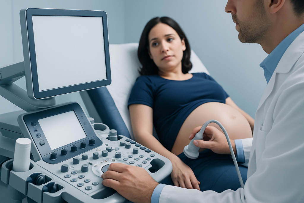
Hydrops fetalis is usually detected on prenatal ultrasound rather than through symptoms the pregnant person feels. On imaging, doctors may see fluid in two or more body spaces, such as around the lungs, around the heart, in the abdomen, or under the skin. The placenta may also look enlarged, and the baby may appear swollen.
Before birth, some pregnancies also show signs that help explain how serious the problem is, such as excess amniotic fluid, reduced fetal movement, or signs that the baby’s heart is under strain. These findings do not point to one specific cause, but they guide the next steps in evaluation.
After birth, a newborn with hydrops may have obvious swelling, breathing difficulty, a pale or weak appearance, or trouble maintaining circulation. Because these babies may need immediate support, delivery is usually planned in a center with newborn intensive care available.
Causes and Risk Factors

Many different conditions can lead to hydrops fetalis, and the cause is sometimes more than one problem at a time. A baby may develop fluid buildup if the heart is unable to pump effectively, if severe anemia reduces oxygen delivery, or if an infection changes how organs and blood vessels function.
Common causes and contributors include:
- Blood group incompatibility between mother and baby, especially in immune hydrops
- Fetal heart defects or rhythm problems
- Severe anemia from inherited blood disorders or blood loss
- Genetic or chromosomal conditions
- Congenital infections, such as parvovirus B19, cytomegalovirus, or syphilis
- Problems with the placenta, twin pregnancy complications, or lymphatic abnormalities
Risk is higher when there is a known family history of genetic disease, a previously affected pregnancy, untreated maternal infection, or a pregnancy with a twin-to-twin complication. In many cases, however, no single clear risk factor is identified until testing is completed.
Diagnosis
Diagnosis begins with ultrasound, which can show the fluid pattern and suggest whether the condition is mild, moderate, or severe. From there, doctors work backward to understand why it developed, because the treatment plan depends on the cause rather than the swelling alone.
Evaluation may include fetal echocardiography to look closely at the heart, detailed anatomy ultrasound, maternal blood tests, testing for infections, and genetic testing such as amniocentesis or other chromosome analysis when appropriate. Doppler studies may also be used to assess blood flow and signs of fetal anemia.
For Myomectomy Recovery for International Patients: The Usual Travel Timeline" class="ahp-ilk">international patients, this stage often involves several coordinated appointments in a short period. That may include imaging, lab work, counseling, and a discussion of whether treatment should happen during pregnancy, at delivery, or after birth. Clear planning matters because some causes can be treated before delivery, while others are best managed in a neonatal intensive care unit.
Treatment Options
Treatment is guided by the underlying cause, the gestational age, and how stable the fetus or newborn is. In some cases, doctors may treat fetal anemia with an intrauterine blood transfusion, while certain rhythm problems may be managed with medication given to the mother under specialist supervision.
If fluid is placing pressure on the baby’s organs, procedures such as drainage may be considered in selected cases, though this depends on the situation and whether the benefit outweighs the risks. When the condition is severe or worsening, early delivery may be recommended if the baby is far enough along to receive effective newborn support.
After birth, treatment may include breathing support, careful fluid management, blood transfusions, treatment for infection, and intensive monitoring. Babies with hydrops often need a neonatal intensive care team that can respond quickly to changes in oxygen, circulation, and organ function.
Because the condition is medically complex, decisions are usually individualized. Families benefit from a team discussion that includes maternal-fetal medicine, neonatology, genetics, cardiology, and other specialties as needed, so that care is aligned with both medical needs and the family’s travel and follow-up plans.
Prevention and Self-care
Not every case of hydrops fetalis can be prevented, especially when the cause is genetic or unrelated to a known risk. Still, some steps may lower risk or improve the chance of early detection. Routine prenatal care gives doctors the best opportunity to notice changes before they become emergencies.
Pregnant people are usually advised to attend all scheduled scans and blood tests, report any reduced fetal movement, and share any relevant history such as previous affected pregnancies, known blood group issues, or family genetic conditions. Vaccination, infection prevention, and prompt evaluation of febrile illness can also be important, depending on local guidance and the individual pregnancy.
When care involves travel to another country, it helps to bring all previous ultrasound reports, lab results, medication lists, and genetic test results if available. That documentation can shorten the time needed to build a plan and may reduce repeated testing.
When to See a Doctor
Hydrops fetalis is usually identified by a healthcare professional, but it should be treated as an urgent finding that needs specialist assessment. If a routine scan shows fluid around the baby, the next step is typically prompt referral to a maternal-fetal medicine team or a high-risk pregnancy center.
Pregnant people should seek medical advice quickly if they notice reduced fetal movement, bleeding, severe swelling, fluid leakage, persistent fever, or signs of illness that could suggest infection. These symptoms do not always mean hydrops, but they deserve timely attention during pregnancy.
After delivery, a newborn with swelling, fast breathing, poor feeding, unusual sleepiness, or color change needs immediate medical evaluation. If families are planning care abroad, it is reasonable to ask whether the receiving hospital has fetal medicine, neonatal intensive care, imaging, and genetic services available on the same campus.
Acibadem Health Point can help international families navigate coordinated diagnosis and treatment through multidisciplinary specialists and JCI-accredited hospitals, with care arranged around the needs of the mother and baby.
Living With the Diagnosis
Once hydrops fetalis is identified, the experience often becomes a series of decisions made under time pressure: what the cause might be, whether the pregnancy can continue safely, and where the baby should be delivered. Families often find it helpful when the care team explains each step in plain language and keeps the plan updated as results come in.
Emotional support matters as much as medical planning. Counseling, social work support, and opportunities to ask repeated questions can help parents process uncertainty, especially when they are far from home or receiving care in an unfamiliar healthcare system. Follow-up after discharge is also important because some babies need ongoing monitoring for heart, blood, developmental, or genetic issues.
With timely investigation and coordinated care, some causes of hydrops fetalis can be treated effectively, while others can at least be managed in a way that supports the safest possible birth and newborn care.
Recovery and Follow-up
Recovery depends on the cause of the hydrops and how the baby responds to treatment. Some newborns improve quickly after transfusion, drainage, or respiratory support, while others require a longer stay in intensive care and repeated testing.
Follow-up may include repeat imaging, blood tests, cardiology review, genetic counseling, hearing or developmental assessment, and feeding support. For mothers, follow-up may also include review of the pregnancy course and planning for future pregnancies if a recurrent cause is identified.
Families are often reassured by having a written summary of the diagnosis, treatments given, and recommended next steps. This is especially useful for those who will continue care in another country, because it helps the next medical team understand what has already been done.
Frequently asked questions
Is hydrops fetalis a disease by itself?
No. It is a sign that fluid is accumulating abnormally before birth because of another medical problem. Doctors focus on finding the underlying cause so treatment can be targeted.
Can hydrops fetalis be seen on ultrasound?
Yes. Ultrasound is the main test used to detect fluid in the fetus and to assess how widespread it is. It also helps guide further testing.
Does hydrops fetalis always mean the baby will not survive?
No, but it is a serious condition that needs urgent evaluation. The outlook depends on the cause, how advanced the pregnancy is, and how the baby responds to treatment.
What tests are commonly done after the diagnosis?
Doctors may order fetal echocardiography, maternal blood tests, infection screening, and genetic testing. Additional imaging or Doppler studies may be used to look for anemia or heart strain.
Can hydrops fetalis be treated before birth?
In some cases, yes. Treatment may include fetal blood transfusion, medication for rhythm problems, or other specialist procedures, depending on the cause and timing.
Should families seek care at a specialized center?
That is often helpful because hydrops fetalis may require coordinated maternal-fetal, neonatal, cardiology, and genetics care. A center with these services can streamline diagnosis, delivery planning, and newborn treatment.
References
- American College of Obstetricians and Gynecologists
- Merck Manual Professional Edition
- National Organization for Rare Disorders
- Centers for Disease Control and Prevention
- Fetal Medicine Foundation
This article is for general information only and is not a substitute for professional medical advice. Please consult a qualified doctor about your individual situation.
More from the Health Library



Related Specialists

Prof. Dr. A. Taner Usta
Gynecology & Obstetrics
Prof. Dr. Ahmet Tayyar
Gynecology & Obstetrics
Prof. Dr. Ahmet Cem Batukan
Gynecology & Obstetrics
Dr. Sevgi Selen
Gynecology & Obstetrics