PET-CT for Cancer Staging: When It Is Useful and When It Adds Little

Key Takeaways
- PET-CT combines metabolic information from PET with detailed body images from CT.
- It is most useful when doctors need to look for spread of certain cancers or clarify findings from other scans.
- PET-CT can add little when cancer is small, very slow-growing, or already well characterized by other tests.
- The scan may be less reliable for some brain, urinary tract, and very early-stage cancers.
- Results are interpreted alongside pathology, blood tests, and other imaging, not by PET-CT alone.
Medically reviewed by the Acıbadem clinical team — June 13, 2026
PET-CT can be a valuable tool in cancer staging because it shows both anatomy and metabolic activity, helping doctors understand how far a cancer may have spread. It is not the best test for every cancer or every situation, so its value depends on the tumor type, the stage being assessed, and the clinical question being asked.
Overview
PET-CT is often described as one scan, but it is really two kinds of imaging working together. The PET part looks for areas of increased metabolic activity, while the CT part shows the body’s structures in detail. In cancer staging, that combination can help doctors map where a tumor is, whether nearby lymph nodes look involved, and whether there may be disease elsewhere in the body.
For patients, the practical question is usually not whether PET-CT is advanced, but whether it answers the right question. A scan that changes treatment planning can be very useful. A scan that repeats information already known from biopsy, ultrasound, MRI, or standard CT may offer little extra value and can sometimes create uncertainty if it shows changes that are not actually cancer.
Staging is the process of finding out how far cancer has spread. PET-CT may be part of that process for some cancers, especially when the treatment team needs a whole-body view before surgery, chemotherapy, radiation, or a combination of therapies. In other situations, it is not the first choice because other tests are more accurate for that cancer type or for that specific body area.
How PET-CT Works in Cancer Staging
Cells use sugar for energy. PET imaging takes advantage of that basic biology by using a small amount of radioactive tracer that behaves like glucose. Many cancer cells use more glucose than normal cells, so they may appear as areas of increased uptake on the scan. The CT portion then shows the exact location and size of the spot that lights up.
That pairing matters because a bright area on PET does not automatically mean cancer. Infection, inflammation, recent surgery, healing tissue, and some benign conditions can also show increased activity. The CT image helps narrow down what the finding might represent, but the final interpretation still depends on the full clinical picture.
Because of this, PET-CT is best understood as a decision-support tool. It helps oncologists, surgeons, and radiologists answer specific questions such as: Is disease present beyond the primary tumor? Are lymph nodes suspicious? Did the earlier scan miss anything important? When the answer to those questions is likely to change treatment, PET-CT can be especially valuable.
When PET-CT Is Most Useful

PET-CT is often most helpful in cancers that commonly spread beyond the original site and in which finding distant disease would change the treatment plan. It can be particularly useful when doctors need a whole-body survey before deciding between surgery and non-surgical treatment, or when there is uncertainty after conventional imaging.
It is also useful when a tumor is known to be metabolically active and tends to show up clearly on PET imaging. In those cases, the scan can help identify involved lymph nodes, distant metastases, or unexpected sites of disease that might otherwise be missed. For some patients, PET-CT also helps guide radiation planning by showing the most active parts of a tumor.
- Clarifying whether suspicious lymph nodes are involved
- Looking for distant spread before major treatment decisions
- Checking unclear findings from CT, MRI, or ultrasound
- Helping plan radiation fields in selected cases
- Evaluating recurrence when symptoms or labs raise concern
International patients often find PET-CT particularly helpful when they are trying to assemble a clear treatment plan across borders. A scan that gives a fuller map of disease can reduce delays between diagnosis, surgical consultation, and the start of treatment, although the decision to order it should still be individualized.
When PET-CT Adds Little Value
PET-CT is not a universal answer. In some cancers, it does not improve staging enough to justify the test, especially if the tumor is usually small, slow-growing, or already well staged by pathology and standard imaging. In those cases, a PET-CT may not change treatment at all.
It can also be less informative in body regions where the tracer naturally collects or where the scan has technical limitations. The brain, urinary tract, and some small lesions can be difficult to assess because normal physiology can hide disease or make interpretation more difficult. Very early-stage cancers may also be too limited for PET-CT to add much beyond the biopsy result and local imaging.
Another reason the scan may add little is timing. If a patient recently had surgery, radiation, infection, or active inflammation, PET-CT can show changes that look suspicious but are not cancer. That does not mean the scan is useless; it means the timing and the clinical question need to be chosen carefully so the result is genuinely helpful rather than confusing.
What the Scan Can and Cannot Show
PET-CT can help reveal whether a cancer is active somewhere in the body, but it cannot tell the whole story on its own. It does not replace tissue diagnosis, and it cannot confirm cancer type or grade by itself. A biopsy remains the standard way to know exactly what a lesion is.
The scan also has limits in detecting very small tumors or microscopic spread. A negative PET-CT does not always mean cancer is absent; it may simply mean the disease is below the resolution of the scan or not metabolically active enough to appear clearly. That is why doctors combine PET-CT with symptoms, exam findings, pathology reports, and other imaging studies.
Patients sometimes worry that one scan should give a final answer. In reality, staging is often assembled piece by piece. PET-CT may be one of the strongest pieces when the tumor type and clinical situation are suitable, but it is still part of a larger medical puzzle.
Diagnosis and What to Expect on the Day
Before the scan, the care team gives preparation instructions based on the reason for the exam. These usually involve fasting for a period of time, limiting strenuous activity, and checking blood sugar in patients with diabetes because glucose levels can affect image quality. The tracer is then given through a small vein, and there is usually a short waiting period before imaging begins.
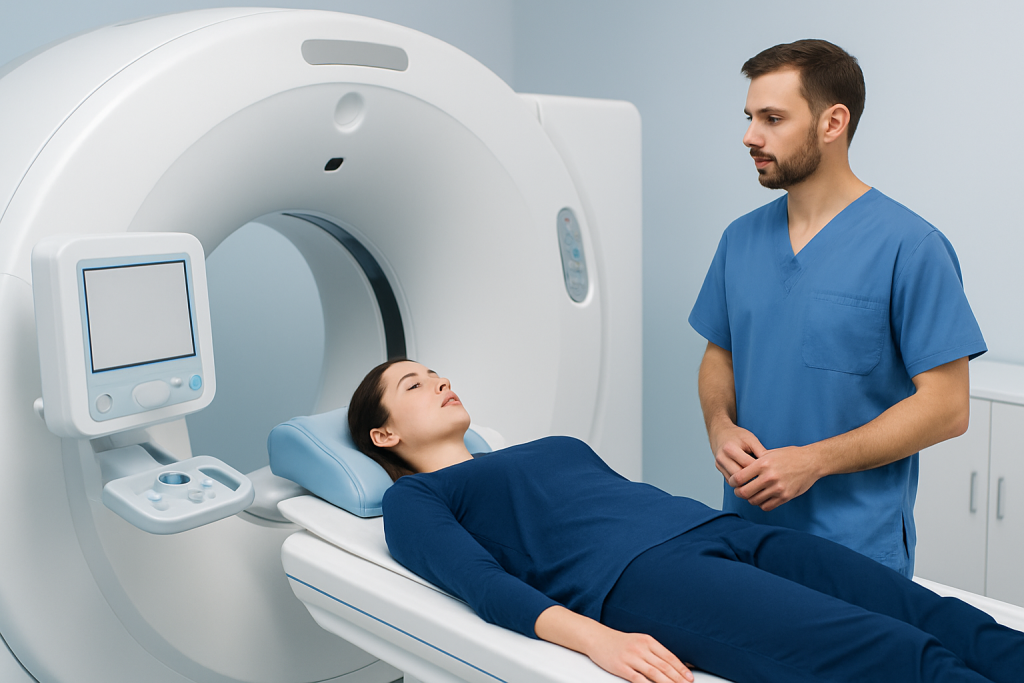
The scan itself is painless. The patient lies still on the table while the scanner moves around the body. Depending on the area being studied, the scan may take longer than a standard CT because both the PET and CT parts must be completed. Most people can return to ordinary activities soon after, unless the physician gives different instructions.
For patients traveling from another country, preparation can be easier when the schedule, fasting instructions, and any lab requirements are organized in advance. Clear coordination matters because even a small mismatch in timing, blood sugar control, or recent treatment dates can affect how useful the scan will be.
Treatment Planning, Prevention & Self-care
PET-CT itself is not a treatment, but it can strongly influence treatment choices. By showing where disease is and is not present, it may help doctors decide whether surgery is appropriate, whether radiation needs to cover a larger area, or whether systemic therapy should be prioritized. In that sense, the scan can prevent unnecessary procedures by revealing disease that would otherwise be missed.
There is no lifestyle habit that replaces staging imaging, but patients can help improve the quality and usefulness of the exam by following instructions carefully. That may include keeping appointments, sharing a full medication list, telling the team about diabetes or kidney issues, and informing them about recent infections, operations, or treatments. These details help the radiology team judge whether the timing is appropriate.
After the scan, self-care is usually simple. Drinking fluids as advised and following any safety instructions about close contact with infants or pregnant people can be enough. If the patient is waiting for results while living abroad, it helps to arrange a direct follow-up plan so the next step is clear, whether that is treatment locally or consultation with the original oncology team.
When to See a Doctor
Patients should discuss PET-CT with their oncologist or specialist when staging results are likely to change treatment. That is often the case before major surgery, when other imaging is unclear, or when there is concern that cancer may have spread beyond the original site. It is also reasonable to ask why the scan is being recommended if another test might answer the same question more efficiently.
After a PET-CT, a doctor should review the findings in context rather than in isolation. A report that mentions an abnormal spot does not automatically mean cancer progression, and a normal scan does not always end the evaluation if symptoms persist. The next step may be observation, another imaging study, or a biopsy depending on the situation.
Acibadem Health Point’s multidisciplinary specialists and JCI-accredited hospitals diagnose and treat cancer for international patients, coordinating imaging and follow-up when a clear staging plan is needed. Patients should seek medical advice promptly if they have new pain, unexplained weight loss, swelling, persistent cough, neurological symptoms, or any new concern during or after cancer workup.
Frequently asked questions
What is the main purpose of PET-CT in cancer staging?
Its main purpose is to show whether cancer is active in the body and whether it may have spread beyond the original site. Doctors use that information to choose the most appropriate treatment plan. The scan is most valuable when the result is likely to change management.
Does a PET-CT scan always find cancer spread?
No. Small tumors, microscopic spread, and some slow-growing cancers may not appear clearly on PET-CT. A negative scan is reassuring, but it does not replace pathology or the rest of the staging workup.
Can inflammation look like cancer on PET-CT?
Yes. Infection, healing tissue, recent surgery, and inflammation can all cause increased tracer uptake. That is one reason PET-CT findings must be interpreted together with the patient’s history and other tests.
Why would a doctor choose CT or MRI instead of PET-CT?
CT or MRI may be better for certain organs, certain tumor types, or situations where very detailed anatomy matters more than metabolic activity. Sometimes they provide the answer more clearly and with less ambiguity. The best test depends on the clinical question.
Is PET-CT useful after treatment has started?
It can be, but timing matters. A scan done too soon after surgery, radiation, or chemotherapy may be difficult to interpret because normal healing can mimic disease. The oncologist chooses the timing to make the result as reliable as possible.
Do patients need special preparation before PET-CT?
Usually yes, especially fasting and blood sugar control for people with diabetes. The exact instructions depend on the center and the reason for the scan. Following the preparation steps carefully helps improve image quality and accuracy.
References
- World Health Organization
- National Cancer Institute
- Radiological Society of North America
- American College of Radiology
- European Society for Medical Oncology
This article is for general information only and is not a substitute for professional medical advice. Please consult a qualified doctor about your individual situation.
More from the Health Library


Related Specialists

Dr. Firat Beğde
Pediatrics
Assoc. Prof. Dr. Özgüç Takmaz
Gynecology & Obstetrics
Prof. Dr. H. Armağan Arıcan
Radiation Oncology
Assoc. Prof. Dr. Serap Aktaş Yıldırım
Anesthesiology