Nuclear Cardiology Stress Testing

Key Takeaways
- It combines a stress test with imaging to assess blood flow to the heart muscle.
- The test may use exercise or a medication to simulate stress when exercise is not possible.
- Results can help identify reduced blood flow, prior heart damage, or a low-risk heart pattern.
- Preparation is usually simple, but medicines, caffeine, and recent meals may matter.
- Most people go home the same day and can resume normal activities unless their doctor advises otherwise.
Nuclear cardiology stress testing is a heart imaging exam that shows how well blood reaches the heart muscle at rest and during stress. It can help doctors understand chest symptoms, check heart function, and guide next steps in care.
Overview
Nuclear cardiology stress testing is a way to see how the heart performs when it is working harder than usual. Unlike a standard treadmill test, it adds an imaging step that tracks a small amount of radioactive tracer as it moves through the heart muscle. This helps doctors compare blood flow at rest and during stress.
For many patients, the test is ordered when symptoms such as chest discomfort, shortness of breath, or unusual fatigue need a clearer explanation. It may also be used after a heart event or procedure, or when a doctor wants to understand whether a blocked artery is limiting blood supply to part of the heart.
For international patients planning care abroad, the exam is often appealing because it is non-surgical, usually completed in one day, and can provide information that helps shape the rest of the treatment plan. The goal is not only to look for disease, but also to better understand how urgently follow-up care may be needed.
Symptoms and Reasons the Test Is Ordered
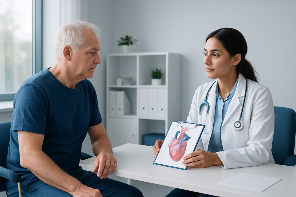
Doctors may recommend nuclear cardiology stress testing when symptoms suggest that the heart may not be receiving enough blood during activity. Common reasons include chest pressure, breathlessness with exertion, reduced exercise tolerance, dizziness during effort, or unexplained fatigue. Some people have no symptoms at all and are tested because of an abnormal ECG, a known history of coronary artery disease, or prior treatment such as stenting or bypass surgery.
The test is also useful when symptoms are difficult to interpret. For example, chest discomfort may come from the heart, the lungs, the stomach, muscles, or anxiety-related causes. Imaging can add useful detail by showing whether blood flow changes under stress in a way that suggests a heart-related cause.
In some cases, the test is ordered as part of a pre-treatment or pre-surgery evaluation. A doctor may want to know whether the heart can tolerate the physical demands of another procedure, or whether medical treatment should be adjusted before travel, surgery, or rehabilitation begins.
Causes and Risk Factors
Nuclear cardiology stress testing is not a diagnosis by itself; it is a tool used to investigate possible causes of reduced blood flow to the heart muscle. The most common concern is coronary artery disease, in which plaque buildup narrows the arteries that supply the heart. During stress, these narrowed vessels may be unable to deliver enough oxygen-rich blood.
Risk factors that make this evaluation more likely to be useful include high blood pressure, high cholesterol, diabetes, smoking, older age, obesity, a family history of heart disease, and a personal history of heart attack, angina, stent placement, or bypass surgery. People with kidney disease or other medical conditions may also need a carefully chosen test plan.
Exercise ability matters as well. If a patient cannot reach an adequate activity level because of joint pain, lung disease, deconditioning, or neurologic limitations, a medication may be used to widen the coronary arteries and create a stress-like effect. This allows imaging to remain informative even when treadmill exercise is not possible.
Diagnosis and How the Test Works
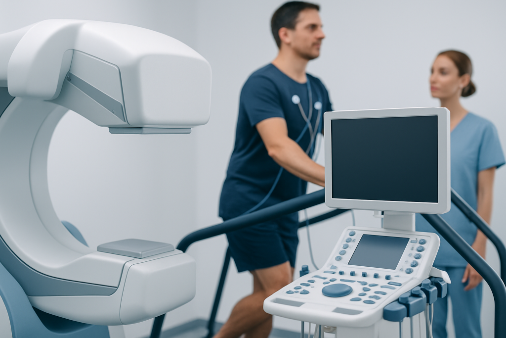
The exam usually has two parts: stress and imaging. First, the patient is connected to monitoring equipment, and the team records heart rate, blood pressure, and ECG activity. Stress may be created by walking on a treadmill or cycling, or by giving a medicine that temporarily increases blood flow demand or changes the way the arteries behave.
A small amount of tracer is injected into a vein during the stress portion, and images are then taken with a special camera. In many centers, the process is repeated at rest so the two sets of images can be compared. The difference between them helps show whether an area of the heart receives less blood only during stress or whether there is evidence of older, fixed damage.
Preparation varies by center, but patients are often asked to avoid caffeine before certain medication-based studies, follow instructions about fasting, and discuss prescription and over-the-counter medicines in advance. The team will explain how long the visit may take, when to stop eating or drinking, and whether any usual medications should be adjusted for the test.
Results are interpreted by a cardiologist or nuclear medicine physician. They are not read in isolation; symptoms, risk factors, ECG findings, and prior test results all help build the full picture. This is especially important for patients traveling from abroad, because the final report may guide whether immediate treatment, follow-up imaging, or routine monitoring is the most appropriate next step after returning home.
Treatment Options and What the Results Can Lead To
Nuclear cardiology stress testing does not treat heart disease directly, but its findings often influence treatment choices. If the images suggest reduced blood flow, a doctor may recommend medication to control blood pressure, lower cholesterol, reduce angina, or improve overall heart protection. Some patients may need additional testing, such as coronary CT angiography or invasive coronary angiography, depending on the pattern seen.
When the scan shows a low-risk pattern, care may focus on reassurance, symptom tracking, and risk-factor control. This can help avoid unnecessary procedures and support a more tailored plan. In other situations, especially when symptoms are concerning or the abnormality is significant, the results may support catheter-based treatment or surgery after careful discussion.
For many international patients, the value of the test lies in its clarity. A single study can help local and traveling physicians agree on whether treatment should be conservative, medical, or procedural, which can make planning across borders much simpler and safer.
Prevention and Self-care
Self-care before and after the test is mostly about following instructions carefully. Patients should tell the care team about all medicines, allergies, asthma or breathing problems, pregnancy or possible pregnancy, recent caffeine intake, and any trouble walking, standing, or lying flat. These details help the team choose the safest version of the exam.
On the day of the test, comfortable clothing and supportive shoes are helpful if exercise is planned. Afterward, most people can return to normal activities unless they were told to rest or avoid certain medicines. Drinking fluids may be encouraged, depending on the instructions given by the care team.
- Follow fasting and caffeine instructions exactly as provided by the clinic.
- Bring a list of current medicines and recent test reports if available.
- Ask whether any heart medications should be paused before the study.
- Arrange transportation if the test may include a medicine that causes temporary dizziness or tiredness.
Beyond the test itself, long-term prevention of heart disease usually includes managing blood pressure, blood sugar, and cholesterol, staying active within personal limits, not smoking, and keeping regular follow-up appointments. The scan can be a useful starting point for those broader goals.
What to Expect During Recovery
Recovery is usually straightforward. If exercise was used, patients may feel tired for a short time, similar to finishing a brisk walk or climb. If a medication was used instead, temporary flushing, chest pressure, headache, or short-lived shortness of breath can occur, but these effects are monitored and usually pass quickly.
The tracer used in nuclear imaging leaves the body gradually, and patients are often advised to drink fluids unless told otherwise. The care team will explain when the final report will be available and whether there are any restrictions after the test, such as avoiding close contact for a short period in special circumstances, though this is uncommon and depends on the type of tracer and local protocol.
For patients who traveled for care, this is also the moment when follow-up planning matters. It can be helpful to leave with a copy of the written report, a summary of findings, and a clear plan for who will review the result after returning home. At Acibadem Health Point, multidisciplinary specialists and JCI-accredited hospitals diagnose and treat this condition for international patients as part of coordinated cardiac care.
When to See a Doctor
Patients should seek medical attention if chest pain is severe, lasting, or accompanied by sweating, nausea, fainting, or shortness of breath. These symptoms may need urgent assessment, especially if they are new or different from previous episodes.
It is also wise to speak with a doctor if exercise feels unusually limited, if symptoms are becoming more frequent, or if a prior heart condition is changing. Nuclear cardiology stress testing may be one piece of the workup, but it should be interpreted in the context of the whole clinical picture.
After the test, patients should contact the care team if they were told to expect specific side effects and those effects last longer than described, or if they are unclear about next steps. A thoughtful review of the results can help determine whether additional testing, medication adjustment, or routine follow-up is the best path forward.
Frequently asked questions
What is nuclear cardiology stress testing used for?
It is used to check how well blood reaches the heart muscle during rest and stress. Doctors often order it when they want to evaluate chest symptoms, assess known coronary artery disease, or guide treatment decisions.
Is the test the same as a regular stress test?
It includes the stress part, but it also adds imaging with a tracer so blood flow can be seen. That extra information can show whether any heart area is under-supplied during exertion or stress-like conditions.
Can someone have the test if they cannot exercise well?
Yes, many patients can have a medication-based stress study instead of treadmill exercise. The medication changes coronary blood flow in a way that helps the images show how the heart responds.
Should medicines be stopped before the test?
Sometimes, but not always. The answer depends on the type of study and the reason for testing, so patients should follow the clinic’s instructions and not stop any medicine on their own.
Is the radiation from the tracer a concern?
The amount used is small and is chosen for medical imaging purposes. As with any scan involving radiation, doctors weigh the expected benefit against the exposure and use it only when it is appropriate.
How long does it take to get results?
Timing varies by center, but many patients receive a report after the images are reviewed by a specialist. If the result could change treatment quickly, the care team may discuss it sooner.
References
- American College of Cardiology
- American Heart Association
- Society of Nuclear Medicine and Molecular Imaging
- National Heart, Lung, and Blood Institute
This article is for general information only and is not a substitute for professional medical advice. Please consult a qualified doctor about your individual situation.