Macular Hole Repair: What Recovery Looks Like Week by Week

Key Takeaways
- Recovery after macular hole repair usually improves in stages, not all at once.
- A gas bubble may be used to support healing, and position instructions can be important.
- Blurred vision, mild discomfort, and a temporary change in depth perception are common early on.
- Follow-up visits help the surgeon check closure of the hole and monitor eye pressure and inflammation.
- Patients traveling for treatment may need to plan for postoperative rest, local accommodation, and return visits.
- Prompt medical advice is important if pain, sudden vision loss, or increasing redness develops.
Medically reviewed by the Acıbadem clinical team — June 13, 2026
Macular hole repair recovery is often gradual, with vision changes, positioning advice, and follow-up visits shaping the healing process. Understanding what may happen week by week can help patients prepare, stay comfortable, and know when to contact their eye specialist.
Overview
Macular hole repair is a procedure used to help close a small break in the central retina, the part of the eye responsible for sharp, detailed vision. For many patients, the surgery is a vitrectomy combined with steps that encourage the tissue to settle and seal over time.
Recovery does not usually follow a single, dramatic turning point. Instead, it tends to unfold in layers: early healing, adjustment to a gas bubble if one is used, gradual visual change, and a series of follow-up visits that confirm the eye is healing as expected. That pace can feel especially important for international patients, who may be planning treatment, hotel stays, and return travel around eye positioning instructions and clinic appointments.
This article explains what recovery may look like week by week, while keeping in mind that each case is different. The exact plan depends on the size and duration of the macular hole, the surgical technique, and the doctor’s preferences for postoperative care.
Symptoms During Recovery
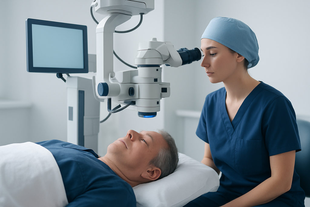
In the first days after surgery, the eye often feels different before it feels better. Blurred vision is expected, and it may be caused by the healing eye itself, prescribed drops, swelling, or the presence of a gas bubble inside the eye. Some patients also notice that straight lines look distorted at first, especially when looking with the operated eye.
Mild soreness, a gritty sensation, tearing, or light sensitivity can occur early on. These symptoms usually improve as the eye settles, although vision may remain hazy for several weeks. If the surgeon used a gas bubble, the patient may also see a dark curved line or a moving edge in the visual field as the bubble shrinks.
Other common temporary changes can include the following:
- Difficulty judging distance or depth
- Seeing colors or objects differently in the operated eye
- Needing more light for reading or moving around
- Fatigue from concentrating on limited vision
Recovery symptoms should generally trend downward over time. New or worsening pain, a sudden drop in vision, or a marked increase in redness should be discussed with an eye doctor promptly.
Causes & Risk Factors

A macular hole forms when the central retina develops an opening, often related to age-related changes in the gel inside the eye, called the vitreous. As the vitreous changes consistency and pulls on the retina, the macula may tear or separate. Surgery is intended to relieve this traction and give the tissue a better chance to close.
Macular holes are more common in older adults, and they may occur without a clear triggering event. Some people notice gradual central blur or distortion before diagnosis, while others discover the problem during an eye examination. A history of eye injury, inflammation, or certain retinal conditions may also play a role in some cases.
Factors that can affect recovery include the size and duration of the hole, whether a gas bubble was placed, and how healthy the retina is overall. Patients who have other eye diseases, such as cataracts or glaucoma, may need more coordinated follow-up.
Diagnosis
Macular hole repair is usually planned after a retina specialist confirms the diagnosis with a detailed eye examination and imaging. Optical coherence tomography, often called OCT, is especially useful because it creates a cross-sectional view of the retina and shows the hole clearly.
Before surgery, the doctor may also check visual acuity, eye pressure, and the overall health of the retina. These measurements help guide the procedure and provide a baseline for recovery visits later on. For patients arranging care from another country, this stage is also when the team typically discusses timing, travel considerations, and how long the patient may need to stay locally after surgery.
After the operation, diagnosis continues in a practical sense through follow-up imaging and examination. The surgeon looks for signs that the hole is closing, inflammation is easing, and pressure inside the eye remains in a safe range.
Treatment Options
Most macular hole repairs involve a vitrectomy, during which the surgeon removes the vitreous gel and reduces traction on the macula. In many cases, a very delicate membrane around the retina is peeled to help the surface relax. A gas bubble is then placed in the eye to support closure while healing occurs.
The gas bubble is a major part of the recovery experience. It can temporarily limit what the patient sees and may require a specific head position for part of the day, especially in the early period after surgery. The doctor’s instructions matter because the bubble needs to rest against the macula in the right way for the best chance of closure.
Medication after surgery commonly includes eye drops to reduce inflammation and lower the risk of infection. Some people may later need cataract treatment if cataracts progress after vitrectomy, which is a known possibility in certain adults. The exact treatment sequence should always be individualized by the eye surgeon.
Prevention & Self-care
There is no guaranteed way to prevent a macular hole once the underlying retinal changes begin, but careful self-care can support a smoother recovery. The most important step is following the surgeon’s instructions exactly, including how often to use drops, how to sleep or position the head, and when to return for review.
Practical planning is especially helpful for patients recovering away from home. Before travel, it is wise to arrange a companion, confirm nearby lodging that is comfortable for resting with the head in the recommended position, and keep all discharge papers and prescriptions in an accessible place. Patients should also ask how soon it is safe to fly, since a gas bubble can make air travel unsafe until the bubble has fully resolved.
Helpful self-care habits during recovery may include:
- Using prescribed eye drops on schedule
- Wearing protective eyewear if advised
- Avoiding eye rubbing or pressure on the eye
- Keeping follow-up appointments even if vision seems to improve slowly
- Asking the team before resuming exercise, driving, or long-distance travel
Rest and patience are both part of the healing process. Vision often improves gradually, and small day-to-day fluctuations are common early on.
When to See a Doctor
Patients should contact their eye surgeon or seek urgent eye care if symptoms change in a way that does not fit the expected recovery pattern. Some discomfort and blurred vision are normal after surgery, but severe pain is not something to ignore.
It is important to get medical advice quickly if there is sudden worsening of vision, a rapid increase in redness, discharge from the eye, fever, flashes of light, a curtain-like shadow, or nausea accompanied by eye pain. These symptoms can signal a complication that needs prompt attention.
Follow-up visits should never be skipped, even when the eye feels comfortable. The surgeon needs to confirm that the macular hole is closing, the gas bubble is behaving as expected, and the eye pressure remains stable. For international patients, this is also the time to verify whether extra in-person review is needed before returning home.
Acibadem Health Point’s multidisciplinary specialists and JCI-accredited hospitals diagnose and treat macular hole cases for international patients, with care plans that are designed to support both surgery and recovery coordination.
Week-by-Week Recovery Timeline
Week 1: This is usually the most instruction-heavy period. Vision may be very blurry if a gas bubble is present, and the eye may feel mildly irritated or tired. Patients often spend much of this week resting, using drops, and following positioning advice carefully. If travel was involved, this is also when questions about sleep setup, meals, and local transport become most practical.
Week 2: Comfort often improves, although sight may still be limited. The gas bubble may shrink enough that patients notice a shifting line in their vision, which can be unsettling but is usually expected. The doctor may check the eye to make sure healing is progressing and to adjust the plan if needed.
Weeks 3-4: The bubble may continue to absorb, and some patients start to see brief improvements in central clarity or distortion. Reading may still be difficult, and vision may not yet be stable enough for driving or work that requires sharp sight. This period often involves learning what the new “normal” feels like while the retina continues to settle.
Weeks 5-8 and beyond: Many patients notice gradual improvement in functional vision, although the final result can take longer. Some people recover central detail more quickly than others, and the outcome depends on how long the hole was present before repair. Follow-up imaging helps the specialist assess closure and guide future care, including any additional treatment if needed.
Frequently asked questions
How long does recovery after macular hole repair usually take?
Recovery is often gradual and may take several weeks to months. Some patients notice early comfort improvement in the first one to two weeks, while vision can continue to change over a longer period. The exact timeline depends on the hole itself and the surgical plan.
Is it normal to see a bubble after surgery?
Yes. If a gas bubble was used, patients commonly notice a moving shadow or line in the eye as the bubble slowly shrinks. This is usually part of the expected healing process and should be discussed with the surgeon during follow-up.
Why do some patients need to keep a special head position?
Positioning helps the gas bubble rest against the macula so the repaired area has support while it heals. The surgeon may recommend a specific posture for a certain number of hours per day, especially early after surgery. Patients should follow those directions closely unless the doctor gives different advice.
When can someone fly after macular hole repair?
Patients should not fly until the eye surgeon confirms it is safe, especially if a gas bubble is still present. Changes in air pressure can be dangerous for an eye with gas inside it. The timing varies, so the medical team should give individualized guidance before any trip home.
Will the vision go back to normal?
Many patients improve, but recovery is different for each person. Some regain useful central vision, while others still have some distortion or blur. The best estimate depends on how long the hole existed before repair and how the retina responds after surgery.
What should a patient do if the eye becomes painful after the first few days?
Pain that is severe, increasing, or paired with worsening redness or vision loss should be reported promptly. Mild discomfort can be expected early on, but changing symptoms may need evaluation. It is better to contact the eye clinic sooner rather than wait.
References
- American Academy of Ophthalmology
- National Eye Institute
- Mayo Clinic
- Royal College of Ophthalmologists
This article is for general information only and is not a substitute for professional medical advice. Please consult a qualified doctor about your individual situation.