

Hyaline Cartilage

Key Takeaways
- Hyaline cartilage provides a low-friction surface in joints and helps support normal movement.
- It is found in joint surfaces, the nose, rib cage, trachea, and areas of growth in children.
- Cartilage damage may develop gradually after injury, overuse, aging, or joint disorders.
- Diagnosis usually relies on medical history, physical examination, and imaging when needed.
- Treatment may include activity modification, rehabilitation, medications, injections, or surgical options depending on severity.
Hyaline cartilage is the smooth, resilient tissue that helps joints move with less friction and also supports parts of the respiratory system and growing bones. When it is damaged or worn, movement may become painful or stiff, so understanding its role can help patients make informed care decisions.
Overview
Hyaline cartilage is the most common type of cartilage in the body, and it plays a quiet but essential role in everyday movement. Its smooth surface covers the ends of bones inside joints, helping them glide with minimal friction while also absorbing part of the force that comes with walking, climbing stairs, or reaching overhead.
Although people often notice it only when it becomes injured or worn, this tissue is important in several parts of the body. It forms the framework of the nose, parts of the ribs, the trachea, and the larynx, and it appears in growing bones during childhood and adolescence. Because hyaline cartilage has a limited blood supply, it heals more slowly than many other tissues, which is why early assessment matters when symptoms persist.
For international patients considering care abroad, understanding the tissue’s function is a practical first step. It helps explain why doctors may focus not only on pain, but also on movement, joint alignment, activity patterns, and the quality of the cartilage surface itself.
Symptoms
Hyaline cartilage itself does not create symptoms when it is healthy, but problems involving it often show up as joint discomfort or reduced smoothness of movement. A person may notice pain with activity, stiffness after resting, swelling around a joint, or a sensation that the joint is catching or moving less freely.
When cartilage in a weight-bearing joint begins to wear, symptoms can develop gradually. Some people feel discomfort during walking or stairs, while others notice that a shoulder, knee, or hip feels less flexible than before. In more advanced cases, daily activities may take longer because the joint is no longer gliding as smoothly as it once did.
Symptoms depend on where the cartilage is located. In the airway, cartilage-related problems may affect breathing or voice quality, while in the nose or rib cage they may be noticed as structural changes or localized tenderness rather than joint pain.
- Joint pain during movement or after exercise
- Stiffness, especially after sitting or waking up
- Swelling or a feeling of fullness around the joint
- Reduced range of motion
- Clicking, catching, or grinding sensations
Causes & Risk Factors

Hyaline cartilage can be affected by several processes, and the cause often depends on the location and the person’s overall joint health. In joints, the most common issue is gradual wear and tear, especially when the joint has been under repeated load for years or after a previous injury has changed the way the joint moves.
Sports injuries, falls, and twists can damage the cartilage surface directly. In some people, joint disease such as osteoarthritis, inflammatory arthritis, or developmental alignment problems places extra stress on the tissue over time. Age is also relevant, because cartilage tends to repair more slowly as the body gets older.
Risk is not determined by one factor alone. Body weight, occupation, repetitive motion, prior surgery, muscle weakness, and poor joint alignment can all contribute to cartilage strain. For patients traveling for assessment, clinicians often ask about the pattern of symptoms, earlier injuries, work demands, and the kinds of activities that aggravate the joint.
- Previous joint injury or surgery
- Repetitive impact or heavy physical work
- Age-related cartilage wear
- Joint misalignment or instability
- Inflammatory or degenerative joint disease
Diagnosis
Diagnosis usually begins with a careful history and physical examination. A clinician will ask when symptoms started, what makes them better or worse, whether there has been an injury, and how the problem affects daily life. Exam findings can suggest whether the pain is coming from cartilage, tendons, ligaments, muscles, or the joint lining.
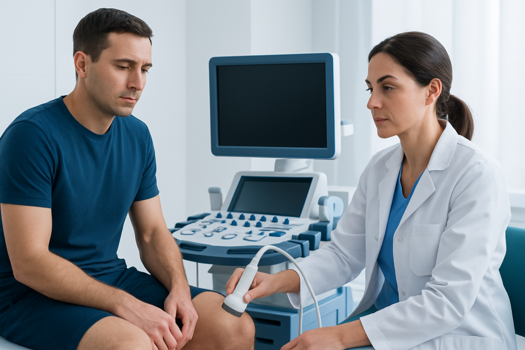
Imaging is often used when cartilage injury is suspected. X-rays can show joint spacing, alignment, and signs of arthritis, but they do not display cartilage directly. MRI is more useful for viewing cartilage, surrounding soft tissues, and other structures in the joint, while ultrasound may help in certain superficial or fluid-related problems.
Not every patient needs every test. The right workup depends on the joint involved, the severity of symptoms, and whether the goal is reassurance, a treatment plan, or preparation for a procedure. In international care settings, doctors may also review any prior images or reports brought from home to avoid repeating tests unnecessarily.
Treatment Options
Treatment depends on whether the cartilage problem is mild irritation, a small focal defect, or a broader condition such as osteoarthritis. For many patients, the first step is reducing the stress placed on the joint while preserving safe movement. This often includes temporary activity modification and a structured rehabilitation plan.
Physical therapy can be especially helpful because strong muscles support the joint and may reduce pressure on the damaged surface. Doctors may also recommend anti-inflammatory medicines or pain-relief strategies when appropriate, always based on the patient’s medical history and overall risk profile. Some patients benefit from assistive devices, bracing, or changes in footwear and movement technique.
For more persistent cartilage damage, procedures may be considered. These can range from injections used to manage symptoms to arthroscopic or reconstructive surgeries aimed at smoothing, repairing, or restoring the damaged area. The best choice depends on age, activity level, the size and location of the defect, and whether the cartilage problem is isolated or part of a larger joint condition.
Patients exploring Herniated Disc Treatment Abroad: Injection, Rehab, or Surgery?" class="ahp-ilk">treatment abroad often find it helpful to ask how the plan will work after they return home. Good care includes clear follow-up instructions, rehabilitation goals, and coordination with a local physician or therapist when needed.
Prevention & Self-care
Healthy cartilage is supported by healthy joints, so self-care focuses on protecting the joint surface from repeated overload. Low-impact exercise, maintaining a body weight that is comfortable for the joints, and strengthening the muscles around the affected area can all reduce stress on hyaline cartilage.
Warm-ups before activity, gradual increases in training, and attention to technique may help limit injury in active people. For those with desk jobs or long travel schedules, regular movement breaks can ease stiffness and prevent a joint from staying in one position too long. When symptoms are already present, it is usually wiser to stay active in a modified way than to stop moving completely.
Patients should also pay attention to warning patterns such as swelling after exercise, pain that lasts longer than expected, or a joint that feels unstable. These signs do not always mean serious damage, but they do suggest that a medical review would be useful before the issue becomes harder to treat.
- Choose low-impact activities when joints are sensitive
- Strengthen supporting muscles with guided exercise
- Use supportive footwear and proper training technique
- Maintain joint-friendly body mechanics during work and sport
- Seek timely advice after a joint injury
When to See a Doctor
A medical review is a good idea if joint pain lasts more than a short period, keeps returning, or begins to limit normal activities. It is also worth seeking care if there is swelling, reduced motion, locking, or a sense that the joint is giving way, because these features may point to cartilage injury or another treatable joint problem.
Urgent assessment is appropriate after a significant injury, especially if the joint is very painful, visibly deformed, or unable to bear weight. Sudden breathing trouble, noisy breathing, or new voice changes should also be checked promptly when cartilage in the airway may be involved.
For people considering evaluation abroad, it is sensible to bring previous scans, operative notes, and medication lists. Acibadem Health Point’s multidisciplinary specialists and JCI-accredited hospitals diagnose and treat this condition for international patients, with care plans designed to support both the in-person visit and the return home.
Frequently asked questions
What is hyaline cartilage made of?
Hyaline cartilage is made mainly of water, collagen fibers, and specialized cells called chondrocytes. This combination gives it a smooth surface and enough resilience to help joints move comfortably.
Can hyaline cartilage repair itself?
It has only limited healing capacity because it does not have a strong blood supply. Small injuries may settle with conservative care, but deeper or larger defects often need medical evaluation.
Is hyaline cartilage the same as articular cartilage?
Articular cartilage is a type of hyaline cartilage found on the ends of bones within joints. It is the smooth joint-lining surface that helps bones move against each other with less friction.
What does cartilage damage feel like?
People commonly describe pain with movement, stiffness, swelling, or a grinding or catching sensation. Some also notice that the joint feels less stable or does not move as smoothly as before.
How is cartilage damage usually treated without surgery?
Non-surgical care often includes activity changes, physical therapy, Knee Pain Relief: Causes and Treatment" class="ahp-ilk">pain relief strategies, weight management when relevant, and sometimes bracing or injections. The exact plan depends on the joint, symptoms, and underlying cause.
Can exercise help protect cartilage?
Yes, the right kind of exercise can support the muscles around a joint and reduce strain on cartilage. Low-impact activities and guided strengthening are usually preferred when symptoms are present.
References
- American Academy of Orthopaedic Surgeons
- Mayo Clinic
- National Institute of Arthritis and Musculoskeletal and Skin Diseases
- Merck Manual Professional Edition
- Cleveland Clinic
This article is for general information only and is not a substitute for professional medical advice. Please consult a qualified doctor about your individual situation.
More from the Health Library



Related Specialists

Prof. Dr. Ahmet Alanay
Orthopedic Surgery & Traumatology
Dr. Sinan Aksu
Orthopedic Surgery & Traumatology
Dr. Sirri Baştürk
Orthopedic Surgery & Traumatology
Prof. Dr. Korhan Özkan
Orthopedic Surgery & Traumatology