Eye Anatomy: Parts and Functions

Key Takeaways
- The eye is a complex organ that focuses light, converts it into signals, and sends those signals to the brain.
- Different eye structures have different jobs, from protecting the eye’s surface to detecting color and movement.
- Many common vision problems involve more than one part of the eye, which is why a full eye examination matters.
- Changes in vision, eye pain, flashes, floaters, or redness should not be ignored, especially if symptoms are sudden.
- Healthy habits and regular eye exams can help protect vision over time.
Medically reviewed by the Acıbadem clinical team — August 19, 2026
Eye anatomy explains how the cornea, lens, retina, and optic nerve work together to create vision. Understanding these parts can help patients recognize symptoms earlier and make informed decisions about eye care.
Overview
The eye is often described as a camera, but that comparison only goes so far. In reality, it is a living, layered organ that takes in light, shapes that light into a sharp image, and passes the information to the brain in a fraction of a second. Every clear image depends on several structures working in sequence, and even small changes in one part can affect sight.
For patients, learning eye anatomy is not just a biology lesson. It helps explain why a dry surface can blur vision, why the lens becomes less flexible with age, and why the retina and optic nerve are so important for preserving sight. It also gives context for what eye specialists look for during an examination, whether care is being planned locally or during an international visit for a second opinion or treatment.
Understanding the eye’s structure can make medical conversations easier. When a doctor mentions the cornea, macula, or vitreous, the terms are less mysterious if the patient already knows how these parts fit together. That knowledge can support calmer, more confident decision-making about tests, treatment, and follow-up care.
The Main Parts of the Eye
The front of the eye includes the cornea, conjunctiva, iris, and pupil. The cornea is the clear outer window that helps focus incoming light. The conjunctiva is a thin membrane that covers the white of the eye and lines the inner eyelids, while the iris is the colored ring that controls pupil size and helps regulate how much light enters.
Behind the iris sits the lens, a flexible structure that fine-tunes focus for near and distant objects. The lens works with the cornea to bend light onto the retina. The retina is the light-sensitive tissue at the back of the eye, and the macula is its central area responsible for detailed central vision, such as reading and recognizing faces.
Other important structures include the vitreous, a gel-like substance that fills the middle of the eye, and the optic nerve, which carries visual signals from the retina to the brain. Around these are the sclera, the tough white outer wall of the eye, and the eyelids and tear film, which protect the surface and keep it moist.
- Cornea: first major focusing surface
- Iris and pupil: control light entry
- Lens: adjusts focus
- Retina and macula: detect and refine visual detail
- Optic nerve: transmits signals to the brain
How Vision Happens
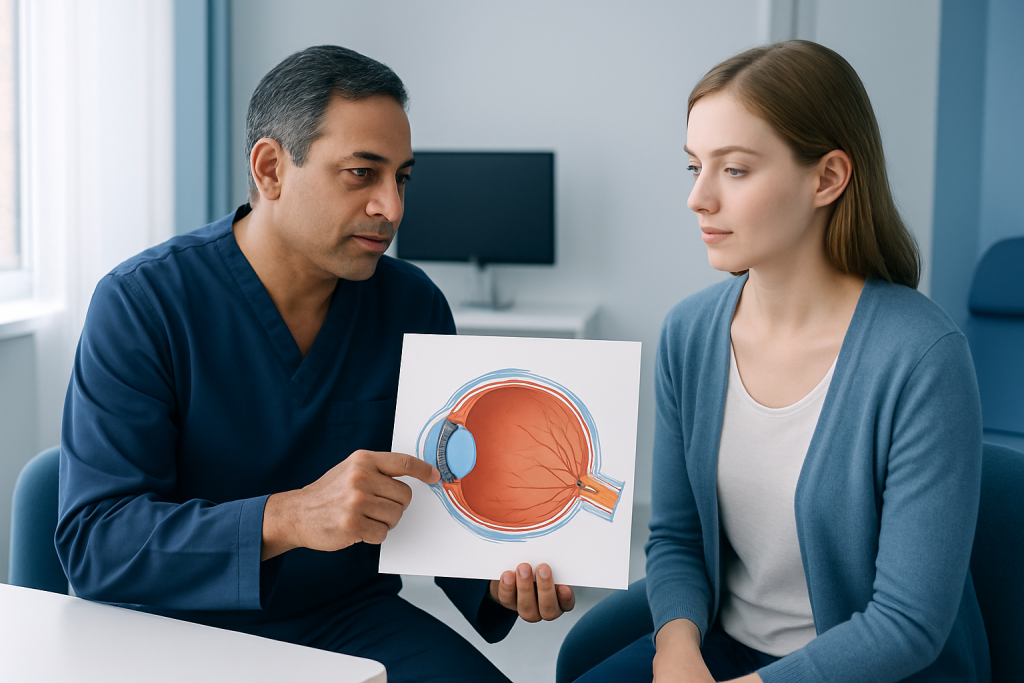
Vision begins when light passes through the cornea and pupil, then is focused by the lens onto the retina. The retina contains specialized cells that convert light into electrical signals. Those signals travel through the optic nerve to the brain, which interprets them as a meaningful image.
Two kinds of retinal cells do most of this work. Rods are better for low-light and peripheral vision, while cones help with color and sharp central detail. Because these cells are arranged differently across the retina, the eye is built for both wide awareness and precise focus.
Clear vision also depends on alignment and coordination. Both eyes need to work together so the brain can combine their input into one stable image. When the eye muscles, focusing system, or visual pathway is disrupted, symptoms can include blur, double vision, eye strain, or trouble judging distance.
Symptoms That Suggest an Eye Problem
Eye anatomy becomes especially relevant when symptoms appear. Some symptoms come from the surface of the eye, such as dryness, burning, watering, or a gritty sensation. Others reflect deeper structures and may affect clarity, brightness, or field of vision.
People should pay attention to new or changing symptoms, especially if they are one-sided, sudden, or persistent. Examples include blurred vision, distortion, flashes of light, new floaters, halos around lights, eye pain, sensitivity to light, or a dark curtain-like shadow in part of the visual field. These signs do not always mean something serious, but they do deserve prompt medical attention.
Because the eye is so interconnected, symptoms may not point to a single structure without a proper exam. A red eye, for instance, could involve the conjunctiva, cornea, eyelids, or deeper parts of the eye. That is why self-diagnosis is unreliable and why an eye examination is the safest next step when symptoms do not settle quickly.
Causes, Aging, and Risk Factors
Many eye conditions begin with gradual changes in anatomy. With age, the lens becomes less flexible, which can affect reading vision. The tear film may become less stable, causing dryness, irritation, and fluctuating blur. In the retina, age-related or vascular changes can affect central and peripheral vision over time.
Some people are more likely to develop eye problems because of family history, diabetes, high blood pressure, autoimmune disease, smoking, prolonged ultraviolet exposure, or previous eye injury. Long-term screen use can also contribute to eye strain and dry-eye symptoms, although it does not usually damage the eye’s internal structures on its own.
Risk is not only about age or heredity. Contact lens habits, poor lubrication of the eye surface, untreated inflammation, and certain medications can also affect comfort or visual quality. A careful history helps doctors decide which structures to examine most closely and which tests are most useful.
Diagnosis and Eye Examinations
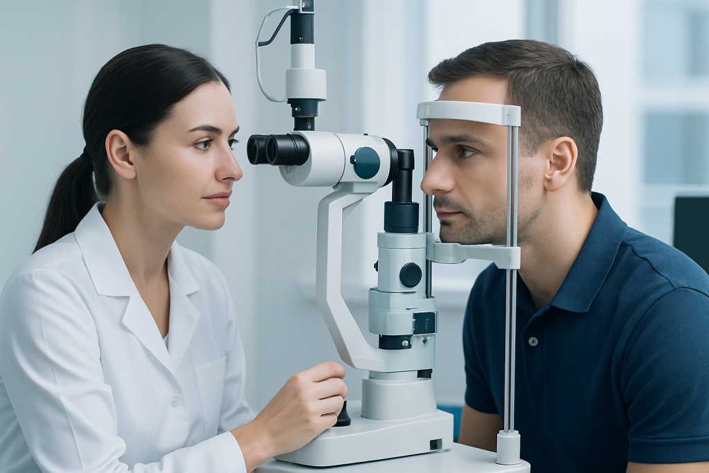
An eye examination often starts with a conversation about symptoms, medical history, medications, and any change in daily function. The doctor then checks vision, eye alignment, pupil response, eye pressure when appropriate, and the health of the front and back of the eye. These steps help identify whether the issue is on the surface, in the lens, within the retina, or along the optic nerve.
Depending on the concern, the specialist may use a slit-lamp exam, dilated retinal examination, optical coherence tomography, visual field testing, or imaging of the eye and orbit. Dilation allows the doctor to inspect the retina and optic nerve more fully, which is important in conditions that may not be visible otherwise.
For international patients, clear records matter. Bringing previous prescriptions, surgery reports, imaging, and a symptom timeline can make the evaluation more efficient, especially when time abroad is limited. If a treatment plan involves follow-up after returning home, the doctor may help outline which findings need monitoring and which symptoms require urgent reassessment.
Treatment Options and Care Pathways
Treatment depends entirely on which part of the eye is affected. Dry eye may be managed with lubrication, lid hygiene, and environmental changes. Refractive errors such as nearsightedness or astigmatism are typically corrected with glasses, contact lenses, or refractive procedures when appropriate. Cataracts, which affect the lens, may eventually require surgery if vision is reduced enough to interfere with daily life.
More serious conditions involving the retina, optic nerve, pressure inside the eye, or inflammation may need prescription treatment, laser procedures, injections, surgery, or close observation. The goal is not only to relieve symptoms but also to protect the anatomical structures that preserve sight. Early care often gives patients more options and may help prevent avoidable vision loss.
When patients travel for care, planning is part of treatment. That includes understanding the expected recovery period, whether additional visits are needed, and how to coordinate local follow-up once back home. Acibadem Health Point’s multidisciplinary specialists and JCI-accredited hospitals diagnose and treat eye conditions for international patients, with attention to continuity of care before, during, and after the visit.
Prevention, Self-care, and When to See a Doctor
Not every eye problem can be prevented, but many can be managed more comfortably with steady habits. Protective eyewear during sports or hazardous work can reduce injury risk. Regular breaks during close work may ease strain, and blinking more often can support the tear film when reading or using screens for long periods.
Sun protection also matters. Sunglasses that block ultraviolet light help shield the front of the eye over time. People with diabetes, hypertension, or a family history of eye disease should keep their overall medical conditions well controlled and follow recommended screening intervals, since the eye can reflect broader health changes.
A doctor should be contacted promptly for sudden vision loss, flashes and floaters that appear together, severe pain, chemical exposure, a very red eye with blurred vision, or any eye injury. A non-urgent appointment is still appropriate for persistent blur, frequent headaches related to reading, ongoing dryness, or a gradual change in one eye that has not improved.
Frequently asked questions
What is the most important part of the eye for vision?
Several parts are essential, but the retina and optic nerve play a central role in turning light into visual information and sending it to the brain. The cornea and lens are also critical because they focus light onto the retina. If any one of these systems is affected, vision can change.
Why does the eye need both the cornea and the lens?
The cornea provides the eye’s main focusing power, while the lens fine-tunes that focus for near and distant objects. Together, they help place light sharply on the retina. If either structure is abnormal, vision can become blurred or distorted.
Can eye anatomy explain dry eye symptoms?
Yes. Dry eye often involves the tear film, eyelids, and surface of the conjunctiva and cornea. When this protective layer is unstable, the eye may feel burning, gritty, watery, or intermittently blurred.
Are floaters and flashes always a sign of a serious problem?
Not always, but new floaters or flashes should be taken seriously, especially if they appear suddenly or with a shadow in the vision. They can sometimes signal a problem involving the retina or vitreous. A prompt eye examination is the safest approach.
How often should someone have an eye exam?
That depends on age, symptoms, and health history. People with diabetes, high blood pressure, a family history of eye disease, or existing vision problems may need more frequent exams. A doctor can recommend the most appropriate schedule.
Can long screen time damage the inside of the eye?
Screen time commonly causes eye strain and dry-eye symptoms, but it usually does not damage internal eye structures by itself. Taking breaks, blinking regularly, and adjusting lighting can help reduce discomfort. Persistent symptoms should still be checked by an eye specialist.
References
- American Academy of Ophthalmology
- National Eye Institute
- World Health Organization
- Mayo Clinic
This article is for general information only and is not a substitute for professional medical advice. Please consult a qualified doctor about your individual situation.
Explore treatments in Turkey — costs, top hospitals & a free quote
JCI-accredited · board-certified surgeons · reply within 24h
Add us as a Preferred Source to see more of our trusted health content across Google Search, AI Overviews and Discover.