

What Your X-Ray, MRI, and Exam Together Reveal Before Orthopedic Treatment
Key Takeaways
- A physical exam shows how a joint, bone, tendon, or muscle behaves in real life, not just on a screen.
- X-rays are best for bones, alignment, arthritis changes, and some fractures.
- MRI is useful for soft tissues such as ligaments, cartilage, tendons, discs, and nerves.
- The right test depends on the symptom pattern, injury history, and what the doctor needs to confirm or rule out.
- Imaging findings must be interpreted together with symptoms and examination results to avoid treating a picture instead of the patient.
Medically reviewed by the Acıbadem clinical team — June 13, 2026
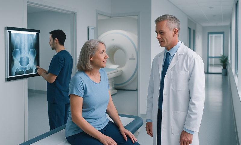
Before orthopedic treatment begins, a careful physical exam and the right imaging tests often tell a more complete story than any one test alone. X-rays and MRI scans answer different questions, helping doctors match symptoms with the safest, most practical treatment plan.
Overview
Orthopedic care often starts with a simple but important question: what is actually causing the pain, stiffness, swelling, or loss of function? A person may arrive with a sore knee, a painful shoulder, or back symptoms after travel, sport, or everyday strain. The exam and imaging tests are then used together to map the problem from more than one angle.
An X-ray, an MRI, and a hands-on clinical exam each reveal different parts of the same story. The exam shows how movement, strength, tenderness, and stability are affected. X-rays highlight bones and alignment. MRI adds detail about soft tissues, nerves, cartilage, and deeper structures that X-rays cannot show well.
For international patients, this combined approach is especially helpful because it supports efficient decision-making. When someone is traveling for orthopedic consultation or treatment, doctors aim to understand whether the condition is best managed with rest, rehabilitation, injections, bracing, or surgery — and that decision is usually strongest when built on a full assessment rather than a single image.
What the Physical Exam Adds
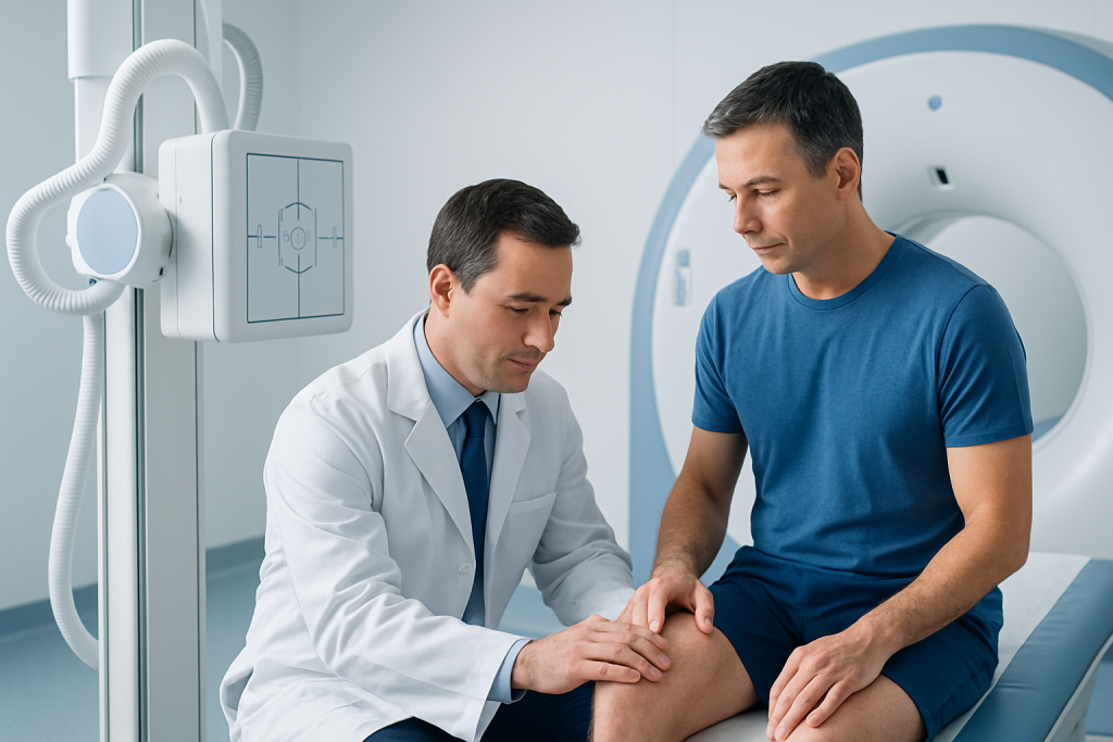
A physical exam is not just a formality before imaging. It often gives the first clues about which structure is affected and how serious the problem may be. Doctors observe posture, walking pattern, swelling, bruising, and how a person uses the body during everyday movements. They also ask when symptoms began, what worsens them, and whether there was an injury, overuse, or gradual decline.
The exam may include gentle range-of-motion testing, strength testing, special maneuvers for joints, and checks for tenderness or instability. A shoulder that hurts only when lifted overhead points to a different concern than a shoulder that is painful even at rest. A knee that “gives way” may suggest ligament or meniscus issues, while a stiff knee after long inactivity may suggest a different pattern entirely.
Just as importantly, the exam can identify warning signs that should prompt more urgent attention, such as marked deformity, inability to bear weight, numbness, or signs that pain is coming from the spine rather than the joint itself. In many cases, the exam narrows the list of possibilities before imaging is even ordered.
How X-Rays Guide Orthopedic Decisions
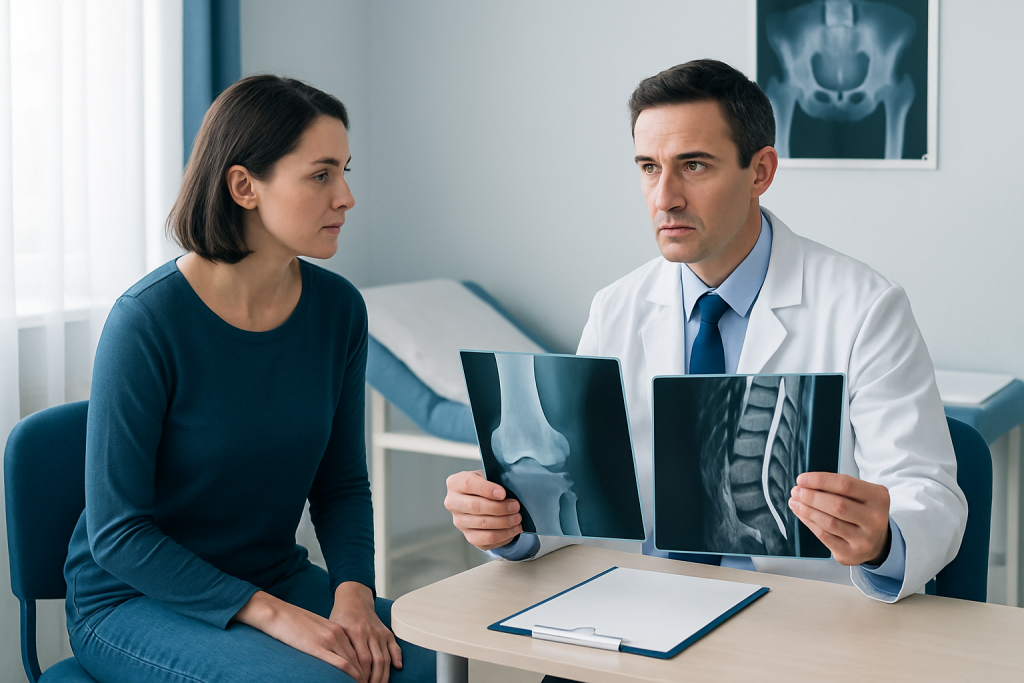
X-rays are often the first imaging test in orthopedics because they are fast, widely available, and very good at showing bones, joint space, alignment, and some fractures. They can reveal arthritis changes, bone spurs, dislocations, healed injuries, and structural differences that may influence treatment. If someone has persistent pain after a fall or an injury, an X-ray may be the quickest way to rule out a break or dislocation.
In chronic conditions, X-rays help show whether a joint has narrowed, worn, or shifted over time. They may also help the doctor compare one side with the other or assess posture and load-bearing alignment. In spine care, X-rays can show curvature, slippage, degenerative changes, or evidence of prior injury.
Still, X-rays have limits. They do not show ligaments, tendons, muscles, or many cartilage injuries in detail. A person can have a very painful soft-tissue injury with a normal X-ray. That is why the result should never be read in isolation; it is only one piece of the orthopedic picture.
When MRI Brings the Missing Detail
MRI is especially helpful when the doctor needs to look beyond bone. It can show cartilage damage, tendon tears, ligament injuries, muscle strains, marrow changes, discs, and nerve-related problems. This makes it valuable for sports injuries, persistent joint pain, spinal symptoms, and cases where the exam suggests a deeper soft-tissue issue.
Unlike X-rays, MRI does not use ionizing radiation. It is often chosen when symptoms have continued despite initial care, when a more detailed map is needed before treatment, or when surgery is being considered and the surgeon wants to understand the full extent of injury. It can also help explain why a joint feels unstable or why pain seems out of proportion to a plain X-ray finding.
That said, MRI is not automatically the best test for every orthopedic problem. Some findings may appear on MRI even in people without symptoms, and not every painful condition needs advanced imaging. The value of MRI comes from matching the image to the physical exam and the patient’s story, not from scanning first and interpreting later.
Why the Three Pieces Must Be Read Together
The most useful orthopedic assessment is usually a conversation between three sources of information: what the patient feels, what the doctor finds on exam, and what the imaging shows. When those three match, the diagnosis becomes much clearer. When they do not match, the doctor may need to look for a different cause of pain or choose a more conservative path.
For example, an MRI may show a meniscus tear, but if the knee exam is stable and symptoms are mild, the tear may not be the main reason for the pain. On the other hand, a normal X-ray does not rule out a ligament injury, tendon problem, or nerve irritation. The goal is to treat the person, not the picture.
This balanced approach helps avoid unnecessary procedures and supports better planning for rehabilitation. It also helps international patients, who may be trying to fit diagnosis, treatment, and travel into a short window, understand which findings are urgent, which are manageable, and what the next step should realistically be.
Typical Causes and Risk Factors Behind Abnormal Findings
Orthopedic findings on exam or imaging can arise from many different situations. A sudden twist, fall, or sports injury may cause a fracture, ligament sprain, or cartilage damage. Repetitive strain, heavy lifting, or years of joint loading can contribute to tendon irritation, wear-and-tear changes, or arthritis. Sometimes the problem begins gradually with no single memorable event.
Age, previous injury, high-impact activity, joint overuse, poor biomechanics, and certain medical conditions can all influence how bones and soft tissues change over time. A person with long-standing knee pain, for instance, may have a combination of alignment issues, meniscal wear, and early arthritis rather than one isolated cause.
Risk factors do not automatically mean disease, and imaging abnormalities do not always cause symptoms. That is why orthopedic doctors look carefully at function, timing, and exam findings before recommending treatment. The wider context matters as much as the scan itself.
Treatment Options and How Findings Shape the Plan
Treatment depends on the final clinical picture, not just on the name of the finding. Many orthopedic problems begin with conservative care such as activity modification, targeted rehabilitation, pain-relief strategies recommended by the doctor, bracing, or time for tissue healing. Imaging results can help decide whether rest alone is reasonable or whether a more active approach is needed.
Rehabilitation often plays a central role. A well-designed program can improve mobility, reduce pain, rebuild strength, and help the body compensate for damage that cannot be fully reversed. If imaging shows a structural problem that may not respond to rehabilitation alone, the doctor may discuss injections, procedures, or surgery. Even then, therapy commonly remains part of recovery.
For international patients, planning matters. Doctors may explain whether treatment can start immediately, whether further imaging is needed before travel home, and what follow-up should look like once care continues in another country. When needed, Acibadem Health Point’s multidisciplinary specialists and JCI-accredited hospitals can diagnose and treat orthopedic conditions for international patients in a coordinated way.
Prevention, Self-care, and Practical Follow-up
Not every orthopedic issue can be prevented, but many can be managed more safely with good habits. Keeping a healthy activity level, strengthening the muscles that support joints, warming up before exercise, using proper lifting technique, and allowing adequate recovery after strain all reduce stress on bones and soft tissues. Footwear, posture, and workstation setup can also matter more than people expect.
After imaging or examination, the most helpful self-care is usually the plan the doctor has tailored to the findings. That may include using a support device correctly, following rehabilitation exercises consistently, and avoiding a quick return to high-impact activity before healing is ready. Overdoing “rest” can also slow recovery, so the balance should be individual and guided.
If care is being received away from home, the patient should leave with a clear summary of the diagnosis, what the scans showed, what was tested on exam, and what should happen next. This makes it easier to continue treatment confidently with a local doctor or rehabilitation team after returning home.
When to See a Doctor
Orthopedic evaluation is worth seeking when pain lasts longer than expected, keeps returning, limits walking or daily tasks, or follows an injury that has not improved. Swelling, instability, repeated locking, reduced range of motion, or pain that wakes a person at night are also reasons to be assessed rather than waiting indefinitely.
Urgent medical review is important if there is visible deformity, inability to bear weight, sudden severe weakness, numbness, loss of bladder or bowel control, or major swelling after trauma. These symptoms do not always mean something serious, but they do deserve prompt attention.
When a condition is not urgent but still unresolved, a thoughtful orthopedic visit can prevent months of uncertainty. The right combination of exam and imaging can clarify the path forward and help the patient choose treatment with more confidence.
Frequently asked questions
Why do doctors need both an exam and imaging before orthopedic treatment?
Because they answer different questions. The exam shows how the body functions, while imaging shows the underlying structures. When both point to the same problem, the treatment plan is usually more precise.
Is an X-ray enough for most orthopedic problems?
Sometimes it is, especially when the concern is a fracture, joint alignment, or arthritis. But X-rays do not show many soft-tissue problems well, so additional testing may be needed if symptoms suggest a ligament, tendon, cartilage, or nerve issue.
What does MRI show that X-ray cannot?
MRI is better for soft tissues and deeper structures, including ligaments, tendons, cartilage, muscles, discs, and nerves. It can help explain pain when the X-ray looks normal or when a more detailed treatment plan is needed.
Can imaging findings be misleading?
Yes. Some findings appear on scans in people without symptoms, and some painful problems do not show clearly on simple imaging. That is why doctors interpret imaging together with symptoms and the physical exam.
Do all orthopedic patients need an MRI before treatment?
No. Many conditions can be diagnosed and treated without MRI. The doctor decides whether it is useful based on the injury, the exam, and whether the result would change the treatment plan.
How should a patient prepare for an orthopedic consultation abroad?
Bringing prior reports, images, medication lists, and a clear symptom timeline helps the doctor compare old and new information. It also makes it easier to plan next steps, especially if part of the recovery will continue after returning home.
References
- American Academy of Orthopaedic Surgeons
- Radiological Society of North America
- National Institute of Arthritis and Musculoskeletal and Skin Diseases
- Mayo Clinic
- World Health Organization
This article is for general information only and is not a substitute for professional medical advice. Please consult a qualified doctor about your individual situation.
More from the Health Library



Related Specialists

Dr. Safiye Sayilir
Physical Medicine & Rehabilitation
Dr. Özgün Barış Çolak
Physical Medicine & Rehabilitation
Prof. Dr. Meltem Vural
Physical Medicine & Rehabilitation
Prof. Dr. Meral Bayramoğlu
Physical Medicine & Rehabilitation