Orbital Bone

Key Takeaways
- The orbital bone forms the eye socket and helps protect the eye and nearby nerves, muscles, and tissues.
- Orbital bone problems are often caused by trauma, but infections, tumors, and congenital differences can also affect the area.
- Symptoms may include swelling, bruising, double vision, pain with eye movement, numbness, or a change in how the eye sits in the socket.
- Diagnosis usually involves an eye exam and imaging, often a CT scan, to assess the bones and surrounding structures.
- Treatment depends on the cause and may include observation, medicines, surgery, or specialist follow-up.
- Prompt evaluation is especially important if vision changes, severe pain, or trouble moving the eye occurs.
The orbital bone is the bony framework that surrounds and protects the eye. When it is injured or develops a problem, it can affect vision, eye movement, comfort, and facial appearance, so timely medical assessment is important.
Overview
The orbital bone is the bony structure that creates the eye socket, also called the orbit. Rather than being a single bone, the orbit is formed by several bones working together to cradle the eye and support the muscles, nerves, blood vessels, and soft tissues around it.
This framework does more than hold the eye in place. It helps shield the eye from injury and gives a stable path for the structures that control vision and eye movement. Because the orbit is so closely connected to the eye and the brain, even a small injury in this area can have effects that feel much larger than a simple bruise.
For people arranging care from another country, orbital bone problems are one of those conditions where good imaging, careful examination, and coordinated specialist input matter early. An eye specialist, facial surgeon, or emergency physician may all be part of the assessment depending on how the problem started.
Symptoms
Symptoms involving the orbital bone often appear after a blow to the face, a fall, a sports injury, or another type of accident. Swelling and bruising around the eye are common, but the pattern of symptoms can vary depending on which part of the orbit is affected and whether the eye itself was also injured.
Some people notice pain when moving the eye, blurred or double vision, or a feeling that the eye is not sitting normally in the socket. Numbness in the cheek, upper lip, or upper teeth can also happen if nearby nerves are affected.
- Bruising or swelling around the eye
- Pain, especially with eye movement
- Double vision or reduced vision
- Numbness in the cheek or upper lip
- A sunken-looking eye or a bulging eye
- Difficulty opening the eye fully
Not every symptom means a serious fracture, but changes in vision, eye position, or eye movement deserve prompt evaluation. In some cases, symptoms develop gradually over hours as swelling increases, so a person may initially feel “fine” and then become more uncomfortable later.
Causes & Risk Factors

Direct trauma is the most common cause of orbital bone injury. This can happen during car accidents, contact sports, falls, workplace injuries, or physical assault. The orbit may fracture because it is a delicate bony ring designed to protect the eye while still remaining light and functional.
Doctors sometimes describe these injuries as “blowout fractures,” which usually involve one of the thinner parts of the orbit. The floor and medial wall of the orbit are especially vulnerable because they are relatively fragile compared with other facial bones.
Other, less common causes can affect the orbital bones or the tissues around them. These include infections that spread from nearby sinuses, bone disorders, tumors, or congenital conditions that change the shape of the orbit. Certain factors may make injury more likely or more complicated.
- Participation in contact or high-impact sports
- Not using seat belts or other protective measures
- Reduced bone strength
- Previous facial injuries or surgery
- Infections involving the sinuses or face
For international patients, the cause is not always obvious from the first history taken in an urgent setting. That is why clinicians often review the mechanism of injury carefully and combine it with imaging to determine whether the orbit, the eye, or both need treatment.
Diagnosis
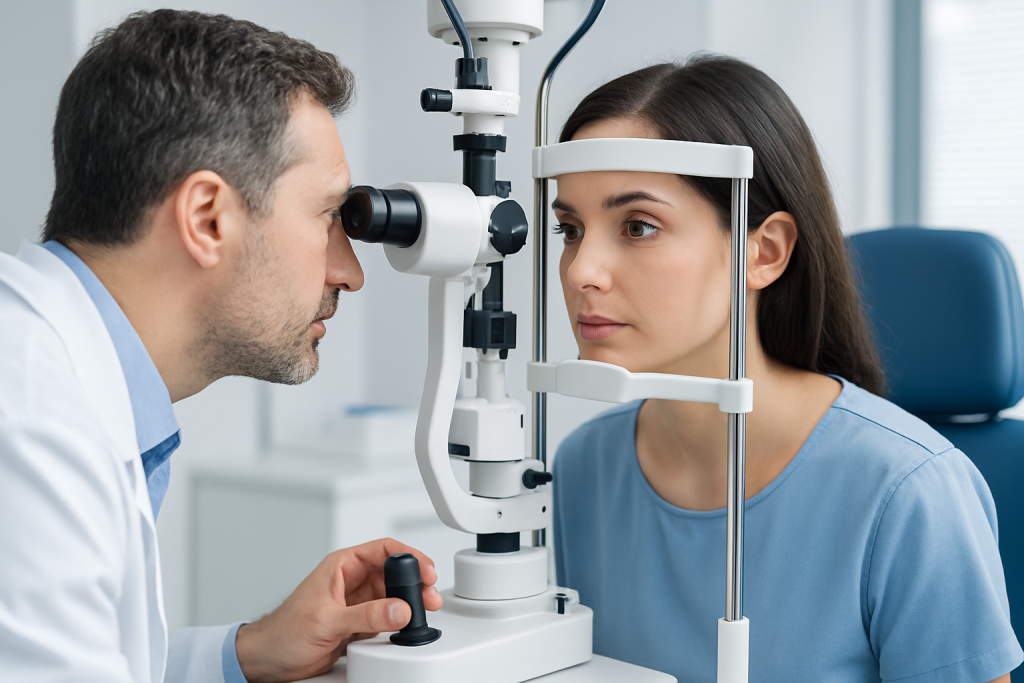
Diagnosis usually begins with a careful physical examination. A doctor will ask how the injury happened, whether vision changed, and whether there is pain, numbness, or double vision. They may check eye movements, pupil response, the position of the eye, and the skin and bones around the socket.
Imaging is often needed to understand the full extent of an orbital bone problem. A CT scan is commonly used because it shows bone detail clearly and can reveal fractures, trapped tissue, bleeding, or sinus involvement. In some situations, other imaging tests may be recommended if soft tissue or nerve concerns are greater.
The evaluation may also include an eye examination to make sure the cornea, retina, optic nerve, and pressure inside the eye are not affected. When symptoms suggest a more complex facial injury, specialists from ophthalmology, maxillofacial surgery, ENT, or neurosurgery may be involved so the plan is coordinated rather than fragmented.
Treatment Options
Treatment depends on the cause, the severity of the injury, and whether the eye is functioning normally. Some orbital fractures are managed without surgery if the eye is stable, vision is preserved, and there is no tissue trapped in the fracture. In these cases, close observation and follow-up imaging or specialist review may be enough.
When surgery is needed, the goal is to restore the shape of the orbit, free any trapped tissue, and reduce the chance of long-term problems such as double vision or a sunken eye. Surgery is generally considered when eye movement is limited, the fracture is large, or the position of the eye has changed in a way that affects function or appearance.
Medicines may be used to manage pain or treat associated issues such as infection, depending on the situation. If swelling is significant, doctors may advise simple measures such as head elevation and avoiding actions that increase pressure in the face, like forceful nose blowing, until the area has been reviewed.
Recovery plans are often individualized. A patient traveling from abroad may need a clear follow-up schedule, written instructions, and coordination with local doctors once they return home. Good communication matters, especially when surgery, monitoring, and later checkups all need to fit around travel plans.
Prevention & Self-care
Not every orbital bone problem can be prevented, but many injuries are avoidable with basic safety habits. Protective eyewear during sports and safe driving practices can lower the risk of facial trauma. Home and workplace fall prevention also plays a meaningful role, especially for older adults.
After an orbital injury, self-care should focus on protecting the area while healing. Rest, elevation of the head when lying down, and following the treatment plan closely can all support recovery. It is usually wise to avoid rubbing the eye, pressing on the socket, or taking part in activities that could cause another hit to the face.
- Use protective eyewear for sports or hazardous work
- Wear a seat belt and follow traffic safety rules
- Reduce fall risks at home, such as loose rugs or poor lighting
- Follow instructions about medicines and activity limits
- Attend all follow-up visits, even if symptoms improve
People recovering away from home should keep copies of imaging reports, operative notes, and discharge instructions. These documents help the next doctor understand what was found and reduce delays if follow-up care is needed in another country.
When to See a Doctor
Medical attention should be sought promptly after any facial injury that causes eye pain, swelling around the orbit, or trouble seeing. Even if the injury seems minor at first, symptoms can evolve as swelling increases or as a small fracture becomes more apparent.
Urgent evaluation is especially important if there is double vision, decreased vision, inability to move the eye normally, marked swelling, a change in eye position, numbness in the face, severe headache, or nausea after the injury. These signs do not always mean a dangerous complication, but they do mean the orbit and the eye should be checked without delay.
If a person already has an orbital condition that is not caused by trauma, they should see a doctor if symptoms persist, worsen, or begin affecting daily activities. For international patients, a specialist team can help decide whether treatment should be done immediately, after more testing, or through staged follow-up once travel is feasible. At Acibadem Health Point, multidisciplinary specialists and JCI-accredited hospitals diagnose and treat orbital conditions for international patients with coordinated care.
Living With an Orbital Bone Condition
Most people think first about the visible injury, but recovery often depends on what is happening beneath the surface. The orbit is a compact space, so even small changes in bone position or swelling can affect comfort, eye movement, or the way the face looks. A careful plan can reduce the chance of missed problems and help the person return to normal activities safely.
Follow-up is important because some issues become clearer after the first few days. Swelling may hide the full picture early on, and doctors may want to reassess vision, eye alignment, or healing once the acute phase settles. This is one reason many specialists prefer a step-by-step review rather than making decisions from a single early snapshot.
When care is organized well, orbital bone problems are often manageable. The goal is not only to heal the bone, but also to protect vision, restore comfortable eye movement, and help the patient feel confident about long-term recovery.
Frequently asked questions
What is the orbital bone?
The orbital bone is part of the bony eye socket that surrounds and protects the eye. It is made up of several bones that support the eye, muscles, nerves, and nearby soft tissues.
Can an orbital fracture heal without surgery?
Yes, some orbital fractures can heal without surgery if the eye is stable and there is no tissue trapped in the break. A doctor usually decides this after an exam and imaging, especially a CT scan.
What symptoms suggest an orbital injury needs urgent care?
Double vision, reduced vision, severe pain, trouble moving the eye, numbness in the face, or a change in eye position should be assessed promptly. These symptoms can signal that the orbit or the eye needs closer review.
Why is a CT scan often used for orbital bone problems?
A CT scan gives detailed pictures of the bones around the eye and helps doctors see fractures, trapped tissue, or nearby sinus involvement. It is one of the most useful tests for understanding orbital trauma.
How long does recovery usually take?
Recovery time varies depending on the type of injury and whether surgery is needed. Some people improve within days to weeks, while others need a longer follow-up period to watch healing and eye function.
Can orbital bone problems affect vision permanently?
They can, especially if the eye itself, the optic nerve, or the muscles around the eye are injured. Early assessment and proper treatment help reduce the chance of lasting problems.
References
- American Academy of Ophthalmology
- Merck Manual Professional Edition
- National Eye Institute
- American Association of Oral and Maxillofacial Surgeons
This article is for general information only and is not a substitute for professional medical advice. Please consult a qualified doctor about your individual situation.