Melanoma Pictures

Key Takeaways
- Melanoma pictures are useful for noticing change, but they cannot replace a medical evaluation.
- The ABCDE pattern and the “ugly duckling” sign are common ways to spot concerning moles.
- A new, changing, or unusually shaped lesion should be checked by a clinician, especially if it bleeds, itches, or grows.
- Early diagnosis matters because melanoma is often treatable when found before it spreads.
- Sun protection and regular skin checks can lower risk and support earlier detection.
Melanoma pictures can help people recognize suspicious skin changes, but images alone cannot confirm whether a spot is cancer. A qualified clinician uses examination, dermoscopy, and sometimes biopsy to determine what a lesion means and what care is needed.
Overview
When people compare melanoma pictures, they are usually looking for changes in size, shape, border, color, or texture. Those changes matter more than any single photo. A spot that is stable over time is usually less concerning than one that evolves from month to month.
For travelers or international patients, this question often comes up before a trip: should they wait until returning home, or seek care during travel? The safest approach is to arrange a skin evaluation promptly if a lesion is changing, because an early visit can make the next steps simpler, no matter where the patient is located.
Dermatology and oncology teams may work together when melanoma is suspected or confirmed. That multidisciplinary approach helps patients move from uncertainty to a plan, whether the goal is a biopsy, surgery, follow-up, or coordinated care after returning to another country.
Symptoms
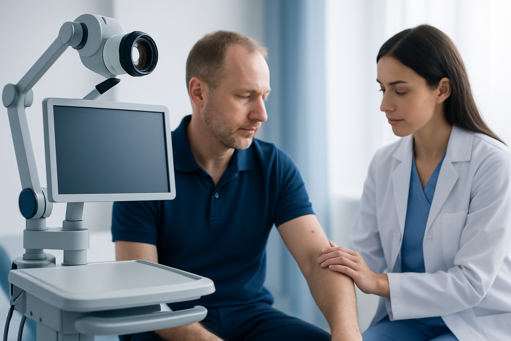
Melanoma pictures may also show a lesion with an unusual surface. Some look shiny or raised; others appear flat and patchy. Subtle features such as asymmetry or blurred borders are easier to miss in a quick glance, which is why a careful self-check in good light is useful.
Not every changing skin mark is melanoma. Seborrheic keratoses, benign moles, eczema, fungal infections, and other skin conditions can look alarming in photographs. The key difference is that melanoma is best evaluated in person, especially when change is recent or persistent.
People with a family history of melanoma, many moles, fair skin, or a history of sunburns should be especially alert to new symptoms. The same is true for anyone who notices a lesion under the nail, on the palm, sole, or scalp, because those locations can be overlooked during routine self-checks.
Causes & Risk Factors
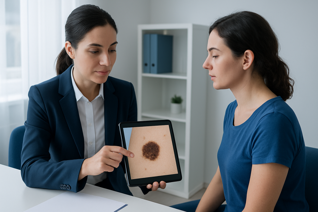
Genetics can also play a role. Some families carry inherited traits that increase the chance of melanoma, which is why a doctor may ask about relatives with skin cancer. People who have had a previous melanoma remain at higher risk of developing another one and usually need regular skin surveillance.
It is worth remembering that melanoma can occur in places that are not strongly sun-exposed. Mucosal areas, palms, soles, and nail beds may be involved, so any persistent new lesion in those areas should be checked rather than assumed to be harmless.
Understanding risk factors is not about creating alarm. It is about making skin checks more intentional and helping patients know which changes deserve a timely specialist opinion.
Diagnosis
Additional tests may be recommended if melanoma is confirmed or if there are signs it may have spread. These can include lymph node evaluation, imaging scans, or lab tests, depending on the stage and the findings from the initial examination. The exact workup is individualized rather than identical for every patient.
It is helpful for patients to bring photographs showing how the lesion has changed over time, if available. A timeline of when a spot first appeared, when it started changing, and whether it bled or itched can give the clinician useful context.
Even when melanoma pictures seem convincing, doctors avoid making a final judgment from photos alone. The combination of direct examination and tissue analysis remains the standard for accurate diagnosis.
Treatment Options
Immunotherapy helps the immune system recognize and attack cancer cells, while targeted therapy is designed for melanomas with certain genetic changes. These treatments are not used for everyone, but they have expanded the options for many patients with advanced disease.
Follow-up after treatment is an important part of care. Doctors may schedule skin exams, lymph node checks, and other visits to watch for recurrence or new skin cancers. Patients who live far from their treatment center often benefit from a clear follow-up plan that can be shared with a local physician.
Before starting any treatment, patients should ask what the goal is, what side effects to expect, and how care will be monitored. Clear information helps people make decisions with more confidence, whether they are being treated locally or after international travel.
Prevention & Self-care
When taking or reviewing melanoma pictures at home, it helps to use natural light and include a reference point such as a ruler or coin if advised by a clinician. Even then, photographs should be treated as a memory aid, not a diagnostic tool.
Self-care also includes not picking, scratching, or repeatedly irritating a suspicious lesion. If a spot is bleeding or painful, that is a reason to have it assessed rather than to try home treatment.
Patients who have already had melanoma should follow the follow-up schedule recommended by their doctor, protect their skin carefully, and report any new or changing lesions promptly. Prevention is not about perfection; it is about reducing avoidable exposure and catching problems early.
When to See a Doctor
Emergency care is not usually needed for a typical suspicious mole, but prompt specialist review is still important. Early assessment can clarify whether a spot is benign, needs monitoring, or should be removed and examined.
Patients who already have a melanoma diagnosis should contact their care team if they notice new symptoms such as rapidly growing lumps, unexplained weight loss, persistent pain, or other concerning changes. These symptoms do not always mean spread, but they do deserve medical review.
When in doubt, it is better to ask. A qualified doctor can sort out what photographs cannot and guide the safest next step.
Frequently asked questions
Can melanoma pictures tell if a spot is cancer?
They can raise suspicion, but they cannot confirm a diagnosis. Many harmless lesions can resemble melanoma in photos, and some early melanomas do not look dramatic. A clinician may need dermoscopy and biopsy to know for sure.
What should people look for in melanoma pictures?
The main clues are asymmetry, uneven borders, multiple colors, increasing size, and change over time. A spot that looks different from a person’s other moles is also worth attention. Any bleeding, itching, or crusting makes an evaluation more important.
Are all melanomas dark brown or black?
No. Some are pink, red, tan, or skin-colored. That is one reason photographs can be misleading and why a changing lesion should be checked in person.
How is melanoma diagnosed after a suspicious picture is noticed?
The usual process starts with a skin examination, often with dermoscopy, followed by biopsy if needed. The tissue sample is then examined under a microscope to confirm whether melanoma is present and to guide treatment planning.
Can melanoma appear in places that are not often exposed to the sun?
Yes. It can occur on the scalp, under the nails, on the palms, soles, and sometimes in mucosal areas. These sites are easy to miss, so regular self-checks should include them.
What can patients do if they are abroad and notice a changing mole?
They should seek a medical opinion promptly rather than waiting for return travel if the lesion is changing or suspicious. Bringing photographs, a written timeline, and previous pathology or dermatology records can help the new doctor assess the situation efficiently.
References
- American Cancer Society
- National Cancer Institute
- World Health Organization
- Skin Cancer Foundation
- American Academy of Dermatology
This article is for general information only and is not a substitute for professional medical advice. Please consult a qualified doctor about your individual situation.