Hydronephrosis: Symptoms, Causes and Treatment

Key Takeaways
- Hydronephrosis is a sign of urine buildup, not a disease on its own.
- It may cause pain, urinary changes, fever, or no symptoms at all.
- Causes include kidney stones, infection, pregnancy, enlarged prostate, and strictures.
- Ultrasound and other imaging tests are commonly used to confirm the diagnosis.
- Treatment depends on the cause and may range from observation to procedures that restore urine flow.
Medically reviewed by the Acıbadem clinical team — August 19, 2026
Hydronephrosis is swelling of one or both kidneys when urine cannot drain normally. It can develop quickly or slowly, and care focuses on identifying the blockage, protecting kidney function, and treating the underlying cause.
Overview
Hydronephrosis describes swelling of a kidney because urine is not draining as it should. The pressure builds up behind a blockage or narrowing somewhere in the urinary tract, which can affect one kidney or both.
It is important to think of hydronephrosis as a warning sign rather than a diagnosis by itself. The real question is why urine is backing up. In some people, the cause is temporary and easy to correct; in others, it reflects a condition that needs prompt attention to protect kidney function.
For international patients, the practical challenge is often timing. A symptom may begin during travel, after a scan done for another issue, or while arranging care across borders. Clear imaging, a focused evaluation, and a plan for follow-up are especially valuable when decisions need to be made efficiently and safely.
Symptoms

Hydronephrosis does not always cause noticeable symptoms. When it does, the signs depend on how quickly the swelling develops, whether one or both kidneys are involved, and what is causing the blockage.
Some people feel a dull ache in the back, side, or lower abdomen. Others notice cramping pain that comes and goes, especially if a stone is moving through the urinary tract. If pressure rises quickly, the discomfort can be more intense.
Possible symptoms include:
- Pain in the flank, side, groin, or lower back
- Nausea or vomiting
- Frequent urge to urinate or difficulty passing urine
- Pain or burning with urination
- Blood in the urine
- Fever or chills when infection is present
In some cases, hydronephrosis is found on an ultrasound or CT scan before a person feels unwell. That is one reason it should never be ignored when it appears on a report, even if the person feels mostly fine at the time.
Causes & Risk Factors
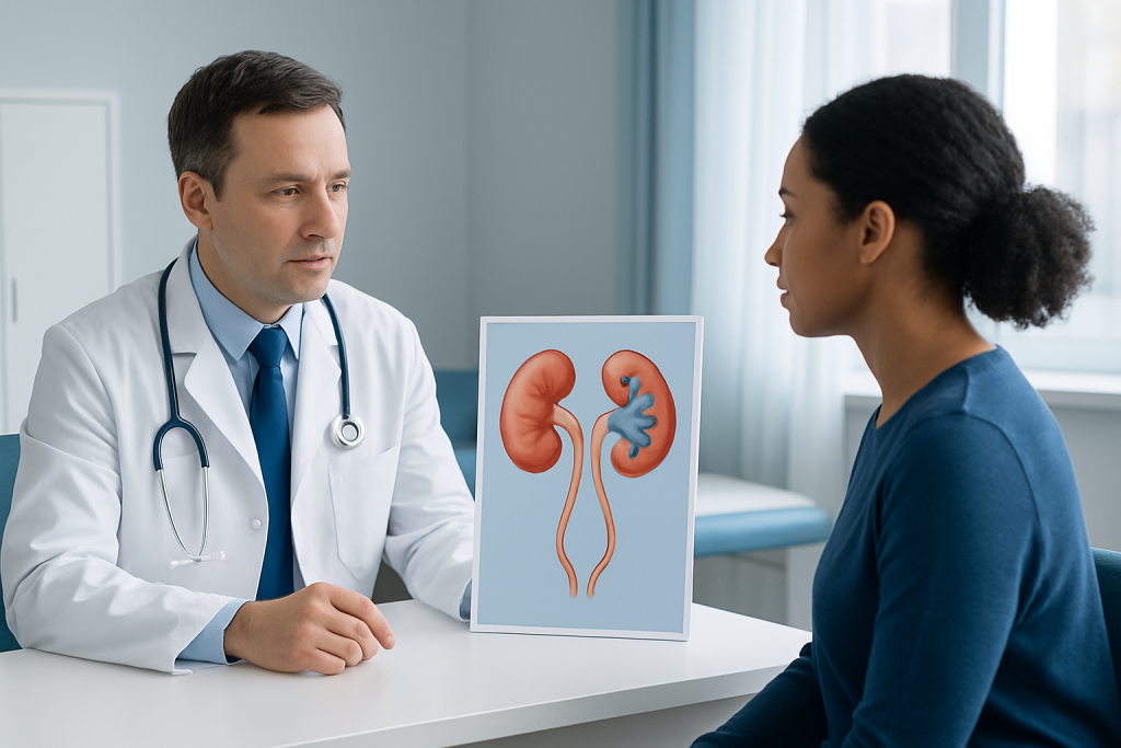
Anything that blocks urine flow can lead to hydronephrosis. The obstruction may be inside the kidney, in the ureter, at the bladder outlet, or related to pressure from nearby structures.
Common causes include kidney stones, urinary tract infections that cause swelling, blood clots, narrowed ureters, enlarged prostate, bladder dysfunction, pregnancy-related pressure on the ureters, pelvic masses, and scar tissue after surgery or inflammation. In children, congenital differences in the urinary tract can also be involved.
Risk tends to be higher in people with a history of stones, recurrent infections, prostate enlargement, pelvic surgery, certain cancers, or known structural urinary tract problems. Pregnancy can be a special situation, because the urinary system changes naturally and some swelling may be mild and temporary, while true obstruction still needs careful assessment.
When traveling or seeking care abroad, it helps to bring prior scan reports, a medication list, and any history of stones or urinary infections. Those details often help specialists move more quickly from symptom relief to the underlying cause.
Diagnosis
Diagnosis usually begins with a medical history and physical examination. A clinician asks about pain, urination changes, fever, previous stones, surgeries, pregnancy, and any prior kidney problems.
Imaging plays the central role. Ultrasound is often the first test because it can show kidney swelling without radiation. Depending on the situation, a CT scan, MRI, or specialized X-ray study may be used to locate a blockage and understand whether the problem is urgent. Blood tests and urine tests are commonly added to check kidney function and look for infection or blood in the urine.
The evaluation is not only about confirming hydronephrosis; it is about identifying the point where urine flow is interrupted. That distinction helps doctors decide whether observation is enough or whether a procedure is needed to relieve pressure on the kidney.
In an international setting, earlier images from another hospital can be very useful. Even if the scan was done before travel, it may shorten the workup and help the treating team compare changes over time.
Treatment Options
Treatment depends on the cause, the severity of the swelling, and whether kidney function is affected. In mild cases, doctors may monitor the condition while treating the trigger, especially if the blockage is expected to resolve on its own.
If a stone is the cause, management may involve pain control, fluids as appropriate, and sometimes a procedure to remove or break up the stone. If infection is present, antibiotics are important, and urgent drainage may be needed if urine cannot pass freely. When the bladder outlet is blocked, such as with prostate enlargement, treatment may focus on relieving that obstruction.
When urine flow needs to be restored quickly, a temporary stent placed in the ureter or a nephrostomy tube draining the kidney may be used. These measures reduce pressure and protect kidney tissue while the longer-term cause is addressed.
Surgery may be recommended for certain strictures, recurrent obstructions, or structural problems. The right plan depends on anatomy, kidney function, age, infection status, and whether the person can safely wait for definitive treatment.
After treatment, follow-up testing is often important. Imaging and kidney function tests help confirm that drainage has improved and that the kidney is recovering as expected.
Prevention & Self-care
Not every case of hydronephrosis can be prevented, especially when it is caused by congenital anatomy, pregnancy-related changes, or an unexpected stone. Still, some steps can lower risk or help detect problems earlier.
Prompt attention to urinary symptoms is one of the most useful habits. Painful urination, flank pain, fever, blood in the urine, or reduced urine output should not be dismissed, particularly if there is a history of stones or infections. Early evaluation can prevent a small issue from becoming a kidney problem.
General self-care may include following the clinician’s advice on fluid intake, taking prescribed medicines as directed, and attending follow-up imaging even if symptoms improve. People recovering after a procedure should ask how to monitor for fever, worsening pain, blocked drainage, or changes in urine.
For those managing care across countries, keeping discharge summaries, scan images, and lab results in one place makes future visits easier. It also helps a new doctor understand what has already been done and what still needs to be checked.
When to See a Doctor
Medical evaluation is advisable whenever hydronephrosis is mentioned on an imaging report, even if the person feels relatively well. The finding may be temporary, but it can also signal a blockage that deserves timely treatment.
Urgent assessment is especially important if hydronephrosis comes with fever, severe pain, vomiting, difficulty passing urine, marked blood in the urine, or reduced kidney function. These features can suggest infection or significant obstruction.
People with one kidney, kidney transplant recipients, pregnant patients, and anyone with known kidney disease should seek prompt care if urinary symptoms develop. In these situations, doctors often take a lower threshold for testing and treatment because the margin for delay is smaller.
Acibadem Health Point’s multidisciplinary specialists and JCI-accredited hospitals diagnose and treat hydronephrosis for international patients, with coordinated care that supports imaging, intervention, and follow-up planning.
Living With Hydronephrosis: Recovery and Follow-Up
Recovery depends on how long the kidney was under pressure and how quickly the underlying cause is corrected. Some people feel better soon after drainage is restored, while others need time and repeat testing before the full picture is clear.
Follow-up matters because symptoms alone do not always show whether the kidney has recovered completely. Repeat ultrasound, blood tests, or additional imaging may be recommended to confirm that urine is flowing normally and that the original problem has not returned.
For patients traveling home after treatment, a written plan is especially helpful. It should explain warning signs, medication instructions, and when to arrange the next scan or specialist visit. Good handoff care makes it easier to continue treatment safely in another country.
Possible Complications
Untreated obstruction can gradually damage kidney tissue. Over time, this may reduce kidney function, especially if both kidneys are affected or if the person already has reduced kidney reserve.
Hydronephrosis can also be complicated by infection. When urine is trapped, bacteria may multiply more easily, and infection can spread faster than usual. That is why fever, chills, and flank pain deserve prompt assessment.
In severe cases, prolonged pressure can lead to permanent loss of kidney function on the affected side. The good news is that many cases improve when the cause is identified early and drainage is restored before lasting damage develops.
What to Expect During Follow-Up
After treatment, doctors usually want to know two things: whether the blockage is gone and whether the kidney is recovering. That may involve another ultrasound, a urine test, a blood creatinine test, or a specialist review.
Some patients need a short-term device, such as a stent or nephrostomy tube, before definitive treatment. If so, follow-up becomes part of the treatment itself, because these devices need monitoring and sometimes replacement or removal on schedule.
A patient-centered follow-up plan should be clear about who is responsible for each step, especially when care is split between countries. This reduces confusion and helps keep treatment on track after the initial visit.
Frequently asked questions
Is hydronephrosis a disease by itself?
Hydronephrosis is usually a sign that urine is not draining properly, rather than a disease on its own. The main goal is to find the reason for the blockage or narrowing and treat that cause.
Can hydronephrosis go away on its own?
Sometimes it can, especially if the blockage is temporary or mild. Even so, it should be checked by a clinician, because ongoing pressure can injure the kidney if the problem persists.
Does hydronephrosis always cause pain?
No. Some people have flank pain or urinary symptoms, but others have no symptoms and learn about the condition from a scan. That is why imaging findings should be reviewed even when a person feels well.
What test is most commonly used to diagnose it?
Ultrasound is often the first test because it shows kidney swelling and does not use radiation. Other tests such as CT, MRI, or lab work may be added to find the cause and assess kidney function.
When is hydronephrosis an emergency?
It needs urgent medical attention if it comes with fever, severe pain, vomiting, inability to urinate, or signs of worsening kidney function. These features can suggest infection or a significant blockage that should be treated quickly.
Will treatment always require surgery?
Not always. Some cases improve with observation, medicines, or treatment of an infection or stone, while others need a stent, drainage tube, or surgery to restore urine flow.
References
- National Kidney Foundation
- Mayo Clinic
- NHS
- American Urological Association
- Merck Manual Professional Edition
This article is for general information only and is not a substitute for professional medical advice. Please consult a qualified doctor about your individual situation.
Explore treatments in Turkey — costs, top hospitals & a free quote
JCI-accredited · board-certified surgeons · reply within 24h
Add us as a Preferred Source to see more of our trusted health content across Google Search, AI Overviews and Discover.