Chest Tube

Key Takeaways
- Chest tubes help the lungs re-expand by draining unwanted air, fluid, or blood from the chest.
- Placement is usually done in a hospital with local anesthesia, sedation, or both, depending on the situation.
- After insertion, the care team monitors drainage, breathing, pain, and signs of complications.
- Most patients improve as the underlying problem is treated and the chest tube is gradually removed.
- Follow-up matters, especially for patients traveling from abroad or recovering after discharge in another country.
A chest tube is a flexible tube used to remove air, fluid, or blood from the space around the lungs so the lungs can expand properly. It is commonly placed in urgent and planned hospital care, and recovery focuses on drainage monitoring, pain control, and safe removal.
Overview
A chest tube is a hollow, flexible tube placed through the chest wall into the pleural space, the thin area between the lungs and the inside of the chest. Its purpose is practical and immediate: it gives trapped air, fluid, pus, or blood a way out so the lung can expand and breathing can become easier.
Doctors may recommend a chest tube after an injury, surgery, infection, or a spontaneous lung collapse. In many cases, it is part of urgent care, but it can also be used in planned treatment when fluid needs to be drained and analyzed. For international patients, this may be one step in a broader hospital journey that includes imaging, specialist review, and follow-up after discharge.
Although the idea of a tube in the chest can sound intimidating, the procedure is routine in experienced hands. The exact approach depends on the reason it is needed, how much material must be removed, and the patient’s overall condition.
Symptoms and situations that may lead to a chest tube
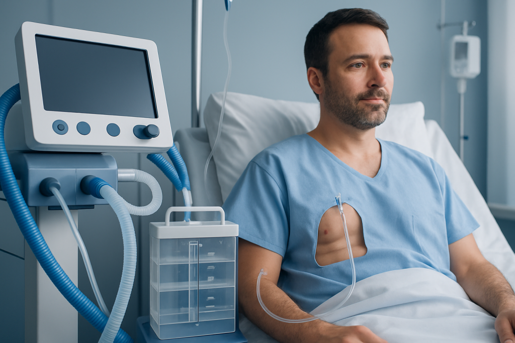
A chest tube is not a diagnosis; it is a treatment tool used when something inside the chest is preventing normal breathing or lung expansion. People may come to medical attention with shortness of breath, chest pain, a feeling of tightness, cough, fever, or reduced oxygen levels. In trauma cases, there may also be obvious injury to the chest or rib fractures.
Common situations include a pneumothorax, which is air in the pleural space, a hemothorax, which is blood in that space, or a pleural effusion, which is excess fluid. Infections such as empyema, where pus collects around the lung, may also require drainage. After some chest or lung operations, a temporary tube is often left in place to remove air and fluid during recovery.
Patients are usually evaluated with a physical examination and imaging before the team decides whether a chest tube is the best option. The symptoms often reflect the underlying problem rather than the tube itself, and urgent treatment aims to relieve pressure and support breathing.
Causes and risk factors

The need for a chest tube is linked to conditions that disrupt the normal balance inside the pleural space. A collapsed lung can happen after trauma, as a complication of certain medical procedures, or sometimes without a clear trigger, especially in taller, younger people or those with lung disease. Fluid buildup may be related to infection, heart failure, cancer, inflammatory disease, or liver or kidney conditions.
Risk factors vary by cause. Smoking, underlying lung disease, recent surgery, severe chest injury, mechanical ventilation, and bleeding disorders can all influence the likelihood of needing drainage or the complexity of care. For some patients, the risk is not only the problem itself but also delayed treatment after the first symptoms appear.
In international care settings, doctors also consider travel-related factors such as long-distance flying after recent lung problems, limited access to follow-up, and the timing of a planned procedure before returning home. These details help shape a safer treatment and discharge plan.
Diagnosis and treatment planning
Before a chest tube is placed, clinicians usually confirm the problem with imaging such as a chest X-ray, ultrasound, or CT scan. Blood tests may be ordered if infection, bleeding, or another systemic issue is suspected. The team also checks breathing status, oxygen levels, and whether the patient needs immediate intervention.
Planning includes choosing the insertion site, the tube size, and the method of pain control. Some patients receive local anesthesia alone, while others need sedation or more intensive monitoring. The medical team explains what will happen, what sensations are expected, and how the drainage system works so the patient and family know what to watch for afterward.
In many hospitals, chest tube care is protocol-driven, but good care is still individualized. The tube may be connected to a water-seal device or suction, depending on the goal of treatment and how the lung responds. The next steps are guided by repeat examinations and imaging.
Treatment options and what the procedure involves
During insertion, the skin is cleaned, numbed, and opened at a carefully selected site between the ribs. The tube is then advanced into the pleural space and secured in place with stitches and dressing. Once connected to a drainage system, it can remove air, blood, or fluid and help restore normal pressure around the lung.
The exact treatment plan depends on why the tube is needed. A small pneumothorax may sometimes be treated with observation or a needle procedure instead of a chest tube, while larger collections of air or fluid more often require tube drainage. If infection is present, antibiotics and sometimes additional procedures are added to address the source of the problem.
Most patients are monitored closely after placement. The care team looks at the amount and appearance of drainage, the patient’s breathing comfort, chest imaging, and whether the lung has re-expanded. The tube is removed when drainage decreases, air leaks stop, and the doctor is confident that the chest has stabilized.
Possible issues include pain, bleeding, infection, tube blockage, accidental displacement, or, more rarely, injury to nearby structures. Careful technique and monitoring reduce these risks, and patients are encouraged to tell staff about worsening pain, new shortness of breath, or changes at the insertion site.
Prevention and self-care
Not every chest tube can be prevented because some are needed after accidents or sudden illness. Still, lowering the chances of the conditions that lead to chest drainage can make a difference. Stopping smoking, managing chronic lung disease, treating infections early, and following surgical instructions carefully are all practical steps.
After a tube is placed, self-care is mostly about cooperation with the care team. Patients are often asked to keep the dressing clean and dry, avoid pulling on the tubing, and report sudden changes in pain or breathing. Gentle movement, as approved by the doctor, may help recovery, but activity limits depend on the underlying condition.
For patients returning home or traveling internationally, discharge instructions deserve extra attention. It helps to clarify who will remove the tube, when follow-up imaging is needed, what symptoms require urgent care, and how to reach the treating team if questions come up after the journey.
- Follow all dressing and drainage instructions exactly as given.
- Avoid smoking and secondhand smoke during recovery.
- Keep scheduled follow-up appointments, including imaging if recommended.
- Know the warning signs that require prompt medical review.
When to see a doctor
Medical attention is needed right away if breathing becomes more difficult, chest pain worsens, oxygen levels drop, or the tube seems to have moved out of place. Fever, increasing redness, swelling, foul-smelling drainage, or sudden bleeding around the site should also be evaluated promptly.
After the tube is removed, patients should still watch for returning shortness of breath, recurrent chest pain, or a feeling that the chest is not fully expanding. These symptoms may suggest that the original problem has returned or that another issue is developing. Prompt reassessment is especially important if the patient is far from the original treating hospital.
If a chest tube is being considered for an international patient, a clear plan for imaging, aftercare, and follow-up is essential before travel begins. Acibadem Health Point’s multidisciplinary specialists and JCI-accredited hospitals diagnose and treat chest tube-related conditions for international patients, helping coordinate care across the hospital stay and the return home.
Frequently asked questions
What is the main purpose of a chest tube?
The main purpose is to drain air, fluid, blood, or pus from around the lung so the lung can expand more normally. This helps relieve pressure and supports breathing while the underlying condition is treated.
Is chest tube insertion painful?
The procedure is usually done with local anesthesia, and some patients also receive sedation. There can still be pressure or soreness, especially afterward, but the team will work to keep discomfort controlled.
How long does a chest tube stay in place?
The length of time depends on why it was placed and how quickly the chest improves. Some tubes are removed after a short period, while others stay longer if drainage or air leakage continues.
Can a person move around with a chest tube?
Many patients can sit up and move with help once the medical team says it is safe. The tube and drainage system must be handled carefully to avoid pulling, kinking, or dislodging it.
What happens after the chest tube is removed?
The doctor may order a follow-up X-ray or exam to make sure the lung stays expanded and that fluid or air is not returning. Patients are usually given clear instructions about activity, wound care, and symptoms that should prompt medical review.
Can a chest tube be managed after hospital discharge?
In some situations, a patient may leave the hospital with a drainage device or with close outpatient follow-up, but this depends on the diagnosis and local practice. If that is planned, the patient should have a very clear aftercare and contact plan, especially when traveling from another country.
References
- Merck Manual Professional Edition
- Mayo Clinic
- NHS
- Cleveland Clinic
- American Thoracic Society
This article is for general information only and is not a substitute for professional medical advice. Please consult a qualified doctor about your individual situation.