Vasa Previa

Key Takeaways
- Vasa previa involves unprotected fetal blood vessels near the cervix, where they can be vulnerable during labor or membrane rupture.
- It is often found during prenatal ultrasound, especially when the placenta or cord placement suggests a higher risk.
- Care usually focuses on close monitoring, activity guidance when needed, and a planned cesarean birth before labor begins.
- Symptoms can be absent until membranes rupture, but vaginal bleeding, contractions, or sudden concerns in pregnancy need prompt medical review.
- Women with a prior cesarean, placenta issues, or multiple pregnancy may have a higher chance of developing vasa previa.
Vasa previa is a rare pregnancy complication in which fetal blood vessels cross or run near the cervix without the protection of the umbilical cord or placenta. Early diagnosis matters because careful monitoring and planned delivery can greatly reduce risk for the baby.
Overview
Vasa previa is a pregnancy condition in which fetal blood vessels travel across, or very close to, the opening of the cervix. These vessels are not protected by the umbilical cord or the placenta, so they can be damaged if the membranes rupture or labor begins.
Although the condition is uncommon, it is important because the baby’s blood volume can be affected quickly if one of these vessels tears. The reassuring part is that when vasa previa is identified before delivery, care can be planned in a way that strongly reduces the chance of an emergency.
For families arranging care from another country, the diagnosis often changes the whole delivery plan. Instead of waiting for labor at home, the team may recommend travel planning, timed follow-up scans, and delivery in a hospital that can respond immediately if needed.
Symptoms and Signs
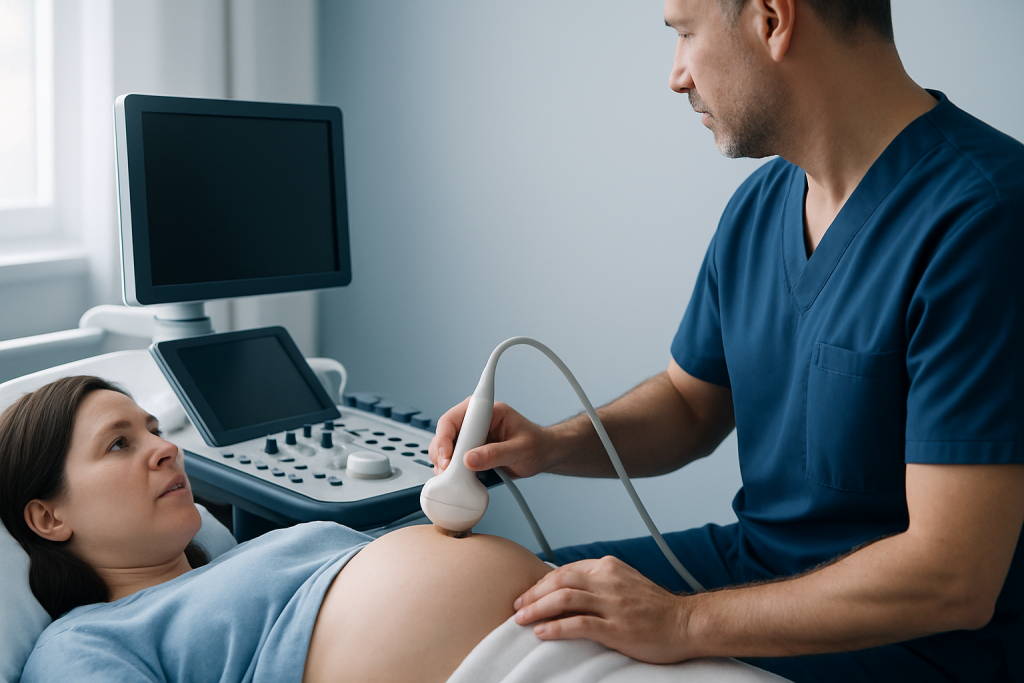
Vasa previa usually does not cause day-to-day symptoms during pregnancy. Many people feel entirely well until the condition is found on an ultrasound scan or until labor-related changes begin.
The most concerning signs are vaginal bleeding after the waters break, a sudden change in the baby’s heartbeat, or bleeding around the time of contractions or membrane rupture. Because fetal vessels are involved, even a small amount of bleeding in this setting deserves urgent medical assessment.
It is also important to distinguish vasa previa from other causes of bleeding in pregnancy. Not every episode of spotting means a serious problem, but any unexplained bleeding, fluid loss, or painful tightening should be discussed promptly with an obstetric team.
- Vaginal bleeding after membrane rupture
- Contractions or labor symptoms before the planned delivery date
- Abnormal fetal heart tracing on monitoring
- Ultrasound finding of vessels near the cervix
Causes & Risk Factors

Vasa previa develops when fetal blood vessels are inserted in an unusual way, such as into the membranes rather than directly into the placenta. These vessels may then pass over the cervix, especially when the placenta has a low position or there is a shared placenta in twin pregnancy.
Several pregnancy features are linked with a higher chance of this pattern. A low-lying placenta earlier in pregnancy, placenta previa, a velamentous cord insertion, assisted reproduction, and multiple pregnancy can all raise suspicion. A history of prior cesarean birth or uterine surgery may also be relevant because it can influence placental implantation.
Risk factors do not mean the condition will definitely occur. They simply help the care team decide who may need a more careful look with targeted ultrasound, especially in the second and third trimesters.
Diagnosis
Diagnosis is usually made before birth with ultrasound. A sonographer or obstetric specialist may use transvaginal ultrasound with color Doppler, which helps map blood flow and shows whether fetal vessels cross the cervical opening.
Routine anatomy scans can suggest the possibility of vasa previa when the placenta is low, the cord insertion looks unusual, or vessels appear near the lower uterus. In many cases, a follow-up scan is then arranged to confirm the findings and to see whether the placenta and vessels move away from the cervix as the pregnancy grows.
When diagnosis is clear, the care plan often includes repeat imaging, fetal monitoring, and delivery planning in a facility prepared for cesarean birth and newborn support. For Myomectomy Recovery for International Patients: The Usual Travel Timeline" class="ahp-ilk">international patients, this is the stage when the treating team may coordinate timing, records, and travel so that the birth occurs where the right specialists are already in place.
Treatment Options
There is no treatment that moves the vessels out of the way once vasa previa is present, so care is centered on prevention of vessel rupture and timely delivery. The exact plan depends on how close the vessels are to the cervix, whether symptoms appear, and how the pregnancy is progressing.
Many pregnancies are managed with increased surveillance and a scheduled cesarean birth before labor starts. Some people are advised to reduce physical strain or avoid certain activities if the obstetric team is concerned about early contractions or bleeding. In selected cases, hospital observation may be recommended later in pregnancy so that labor can be recognized and managed quickly.
Supportive care may also include counseling, repeat ultrasound, and planning for neonatal care after delivery. If bleeding, contractions, or ruptured membranes occur, the medical team may recommend urgent delivery rather than waiting.
- Planned cesarean delivery before labor
- Ongoing ultrasound and fetal monitoring
- Hospital-based observation in higher-risk situations
- Individualized activity guidance from the obstetric team
Prevention & Self-care
Vasa previa itself cannot usually be prevented, but earlier recognition can make a major difference. Attending scheduled prenatal scans and following through on recommended follow-up appointments are the most practical steps a pregnant person can take.
Self-care is mostly about staying alert to changes and communicating clearly with the care team. Any vaginal bleeding, fluid leakage, contractions, or reduced fetal movement should be reported without delay, even if the symptoms seem mild at first.
When a diagnosis has already been made, the safest approach is to follow the obstetric plan closely, including advice about travel, rest, and the timing of delivery. For those coming from abroad, carrying copies of imaging reports and having a clear hospital contact plan can make the final weeks of pregnancy less stressful and more organized.
When to See a Doctor
Medical review is needed right away if there is vaginal bleeding in pregnancy, especially after the waters break or if contractions start. Sudden abdominal pain, fluid leakage, or a change in fetal movement also warrants prompt assessment.
Even when symptoms are mild, it is sensible to contact the obstetric team if a scan has suggested low-lying placenta, velamentous cord insertion, or vessels near the cervix. These findings do not confirm vasa previa on their own, but they do justify a closer look.
People who are traveling for maternity care should seek a hospital where ultrasound, obstetric review, and emergency cesarean capability are available in the same setting. Acibadem Health Point’s multidisciplinary specialists and JCI-accredited hospitals diagnose and treat this condition for international patients, with coordinated care that supports both mother and baby.
Living with the Diagnosis and Planning Birth
A diagnosis of vasa previa often changes the emotional rhythm of pregnancy. Instead of an unpredictable waiting period, care becomes more structured, with scan dates, delivery planning, and clear thresholds for what should trigger hospital evaluation.
That structure is usually helpful. Many families feel more at ease once they understand why the team is recommending a planned cesarean birth and close monitoring rather than expecting labor to begin naturally.
Good communication matters, especially for patients who may be crossing borders for treatment. Written summaries, ultrasound images, and a clear plan for who to call if symptoms start can help the final weeks of pregnancy feel safer and more manageable.
Frequently asked questions
What is vasa previa in simple terms?
Vasa previa means that fetal blood vessels run near or across the cervix without the usual protection of the placenta or umbilical cord. The concern is that these vessels can be injured if labor starts or the membranes rupture. When it is found before birth, doctors can plan delivery carefully to lower risk.
Can vasa previa be seen on ultrasound?
Yes, it is often identified on ultrasound, especially when color Doppler is used to trace blood vessels near the cervix. A transvaginal scan may be needed for a clearer view. Follow-up imaging is common if the first scan raises concern.
Does vasa previa cause symptoms during pregnancy?
It often does not cause obvious symptoms until later in pregnancy or until the waters break. Some people first learn about it after an ultrasound, not because of how they feel. Any bleeding, fluid loss, or contractions in pregnancy should still be assessed promptly.
How is vasa previa treated?
Treatment focuses on protecting the vessels until delivery and preventing labor from starting unexpectedly. This usually means careful monitoring and a planned cesarean birth before labor begins. Some pregnancies also need hospital observation if the risk of early delivery is higher.
Is vaginal birth possible with vasa previa?
In most cases, vaginal birth is not recommended because labor or membrane rupture can damage the fetal vessels. A planned cesarean is usually the safest delivery method. The timing is individualized by the obstetric team.
Who is at higher risk of vasa previa?
Risk is higher when the placenta is low-lying, the cord inserts abnormally, there is a shared placenta in twins, or conception occurred with assisted reproduction. A prior cesarean or uterine surgery may also be relevant. These factors do not mean the condition will occur, but they help guide screening.
References
- American College of Obstetricians and Gynecologists
- Royal College of Obstetricians and Gynaecologists
- Mayo Clinic
- Cleveland Clinic
- National Health Service
This article is for general information only and is not a substitute for professional medical advice. Please consult a qualified doctor about your individual situation.
More from the Health Library



Related Specialists

Prof. Dr. A. Taner Usta
Gynecology & Obstetrics
Prof. Dr. Ahmet Tayyar
Gynecology & Obstetrics
Prof. Dr. Ahmet Cem Batukan
Gynecology & Obstetrics
Dr. Sevgi Selen
Gynecology & Obstetrics