Ultrasound Tech: What They Do and How to Become One

Key Takeaways
- Ultrasound uses sound waves, not radiation, to produce images in real time.
- It can help evaluate many parts of the body, including pregnancy, the abdomen, blood vessels, and soft tissues.
- Preparation depends on the area being examined and may include fasting or a full bladder.
- Ultrasound is usually quick, noninvasive, and performed by trained imaging professionals.
- Results are interpreted by a doctor who combines them with symptoms and other tests.
Medically reviewed by the Acıbadem clinical team — August 19, 2026
Ultrasound technology uses sound waves to create real-time images of organs, tissues, and blood flow without radiation. It is widely used because it is versatile, comfortable for most patients, and often one of the first steps in diagnosis and follow-up care.
Overview
Ultrasound technology, often called sonography, helps clinicians look inside the body without making an incision and without using radiation. A handheld device called a transducer sends sound waves into the body and receives the returning echoes, which are translated into images on a screen.
For many patients, ultrasound is the first imaging test considered because it is accessible, gentle, and useful across a wide range of medical questions. It can help assess abdominal pain, monitor pregnancy, examine the thyroid or breast, evaluate veins and arteries, and guide certain procedures in real time.
In international care settings, ultrasound also plays a practical role because it is quick, repeatable, and often available soon after a consultation. That makes it useful when a patient is traveling for care and needs an efficient way to move from symptoms to diagnosis and treatment planning.
How Ultrasound Works
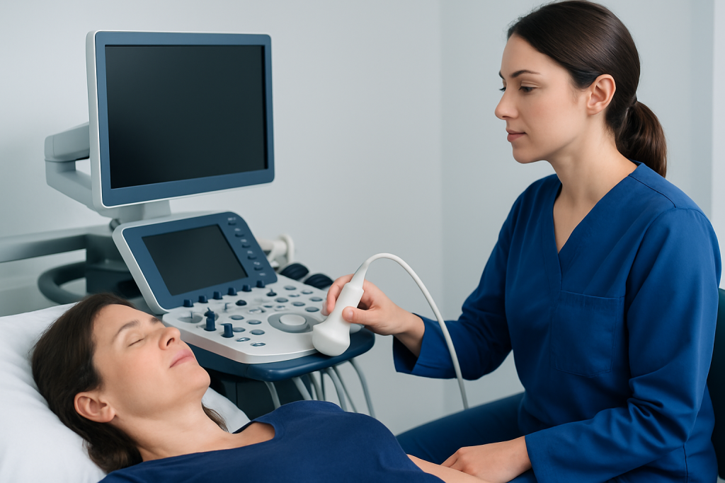
Ultrasound machines convert electrical energy into high-frequency sound waves that are too high for the human ear to hear. As those waves travel through tissues, some are reflected back differently depending on the type of tissue they encounter, such as fluid, soft tissue, or dense structures.
The transducer captures those echoes and the machine builds a moving image in real time. Because the image updates immediately, clinicians can observe motion such as blood flow, a heartbeat, swallowing, breathing movement, or the way a structure responds during the exam.
Different ultrasound techniques can answer different questions. Standard two-dimensional imaging is common, while Doppler ultrasound measures blood flow and color Doppler can show direction and speed of circulation. In some cases, the exam is used to guide a needle or other instrument with precision.
Common Uses

Ultrasound is used in many areas of medicine because it is adaptable. In pregnancy care, it helps confirm gestational age, check fetal growth, and assess the placenta and amniotic fluid. In abdominal imaging, it can evaluate the gallbladder, liver, kidneys, pancreas, bladder, spleen, and reproductive organs.
It is also useful for looking at the neck, thyroid, breast tissue, muscles, tendons, joints, and soft-tissue lumps. Vascular ultrasound can assess veins for clots and arteries for narrowing or poor circulation. In emergency and procedural settings, clinicians may use ultrasound to guide drainage, injections, biopsies, or line placement.
Because ultrasound provides a live image, it is often chosen when a symptom needs a focused answer rather than a broad whole-body scan. The exam is commonly selected after a clinical visit where the doctor wants to match the imaging request to the patient’s story and physical findings.
What to Expect During the Exam
Most ultrasound examinations are performed in a quiet imaging room and usually take place while the patient lies on an exam table. A technologist or sonographer applies a warm gel to the skin so the transducer can glide smoothly and transmit sound waves effectively.
The transducer is then moved over the area being studied, sometimes with light pressure. The room may be dimmed so the screen is easier to view, and patients may be asked to hold their breath briefly or change position to improve image quality. The exam is generally painless, although some pressure may feel uncomfortable if the area is tender.
Many people are surprised by how simple the process is. Once the images are captured, the gel is wiped away and patients can usually return to normal activities right away unless their doctor has given different instructions based on the reason for the test.
Preparation and Safety
Preparation depends on the body part being examined. Some ultrasounds require fasting for several hours, especially when the gallbladder or upper abdomen is being studied. Others may call for a full bladder, which can help move the uterus or pelvic organs into clearer view.
It is helpful to bring prior imaging reports, a list of current medications, and any relevant medical records if the patient is traveling for care. A patient should also tell the imaging team if they are pregnant, have had recent surgery, wear a cast or dressing, or have any discomfort that could affect positioning during the exam.
Ultrasound is widely considered safe because it does not use ionizing radiation. For that reason, it is commonly used in pregnancy and repeated when needed for follow-up, provided the test is medically appropriate and performed by trained professionals.
Understanding the Results
The technologist usually captures the images, but the interpretation is made by a radiologist or another qualified doctor. The report may describe the size, shape, texture, and appearance of an organ or structure, along with any visible fluid collection, mass, inflammation, or blood-flow changes.
Results are not meant to stand alone. A doctor combines the ultrasound findings with symptoms, physical examination, lab tests, and prior studies to decide whether the picture suggests a normal variation, a condition that needs monitoring, or a problem that needs treatment.
In some cases, the exam gives an immediate answer. In others, it narrows the possibilities and helps guide the next step, such as more imaging, a specialist visit, or a procedure. This stepwise approach can be especially valuable for patients arranging care from another country, because it helps keep the diagnostic path organized and efficient.
After the Exam: Recovery, Follow-up, and Patient Tips
Most people recover from ultrasound with no downtime at all. There is no needle involved in standard external ultrasound, and any gel or residual moisture can be wiped off after the exam. If a transvaginal, transrectal, or procedure-guiding ultrasound is planned, the care team will explain what to expect beforehand.
Patients can help make the experience smoother by arriving a little early, bringing the correct paperwork, and following preparation instructions closely. If language is a concern, it may help to ask in advance about interpreter support so that instructions and results are understood clearly.
- Keep the appointment notes and prior reports together in one folder.
- Ask which body area is being examined and why the test was ordered.
- Clarify when and how results will be shared.
- Follow any special eating, drinking, or bladder instructions carefully.
For international patients, planning the scan, interpretation, and follow-up discussion in one coordinated pathway can reduce delays and help the next decision feel more manageable. Acibadem Health Point’s multidisciplinary specialists and JCI-accredited hospitals diagnose and treat this condition for international patients.
When to See a Doctor
A doctor should be consulted if symptoms persist, worsen, or need clarification after an initial ultrasound. Persistent abdominal pain, new swelling, unexplained lumps, pelvic symptoms, breathing concerns, or changes in circulation deserve proper medical assessment rather than guesswork.
It is also important to speak with a clinician if the result is unclear, if the imaging report mentions a finding that needs follow-up, or if symptoms do not match the test result. Ultrasound is a valuable tool, but it works best when used as part of a broader medical evaluation.
Patients who are traveling for treatment should ask how soon they should review the report, whether another test is needed before returning home, and which doctor will coordinate ongoing care. Clear planning makes follow-up safer and helps avoid gaps once the patient leaves the hospital system.
Frequently asked questions
What is ultrasound tech used for?
Ultrasound technology is used to create real-time images of organs, blood vessels, and soft tissues. It helps doctors evaluate symptoms, monitor pregnancy, and guide certain procedures.
Is an ultrasound painful?
Most ultrasound exams are not painful. Patients may feel mild pressure from the transducer, especially if the area being examined is tender or if a full bladder is needed.
Do I need to fast before an ultrasound?
Sometimes, yes, but only for certain exams such as upper abdominal or gallbladder imaging. The imaging team will give specific instructions based on the area being studied.
How long does an ultrasound take?
Many ultrasound exams take about 15 to 45 minutes, although the exact time depends on the area being examined and how detailed the study needs to be. Complex or guided procedures may take longer.
Is ultrasound safe during pregnancy?
Ultrasound is widely used during pregnancy because it does not use ionizing radiation. It should still be performed only when medically appropriate and by trained professionals.
Will I get the results right away?
Sometimes a preliminary impression is available quickly, but the final report is usually reviewed by a radiologist or doctor. The timing depends on the facility and the reason for the test.
References
- Radiological Society of North America
- American College of Radiology
- U.S. Food and Drug Administration
- World Health Organization
This article is for general information only and is not a substitute for professional medical advice. Please consult a qualified doctor about your individual situation.
Explore treatments in Turkey — costs, top hospitals & a free quote
JCI-accredited · board-certified surgeons · reply within 24h
Add us as a Preferred Source to see more of our trusted health content across Google Search, AI Overviews and Discover.