Skin Cancer Pictures

Key Takeaways
- Pictures can help people learn the visual patterns of common skin cancers, but they cannot confirm a diagnosis.
- Changes in size, shape, color, border, or healing are more important than any single image.
- Melanoma, basal cell carcinoma, and squamous cell carcinoma may each look different, and some do not match textbook examples.
- Dermatology examination, dermoscopy, and biopsy are the main ways skin cancer is diagnosed.
- Early medical review is especially important for new, changing, bleeding, or non-healing skin lesions.
Skin cancer pictures can be a useful starting point for noticing changes in moles, spots, or sores that do not look right. Because many harmless skin findings can resemble cancer, the safest approach is to use images as a guide and have any concerning change checked by a qualified doctor.
Overview
Looking at skin cancer pictures can help a person notice when a spot deserves closer attention. The value of images is not to replace a medical visit, but to make changes on the skin easier to recognize, especially when a person has many moles, freckles, sun spots, or long-standing marks that blend into the background.
Skin cancer does not always follow one neat visual pattern. Some lesions are dark, some are pink or red, some look pearly, and some simply fail to heal. That is why a cautious approach works best: compare a spot with the skin around it, track whether it changes over time, and ask a doctor to examine anything that appears new, unusual, or persistent.
For international patients planning care from abroad, pictures often play an early role in deciding whether a lesion should be assessed in person soon. A first opinion may start with photographs, but proper diagnosis usually requires a skin examination and sometimes a biopsy. Images are most useful when they prompt timely evaluation rather than self-diagnosis.
What skin cancer can look like
In photographs, melanoma is often discussed first because it can be more aggressive and because its appearance can be varied. Some melanomas are irregularly pigmented with shades of brown, black, tan, blue, gray, or even red. Others are amelanotic, meaning they may not look dark at all and can resemble a pink bump, a sore, or an inflamed patch.
Basal cell carcinoma often appears as a pearly or shiny bump, a sore that keeps crusting and reopening, or a flat pink area that slowly enlarges. It may have visible small blood vessels on the surface. Squamous cell carcinoma more often looks like a scaly, crusted, thickened, or wart-like lesion, and it can become tender or bleed more easily than expected.
A helpful visual clue is not just what a lesion looks like, but how it behaves. A changing mole, a spot that becomes raised, a patch that does not heal after several weeks, or a lesion that bleeds with minimal irritation should all be taken seriously. Harmless findings can look odd too, so the goal is not to guess from an image, but to notice when the skin is sending a signal that needs review.
- Warning pattern: change over time
- Warning pattern: asymmetry or uneven borders
- Warning pattern: color variation in one lesion
- Warning pattern: bleeding, crusting, or non-healing
Symptoms and visual warning signs
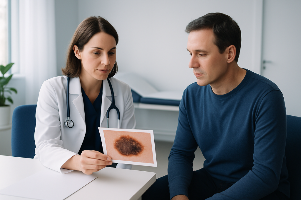
Many people first become concerned when a spot looks different from their other moles or freckles. The classic ABCDE approach for melanoma can be a practical memory aid: asymmetry, border irregularity, color variation, diameter that is larger or increasing, and evolution, meaning change over time. Evolution is especially important because a stable mole is less concerning than one that is clearly changing.
Other visual signs are also worth noticing. A new spot that appears after childhood, a lesion with an unusual color for that person’s skin tone, or a patch that seems to lose its normal skin lines may deserve an examination. Some skin cancers are itchy or sore, but many are painless, so the absence of discomfort does not rule them out.
Patients who are reviewing skin cancer pictures online sometimes worry about every mole that looks imperfect. That reaction is understandable, but most skin marks are benign. A good rule is to pay the most attention to lesions that are new, growing, changing, bleeding, crusting, or failing to heal rather than to every spot that looks a little different.
Causes and risk factors
Ultraviolet exposure from the sun or tanning beds is one of the most important risk factors for skin cancer. The risk rises over time with repeated exposure, sunburns, and outdoor work or recreation without adequate protection. People with fair skin can be at higher risk, but skin cancer can occur in all skin tones, and darker skin can sometimes delay recognition because early lesions may be less obvious.
Other risk factors include a personal or family history of skin cancer, many moles, atypical moles, weakened immune function, and a history of radiation exposure or certain genetic conditions. Age can also play a role, although younger adults are not exempt. A person’s pattern of sun exposure matters as much as the total amount, especially if there were intense burns during childhood or early life.
International patients often ask whether a spot that developed after a holiday or move to a sunnier climate is concerning. The answer depends on the lesion’s behavior, not only on the travel history. A new or changing lesion after increased sun exposure should be assessed, particularly if it differs from the person’s other skin findings.
How doctors diagnose it
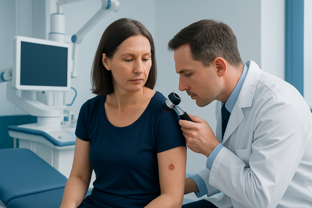
A doctor usually begins with a visual skin examination and questions about how long the spot has been present, whether it has changed, and whether there is any personal or family history of skin cancer. Dermoscopy, a handheld magnifying tool with a light, may help reveal patterns under the skin surface that are not visible to the naked eye. This can improve decision-making, but it still does not replace tissue diagnosis when suspicion remains.
If a lesion looks concerning, the next step is often a biopsy. During a biopsy, a small sample of the skin, or sometimes the whole lesion, is removed and examined under a microscope. This is the most reliable way to confirm whether a lesion is cancerous and, if so, what type it is.
When a patient is traveling for care, it can be helpful to bring clear photos showing how the lesion looked at different times, along with any prior reports if a previous doctor has already examined it. Those details can help the specialist judge whether the lesion has been stable or evolving, which is often just as important as the current appearance.
Treatment options
Treatment depends on the type of skin cancer, its size, its location, and whether it has spread beyond the skin. For many localized skin cancers, surgery is the main treatment. In some cases, the lesion can be removed with a margin of healthy tissue to lower the chance of recurrence. On the face, ears, or other cosmetically sensitive areas, surgeons may use techniques that aim to preserve as much normal skin as possible.
Some patients may be treated with Mohs micrographic surgery, a method that removes skin cancer in stages while checking the edges during the procedure. This can be especially useful when tissue preservation matters or when the cancer has a higher chance of coming back. Other options may include topical therapies, cryotherapy, curettage and electrodessication, radiation therapy, systemic treatment, or immunotherapy, depending on the diagnosis and stage.
For international patients, treatment planning often includes more than the procedure itself. It may involve arranging pathology review, discussing recovery before travel, and making sure follow-up is feasible once home. A good plan includes clear wound care instructions, expected healing time, and a schedule for future skin checks.
Prevention and self-care
Sun protection remains the most practical everyday step. Using broad-spectrum sunscreen, wearing protective clothing, seeking shade, and avoiding tanning beds can reduce ultraviolet damage over time. These habits matter for children and adults alike, because skin damage accumulates across the years rather than appearing all at once.
Self-examination can also help. People can check their skin monthly in good light, looking at the face, scalp, ears, back, chest, arms, legs, palms, soles, and between the toes. Mirrors or help from a family member can make it easier to notice spots in hard-to-see areas. A simple photo log can be useful when a lesion is being watched by a doctor, but self-monitoring should not delay evaluation if a spot changes.
After a skin cancer treatment, following wound care instructions and attending follow-up visits are important parts of recovery. Patients traveling from another country should clarify how they will receive pathology results, whom to contact if healing is not as expected, and when the next skin surveillance visit should occur. Acibadem Health Point’s multidisciplinary specialists and JCI-accredited hospitals diagnose and treat skin cancer for international patients, which can make coordinated follow-up easier to arrange.
When to see a doctor
A skin lesion should be checked if it is new and persistent, changing in appearance, bleeding without clear cause, crusting repeatedly, or not healing within a reasonable period. The same applies to a mole that looks unlike the person’s other moles or to a spot that has become noticeably different in shape, color, or texture.
Prompt review is also sensible when a lesion appears in a high-risk person, such as someone with a history of skin cancer, significant sun exposure, many atypical moles, or a weakened immune system. If there is uncertainty, it is generally safer to book an assessment than to continue watching it indefinitely.
Skin cancer pictures can be a helpful educational tool, but they are best used as a prompt for professional assessment, not as a final answer. A qualified doctor can determine whether the lesion is harmless, needs close observation, or should be removed and tested.
Living with uncertainty while waiting for review
Waiting for an appointment can be stressful, especially when a person has seen photographs online that seem worrying. It can help to remember that many skin conditions share overlapping features, and the visible appearance alone cannot tell the full story. Keeping notes about when the spot first appeared and whether it has changed can make the consultation more efficient and less stressful.
Patients traveling for care may also want to prepare practical questions in advance: What is the likely diagnosis? Is a biopsy needed? If treatment is required, how long is recovery and when is it safe to travel? Asking these questions early helps create a realistic plan and reduces avoidable delays.
With careful evaluation, most concerning skin findings can be sorted out clearly. The main goal is simple: identify the spots that need action and avoid unnecessary worry about the ones that do not.
Frequently asked questions
Can skin cancer pictures tell if a spot is cancerous?
Not reliably. Pictures can help a person notice patterns that deserve medical review, but only a clinician examination and, when needed, a biopsy can confirm the diagnosis.
What does melanoma usually look like in photos?
Melanoma may look like an irregular mole with uneven borders, mixed colors, or a spot that changes over time. Some melanomas are not dark and may appear pink, red, or skin-colored.
Are basal cell carcinoma pictures easy to recognize?
Basal cell carcinoma can sometimes look like a pearly bump, a shiny patch, or a sore that heals and reopens. However, it can also look subtle, so a medical exam is still important.
What should a person do if a mole changes shape or color?
A changing mole should be checked by a doctor, especially if the change is recent or continues to progress. It is better to arrange an assessment sooner rather than wait for it to settle on its own.
Can skin cancer happen on darker skin?
Yes. Skin cancer can affect all skin tones, and it may be easier to miss on darker skin because early changes can be less obvious. Any new, persistent, or unusual lesion should be examined.
Is a non-healing sore always skin cancer?
No, many non-healing sores are caused by other skin problems. Still, a sore that does not improve after several weeks, crusts repeatedly, or bleeds easily should be evaluated.
References
- American Academy of Dermatology
- National Cancer Institute
- World Health Organization
- American Cancer Society
- Skin Cancer Foundation
This article is for general information only and is not a substitute for professional medical advice. Please consult a qualified doctor about your individual situation.