Ple

Key Takeaways
- Pleural effusion means fluid has collected between the lungs and chest wall.
- Shortness of breath, chest discomfort, and a dry cough are common warning signs.
- Many different conditions can cause it, including infection, heart failure, liver disease, and cancer.
- Diagnosis often includes imaging and sometimes a procedure to sample the fluid.
- Treatment depends on the cause and may involve draining fluid, medicines, or treating the underlying illness.
Pleural effusion is the buildup of extra fluid in the space around the lungs, which can make breathing feel harder and more uncomfortable. With the right tests, clinicians can identify the cause and guide treatment that focuses on both relief and the underlying condition.
Overview
Pleural effusion is the medical term for fluid that collects in the pleural space, the thin area between the lungs and the inside of the chest wall. A small amount of lubrication is normal there; the problem begins when the balance of fluid production and absorption is disrupted and the volume grows enough to interfere with comfortable breathing.
For many people, pleural effusion is not a disease on its own but a clue that something else is happening in the body. That is why care is usually focused on two goals at the same time: easing symptoms and finding the underlying reason the fluid appeared. In an international-patient setting, this often means coordinating imaging, specialist review, and if needed a same-day or next-day procedure, so the care plan is clear before travel or follow-up begins.
Effusions can be small and discovered incidentally on an X-ray, or large enough to cause noticeable shortness of breath. The amount of fluid does not always match how unwell a person feels, which is one reason professional assessment matters even when symptoms seem modest.
Symptoms
The first sign is often breathlessness that feels worse with activity or when lying flat. Some people notice a dry cough, a sense of chest pressure, or discomfort that deepens with a full breath. If the effusion is large, the affected side of the chest may feel heavy or tight.
Symptoms can develop gradually or more suddenly, depending on the cause. When fluid builds up because of infection or inflammation, fever, chills, or general fatigue may also appear. When the cause is a chronic condition such as heart failure, symptoms may come with swelling in the legs, reduced exercise tolerance, or waking at night feeling short of breath.
Not every pleural effusion causes obvious symptoms. Small collections may be discovered during evaluation for another issue, which is one reason chest imaging is often part of the diagnostic pathway when breathing changes do not have a simple explanation.
Causes & Risk Factors
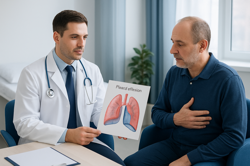
Pleural effusion can develop for many reasons. Common causes include heart failure, pneumonia, tuberculosis, pulmonary embolism, Kidney Disease Treatment" class="ahp-ilk">kidney disease, liver disease, and cancers that affect the lungs or pleura. Autoimmune conditions, such as rheumatoid arthritis or lupus, can also be involved. In some cases, the fluid is related to recent surgery, trauma, or blood clots.
Clinicians often classify pleural fluid as transudative or exudative, a distinction that helps narrow the cause. Transudative effusions are usually linked to pressure changes in the circulation, while exudative effusions often reflect inflammation, infection, or direct irritation of the pleura. This classification is one of the reasons fluid sampling can be so useful.
Risk increases when a person already has a condition that affects the heart, lungs, liver, kidneys, or immune system. Smoking history, recent infection, certain medications, and prior cancer treatment may also influence the likelihood of pleural fluid developing. For travelers seeking care abroad, it helps to bring a list of previous diagnoses, medications, imaging reports, and recent test results, since these can shorten the path to a precise explanation.
Diagnosis
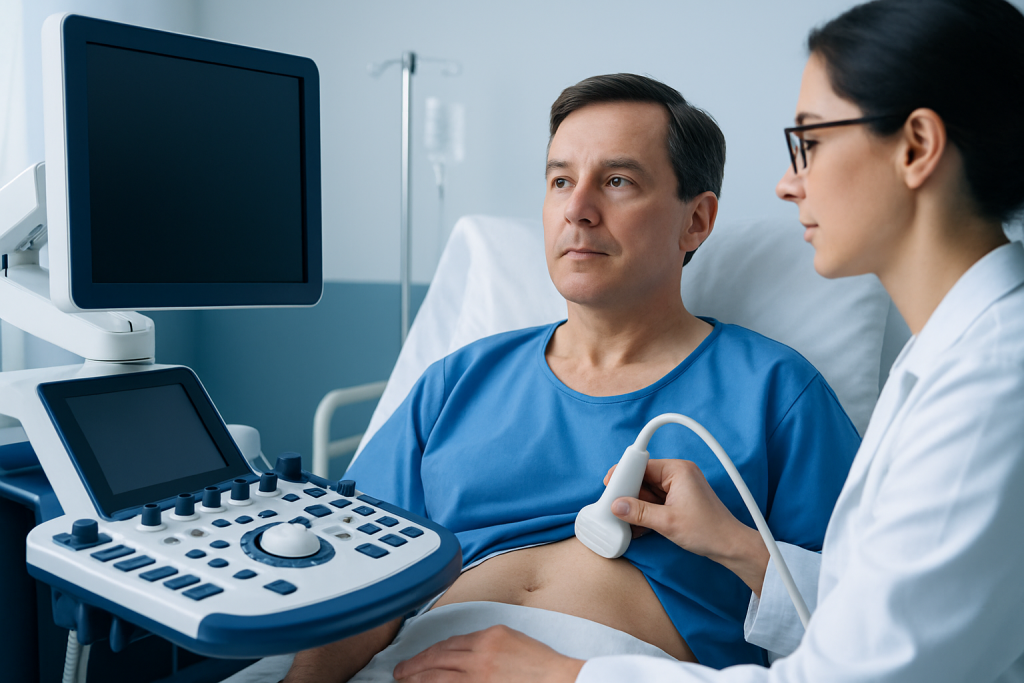
Evaluation usually begins with a medical history, physical examination, and imaging. A chest X-ray may show fluid, while ultrasound can better estimate its size and guide procedures. CT scanning may be used when clinicians need more detail about the lungs, pleura, or surrounding structures.
If the cause is not already obvious, thoracentesis may be recommended. During this procedure, a clinician removes some fluid with a needle and sends it to the laboratory for analysis. Testing can look at protein levels, cell counts, infection markers, pH, glucose, and the presence of abnormal cells, all of which help determine why the fluid formed.
Additional blood tests or specialized studies may be ordered depending on the suspected cause. For example, heart-related effusions may prompt cardiac testing, while infection-related cases may need microbiology studies. The aim is not simply to confirm fluid is present, but to understand what is driving it so treatment can be targeted.
Treatment Options
Treatment depends on the cause, the size of the effusion, and how much it affects breathing. If the effusion is small and symptoms are minimal, clinicians may monitor it while treating the underlying condition. When breathing is affected, draining the fluid can bring quicker relief.
Thoracentesis can relieve pressure and also provide diagnostic information. In some recurrent or complex cases, a chest tube, pleurodesis, or an indwelling pleural catheter may be considered to control repeated fluid buildup. These options are chosen based on the underlying diagnosis, expected course, and how likely the effusion is to return.
Medicine may also play an important role. Antibiotics are used for bacterial infections, diuretics may help in heart-failure-related effusions, and treatment for cancer, inflammation, or clotting disorders may be needed when those are the cause. The most effective plan is usually individualized rather than based on the fluid alone.
Prevention & Self-care
Not every pleural effusion can be prevented, but many can be reduced in likelihood by managing the conditions that commonly lead to it. That includes following treatment plans for heart failure, liver disease, kidney disease, autoimmune disorders, and lung infections as advised by a clinician.
Self-care is mainly about steady, practical habits that support recovery and help symptoms stay visible rather than ignored. Resting when breathless, avoiding smoking, keeping follow-up appointments, and tracking changes in breathing or swelling can all be helpful. If a clinician has recommended fluid restriction, salt control, or medications, it is best to follow those instructions consistently rather than adjusting them on one’s own.
- Keep a written list of symptoms, medication changes, and recent illnesses.
- Carry copies of imaging and procedure reports when traveling for care.
- Ask whether follow-up imaging will be needed after treatment.
- Seek prompt review if breathlessness returns or worsens.
For patients receiving care away from home, planning the return journey around recovery time is important. A stable discharge plan, clear instructions for wound care or catheter care if applicable, and a contact path for follow-up can make the post-treatment period much smoother.
When to See a Doctor
Medical evaluation is advisable if breathing becomes more difficult, chest discomfort appears, or cough persists without a clear explanation. It is especially important to seek care when symptoms are new, worsening, or occurring alongside fever, swelling, weight loss, or a known heart, lung, liver, or cancer condition.
Urgent attention is appropriate if shortness of breath is severe, if the lips or fingertips look bluish, or if chest pain is intense or sudden. These symptoms do not always mean pleural effusion is the cause, but they do need timely assessment.
Because pleural effusion is often a sign of another medical issue, getting checked early can simplify the workup and help prevent complications. In complex or recurrent cases, Acibadem Health Point’s multidisciplinary specialists and JCI-accredited hospitals diagnose and treat pleural effusion for international patients with coordinated care from evaluation through follow-up.
Living With the Diagnosis
When pleural effusion is linked to a longer-term condition, follow-up becomes part of everyday care rather than a one-time event. Clinicians may watch for recurrence, adjust medicines, or repeat imaging to see whether the fluid is resolving. This can feel like a lot of moving pieces, but the plan usually becomes more straightforward once the cause is identified.
Patients often do best when they understand what improvement should look like and what changes should trigger a call to the care team. Breathing patterns, exercise tolerance, and the return of chest heaviness are useful signals. For people recovering abroad, receiving written instructions in clear language can be especially helpful during the journey home and the first weeks after treatment.
Most importantly, pleural effusion should be treated as a measurable medical finding, not a vague symptom to be tolerated. With proper evaluation and targeted care, many people experience relief and a clearer path forward.
Frequently asked questions
Is pleural effusion the same as fluid in the lungs?
Not exactly. Pleural effusion is fluid in the space around the lungs, not inside the air sacs of the lungs themselves. The location matters because it affects both symptoms and treatment choices.
Can pleural effusion go away on its own?
Sometimes a small effusion related to a temporary issue may improve as the underlying problem is treated. However, many cases need medical evaluation to identify why the fluid formed in the first place.
Is pleural effusion always serious?
It is not always an emergency, but it should never be ignored. The seriousness depends on the cause, the amount of fluid, and how much it affects breathing.
Does drainage cure pleural effusion?
Drainage can relieve pressure and help with diagnosis, but it does not always cure the problem. If the underlying cause remains active, the fluid may return unless that condition is treated too.
What tests are most commonly used to diagnose it?
Chest imaging is usually the first step, followed by fluid sampling if needed. Blood tests and sometimes CT scanning or other specialist tests help identify the cause.
Can someone travel after treatment for pleural effusion?
Many people can travel after they are medically stable, but the timing depends on the cause, treatment received, and whether follow-up is needed. The care team should give individualized guidance before any trip home.
References
- Mayo Clinic
- National Heart, Lung, and Blood Institute
- Merck Manual Professional Edition
- American Thoracic Society
- Cleveland Clinic
This article is for general information only and is not a substitute for professional medical advice. Please consult a qualified doctor about your individual situation.