Papilledema

Key Takeaways
- Papilledema is a sign, not a diagnosis, and it often points to increased pressure around the brain.
- Symptoms may include blurred vision, brief episodes of visual dimming, headaches, or double vision, but some people have few eye symptoms at first.
- Diagnosis usually involves a dilated eye exam and tests such as brain imaging, lumbar puncture, or visual field testing.
- Treatment focuses on the cause and may include medication, weight management, surgery, or other specialist care.
- Protecting vision depends on prompt assessment and regular follow-up, especially if symptoms change.
Papilledema is swelling of the optic disc, the point where the optic nerve enters the eye, and it usually signals raised pressure inside the head. Because the underlying cause may affect vision or brain health, timely evaluation by an eye or neurological specialist is important.
Overview
Papilledema describes swelling of the optic disc, the area where the optic nerve connects to the back of the eye. It happens when pressure surrounding the optic nerve is elevated, often because the pressure inside the skull is higher than normal.
This finding matters because papilledema is usually a clue to an underlying condition rather than a disease on its own. Some causes are temporary and treatable, while others need urgent attention from a neurologist, ophthalmologist, or both.
For patients who are considering care far from home, papilledema is one of those conditions where the plan is usually straightforward but carefully coordinated: confirm the cause, protect vision, treat what is driving the pressure, and arrange follow-up so improvement can be tracked over time.
Symptoms
Symptoms can vary widely. Some people notice changes in vision first, while others discover papilledema during an eye examination done for headaches or another reason. In early stages, vision may still seem fairly normal even when swelling is present.
Common symptoms and warning signs may include:
- Blurred or intermittently dim vision
- Brief episodes of vision loss or graying out, often when changing position
- Headache, sometimes worse in the morning or when straining
- Double vision, especially if a nearby nerve is affected
- Ringing in the ears that seems to match the heartbeat
- Nausea or vomiting in some cases
Because papilledema can develop with subtle or nonspecific complaints, an eye exam is often the first place the pattern is recognized. That is one reason persistent headaches with visual changes deserve a professional assessment rather than watchful waiting alone.
Causes & Risk Factors

Papilledema usually reflects raised intracranial pressure, but the reason for that pressure can differ. A blocked pathway for cerebrospinal fluid, a mass or growth in the brain, bleeding, severe hypertension, inflammation, or infection may all contribute depending on the clinical picture.
One common cause in otherwise healthy adults is idiopathic intracranial hypertension, a condition in which pressure is elevated without a clear structural cause on initial testing. It is seen more often in certain groups, including people with higher body weight and some women of childbearing age, though it can occur in others as well.
Risk factors are not the same as causes, but they help guide evaluation. A recent medication change, history of clotting problems, pregnancy, autoimmune disease, or symptoms suggesting a nervous system disorder may influence the workup. The key point is that papilledema always deserves a search for the reason behind it.
Diagnosis
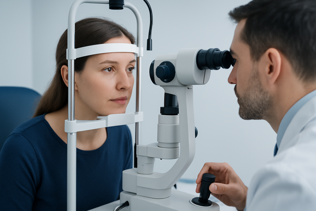
Diagnosis begins with a careful eye examination. An ophthalmologist typically looks at the optic disc with dilation and may use imaging tools such as optical coherence tomography to document swelling and monitor it over time.
Because the source of the problem is often outside the eye, the next steps may include neuroimaging, usually MRI or CT depending on the situation. These tests help look for structural causes, bleeding, or signs of increased pressure that could explain the optic nerve swelling.
In many cases, a lumbar puncture is performed after imaging to measure cerebrospinal fluid pressure and analyze the fluid if needed. Visual field testing may also be used to check whether the swelling is affecting peripheral vision, which can be missed in everyday life. The exact order of tests depends on the person’s symptoms, examination findings, and urgency.
Treatment Options
Treatment is directed at the cause rather than the swelling itself. If a tumor, clot, infection, or other condition is found, specialists treat that specific problem first because lowering the underlying pressure is what protects the optic nerve.
When idiopathic intracranial hypertension is the diagnosis, treatment may include weight management, medicines that reduce cerebrospinal fluid pressure, and close monitoring of vision. In selected situations, procedures such as optic nerve sheath fenestration, cerebrospinal fluid shunting, or venous sinus stenting may be discussed by the care team.
The most appropriate choice depends on how severe the papilledema is, how much vision has changed, and how quickly pressure needs to be controlled. For international patients, this often means care from more than one specialist, with the plan tailored to both immediate treatment and safe follow-up after returning home.
Prevention & Self-care
Not every case of papilledema can be prevented, because some causes are unpredictable. Even so, people can support better outcomes by paying attention to new headache patterns, visual dimming, or double vision and arranging medical review early.
Self-care is mainly about reducing strain on the eyes and avoiding delays in treatment. It helps to keep appointments, take prescribed medicines as directed, and report any change in vision promptly. If a clinician recommends weight management, gradual and sustainable changes are generally more useful than short-term crash approaches.
Practical steps that may help during recovery include:
- Using consistent follow-up with the eye or neurology team
- Tracking symptoms such as headache frequency or visual episodes
- Avoiding activities that worsen symptoms until cleared by a doctor
- Bringing prior scans, records, and medication lists to every appointment
Patients traveling for care may find it useful to ask for written instructions and a clear follow-up plan before leaving. That makes it easier to continue monitoring once they are back home.
When to See a Doctor
Medical evaluation should not be delayed if papilledema is suspected, especially when headache is paired with blurred vision, transient vision loss, or double vision. Even if symptoms seem mild, optic disc swelling can still indicate a problem that needs prompt attention.
Immediate medical care is especially important when symptoms appear suddenly, worsen quickly, or are accompanied by confusion, weakness, fainting, fever, or severe vomiting. These features can point to conditions that require urgent assessment.
People already diagnosed with papilledema should contact their doctor sooner rather than later if vision changes, headaches intensify, or new neurological symptoms appear. Timely follow-up is one of the best ways to preserve vision and avoid missing the underlying cause.
At Acibadem Health Point, multidisciplinary specialists and JCI-accredited hospitals diagnose and treat papilledema for international patients, with coordinated evaluation that can include eye care, neurology, imaging, and follow-up planning.
Living With Papilledema
Living with papilledema is often less about the eye swelling itself and more about understanding the condition that caused it. Many patients need repeated checks of vision and optic nerve appearance while treatment is adjusted, and that monitoring can continue even after symptoms improve.
It can be helpful to think of recovery as a sequence: confirm the cause, stabilize pressure, protect the optic nerve, and verify that the eyes are responding well. When care takes place across countries, this sequence works best if records are organized and shared clearly between the treating team and the local physician.
Some people regain normal vision once the pressure is corrected, while others may have lingering visual field changes. That is why papilledema is taken seriously even when the person feels otherwise well; the aim is to act before lasting damage develops.
Frequently asked questions
Is papilledema the same as optic neuritis?
No. Papilledema is swelling of the optic disc caused by increased pressure around the brain or optic nerve, while optic neuritis is inflammation of the optic nerve itself. The two conditions can affect vision differently and are evaluated and treated in different ways.
Can papilledema be present without symptoms?
Yes. Some people have papilledema with few or no obvious eye symptoms at first, which is why it is often found during a dilated eye exam. Headache or other neurological symptoms may be the main clue.
Does papilledema always mean a brain tumor?
No. A brain tumor is only one possible cause, and many other conditions can raise pressure in the head. Doctors usually use imaging and sometimes a lumbar puncture to identify the reason.
Can papilledema improve after treatment?
Yes, it often improves when the underlying cause is treated and pressure is reduced. The degree of recovery depends on how long the swelling has been present and whether the optic nerve has been damaged.
What tests are commonly used to follow papilledema over time?
Doctors often repeat eye exams, visual field testing, and sometimes optical coherence tomography to monitor the optic nerve. These tests help show whether the swelling is improving, stable, or worsening.
When should someone seek urgent help for papilledema symptoms?
Urgent medical care is needed if vision changes are sudden, severe, or getting worse, or if symptoms come with confusion, weakness, fever, or repeated vomiting. Rapid assessment helps protect both vision and overall neurological health.
References
- American Academy of Ophthalmology
- National Eye Institute
- Mayo Clinic
- Merck Manual Professional Edition
- NINDS (National Institute of Neurological Disorders and Stroke)
This article is for general information only and is not a substitute for professional medical advice. Please consult a qualified doctor about your individual situation.
More from the Health Library



Related Specialists
Psk. Öykü Gönültaş
Clinical Psychology
Dt. Sebiha Nihal Yılmaz
Oral and Maxillofacial Surgery
Assoc. Prof. Dr. Türkmen Bahadır Arıkan
General Surgery
Assoc. Prof. Dr. Emrah Erdoğan
Cardiology