Nevus

Key Takeaways
- Most nevi are benign and remain stable for years without causing symptoms.
- A changing mole deserves medical assessment, especially if it grows, bleeds, itches, or looks different from nearby spots.
- Doctors use skin examination and sometimes dermoscopy or biopsy to evaluate suspicious lesions.
- Treatment is usually not needed unless a nevus is uncomfortable, cosmetically bothersome, or concerning for skin cancer.
- Sun protection and regular self-checks can help people notice changes early.
A nevus is a common skin growth often called a mole, and most are harmless. Understanding the different types, changes to watch for, and available treatment options can help people seek care at the right time.
Overview
A nevus is a localized skin growth that develops when pigment-producing cells or other skin cells cluster together. Many people know nevi as moles, and they can appear anywhere on the body, including areas that are rarely seen and therefore easy to forget during routine skin checks.
Most nevi are harmless and do not need treatment. Still, each one has its own pattern and history, which is why doctors pay attention to whether a lesion is stable or evolving. For international patients who notice a new or changing spot while living abroad or traveling, the practical question is often simple: does this need a routine visit, or should it be evaluated sooner?
That decision is based less on the word “mole” and more on the lesion’s appearance, growth pattern, and any symptoms it may cause. A careful skin assessment can usually clarify whether observation is enough or whether further testing is appropriate.
Symptoms and Common Types
Nevi can differ widely in color, size, shape, and texture. Some are flat and evenly colored, while others are raised, hair-bearing, or slightly rough. They may be brown, tan, black, pink, or skin-colored depending on the type and the person’s skin tone.
Common presentations include acquired melanocytic nevi, which often develop in childhood or young adulthood, and congenital nevi, which are present at birth or appear soon after. Some nevi are dysplastic or atypical, meaning they look somewhat irregular under inspection and may deserve closer follow-up even if they are not cancerous.
Most nevi do not hurt. A lesion that remains unchanged, symmetric, and uniform in color is often observed rather than removed. Symptoms that deserve attention include:
- Sudden growth or change in shape
- Uneven color or several shades within one spot
- Itching, tenderness, crusting, or bleeding
- New elevation or a noticeable change in texture
- A lesion that looks clearly different from the person’s other moles
Causes & Risk Factors

Nevi form when pigment cells, called melanocytes, grow in a clustered pattern rather than being spread evenly through the skin. Why a specific nevus appears on one person and not another is influenced by a mix of genetics, sun exposure, and normal variation in skin development.
Some people naturally develop more nevi than others. Family history can play a role, as can fair skin, frequent sun exposure, and a history of blistering sunburns. Certain types of nevi are also more common during childhood, adolescence, and early adulthood, when skin changes are still actively unfolding.
Important risk factors for closer observation include a large number of moles, atypical moles, a personal or family history of skin cancer, and a lesion in a location that is difficult to monitor, such as the scalp, back, or behind the legs. These factors do not mean cancer is present; they simply make regular review more useful.
Diagnosis
Diagnosis usually begins with a skin examination. A dermatologist or other qualified clinician looks at the lesion directly and compares it with the person’s overall pattern of skin markings. This broader view is important because a mole that seems ordinary in isolation may stand out when compared with the rest of the skin.
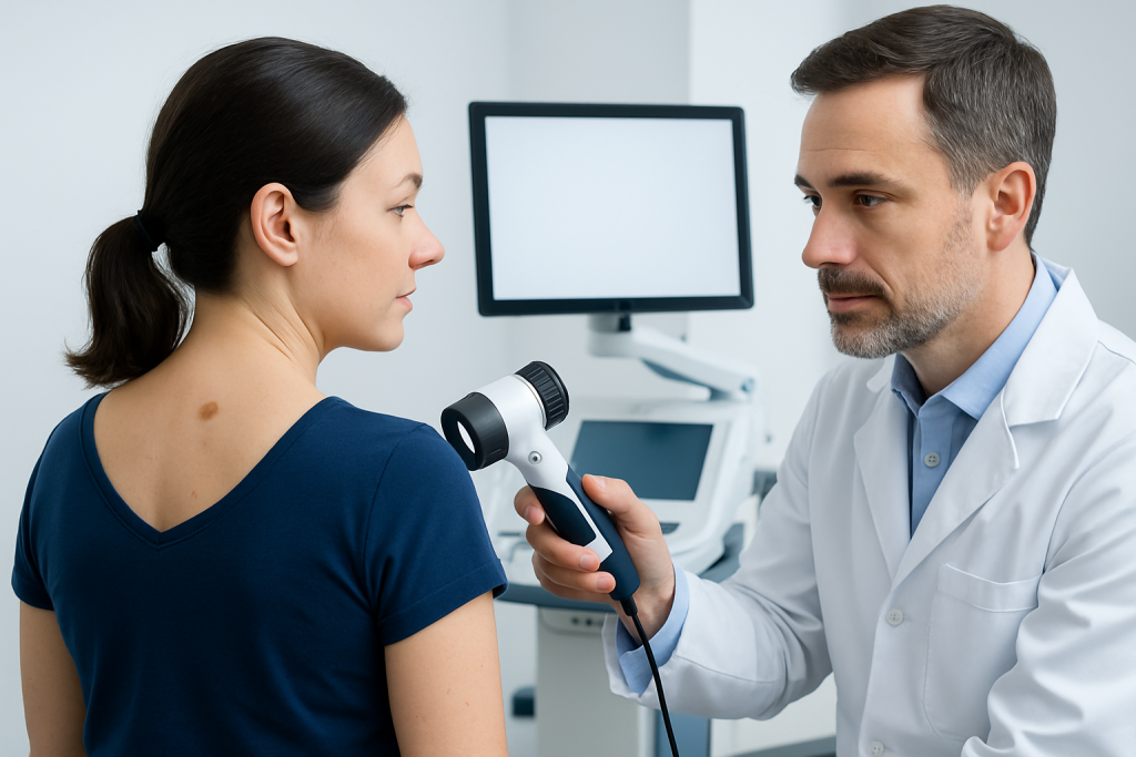
Many clinics use dermoscopy, a noninvasive tool that allows closer inspection of surface and subsurface patterns. Dermoscopy can help a doctor distinguish a benign nevus from a lesion that needs biopsy or continued monitoring. In some cases, photographs are taken so future changes can be measured more accurately.
If the lesion appears suspicious or does not fit a benign pattern, a biopsy may be recommended. This involves removing all or part of the spot so a pathology specialist can examine it under a microscope. For patients arranging care from another country, this step can help create a clear diagnosis and a practical follow-up plan before they return home.
Treatment Options
Many nevi need no treatment at all. When a lesion is clinically benign, observation is often the most appropriate approach. Removal is usually considered if the nevus is irritated by clothing or shaving, causes self-consciousness, or becomes hard to monitor because of its size or location.
When removal is chosen, the method depends on the type of nevus and the reason for treatment. A doctor may use surgical excision, shave removal, or another technique suited to the lesion’s depth and appearance. If there is any concern for skin cancer, the goal is complete removal with tissue sent for histologic examination.
It is important to note that not every mole can or should be removed in the same way. A qualified clinician will recommend an approach that balances diagnosis, appearance, scarring, and long-term monitoring. Patients traveling for care may also benefit from discussing wound care, stitch removal, and pathology follow-up before leaving the country.
Prevention & Self-care
People cannot prevent every nevus from forming, but they can reduce unnecessary skin stress and make changes easier to notice. Daily sun protection is one of the most useful habits, especially for people with many moles or a family history of skin cancer.
Self-checks can be simple and effective. Looking for a familiar pattern, rather than trying to memorize every mark, helps people notice what is new or different. A monthly check in good light, along with occasional photos of hard-to-see areas, can be especially helpful for lesions on the back, scalp, and legs.
Practical self-care steps include:
- Use broad-spectrum sunscreen and protective clothing
- Avoid intentional tanning and minimize sunburns
- Track changing lesions with photos if advised by a doctor
- Ask a partner or family member to check hard-to-see areas
- Keep scheduled follow-up visits if a doctor wants observation over time
When to See a Doctor
A medical visit is a good idea whenever a nevus changes in size, shape, color, or symptoms. Even when the change turns out to be harmless, evaluation is worthwhile because the exam can separate ordinary variation from a lesion that needs closer attention.
Prompt assessment is especially sensible if a mole bleeds without clear injury, develops a crust that does not heal, looks very different from the person’s other spots, or appears new in adulthood and begins changing soon after. People with many moles, atypical moles, or a family history of skin cancer may also benefit from routine dermatology follow-up.
International patients who are deciding whether to seek care locally or while traveling should choose the option that allows timely inspection, clear documentation, and biopsy if needed. Acibadem Health Point’s multidisciplinary specialists and JCI-accredited hospitals diagnose and treat nevus-related concerns for international patients with a coordinated, patient-centered approach.
Living With a Nevus: What Follow-up Often Looks Like
For most people, living with a nevus means learning which spots are stable and which ones deserve a second look. Dermatology visits are often brief, but they can be valuable because they create a baseline for future comparisons. That baseline is especially useful for patients who split their time between countries and may see different doctors over time.
If a lesion has been photographed or mapped, the follow-up plan may be as simple as returning in a few months or bringing updated images to the next visit. When a nevus is removed, follow-up focuses on wound care, pathology results, and whether any further monitoring is needed. Clear communication helps patients avoid unnecessary worry and know exactly what to watch for next.
In many cases, the reassuring answer is that the nevus is benign and can simply be observed. Even then, keeping an eye on skin changes remains wise, because a person’s skin pattern can evolve with age, sun exposure, and overall health.
Frequently asked questions
What is a nevus?
A nevus is a common skin growth formed by clustered skin cells, often called a mole. Most nevi are benign and do not require treatment unless they change or become bothersome.
How can someone tell if a mole needs medical review?
A mole should be checked if it changes in size, shape, color, or symptoms such as itching or bleeding. It is also wise to seek review if it looks very different from other moles on the body.
Does every nevus need to be removed?
No. Many nevi are simply observed if they look benign and cause no symptoms. Removal is usually reserved for lesions that are suspicious, irritated, or cosmetically unwanted.
What tests are used to evaluate a nevus?
A doctor usually starts with a skin exam and may use dermoscopy for closer inspection. If a lesion is concerning, a biopsy may be done so the tissue can be examined under a microscope.
Can sun exposure affect nevi?
Sun exposure can influence how moles develop and may increase the chance of skin damage over time. Using sun protection and avoiding sunburns are helpful habits for skin health overall.
Should people with many moles have regular skin checks?
Yes, regular checks can be useful for people with many moles, atypical moles, or a family history of skin cancer. A dermatologist can decide how often follow-up is needed based on the person’s individual risk.
References
- American Academy of Dermatology
- Mayo Clinic
- National Cancer Institute
- DermNet NZ
- British Association of Dermatologists
This article is for general information only and is not a substitute for professional medical advice. Please consult a qualified doctor about your individual situation.