Nevus/nevi

Key Takeaways
- Nevi are common skin growths made of pigment cells or related skin cells, and most do not cause harm.
- Changes in size, shape, color, or symptoms such as itching or bleeding should be checked by a clinician.
- Family history, sun exposure, and having many moles can increase the need for regular skin monitoring.
- Diagnosis is usually clinical, often with dermoscopy, and biopsy may be used when a lesion looks suspicious.
- Treatment is not always necessary; removal is considered for symptoms, irritation, cosmetic reasons, or diagnostic certainty.
Nevus/nevi are common skin growths often called moles, and most are harmless. Understanding their types, how they change, and when they deserve medical review can help patients make calm, informed decisions about skin care.
Overview
Nevi, commonly known as moles, are among the most familiar findings on the skin. They can appear flat or raised, light or dark, solitary or scattered, and they may show up in childhood or later in adult life. Most nevi are benign and remain stable for years.
From a dermatology perspective, the important task is not to fear every mole, but to understand which ones are simply part of a person’s skin pattern and which ones deserve closer attention. That balance is especially useful for people who travel for care, because a skin check can often be done quickly and then followed by clear home monitoring advice after returning to their own country.
Nevi can be present at birth or develop over time. Some are related to pigment-producing cells called melanocytes, while others are made up of different skin structures. Their appearance, location, and behavior help clinicians decide whether reassurance, observation, or treatment is the right next step.
Symptoms and common appearances
Most nevi are noticed because they look different from the surrounding skin. They are usually tan, brown, black, pink, or skin-colored, and they may be round, oval, or slightly irregular. Some remain small and flat, while others become raised, hairy, or textured over time.
In many cases, a nevus causes no symptoms at all. When symptoms do occur, they may include mild itching, rubbing from clothing, or irritation from shaving. Bleeding, crusting, pain, or a sudden change in appearance should not be ignored, even if the lesion has been present for years.
Features that commonly prompt medical review include:
- A new mole appearing in adulthood
- Uneven borders or asymmetry
- More than one color within the same lesion
- Rapid growth or elevation
- Persistent itching, tenderness, or bleeding
It is also helpful to know that people with many moles may find it difficult to notice small changes on their own. In that situation, photographs, skin mapping, or scheduled checks can make follow-up much easier and less stressful.
Causes and risk factors
Nevi form when certain skin cells grow in clusters rather than spreading evenly through the skin. The exact reason a particular mole appears is not always clear, but genetics, hormone changes, and sun exposure can all play a role. Some nevi are present from birth, while others develop during childhood, adolescence, or adulthood.
Risk factors for having more nevi or for needing closer observation include fair skin, a personal or family history of many moles, frequent sunburns, heavy ultraviolet exposure, and a history of atypical or dysplastic moles. People with a personal or family history of skin cancer should be especially attentive to new or changing lesions.
International patients sometimes arrive with old records or mobile-phone photos of a mole they have been watching for months or years. Those images can be very useful, because comparison over time often helps the dermatologist decide whether a lesion has truly changed or simply looks different under different lighting.
Types of nevi
There are several kinds of nevi, and the appearance of a lesion often gives the first clue about its type. Common acquired nevi are the everyday moles many people develop during life. Congenital nevi are present at birth and vary greatly in size and appearance.
Atypical or dysplastic nevi have features that are less uniform than ordinary moles. They may be larger, more irregular, or more varied in color. These lesions are not cancer, but they deserve careful observation because they can be harder to interpret by eye.
Other examples include blue nevi, which appear bluish due to the way pigment is placed deeper in the skin, and intradermal nevi, which are often raised and skin-colored or lightly pigmented. Only a clinician can tell the difference reliably, and in practice, the most important question is whether the lesion is stable and reassuring or changing and uncertain.
Diagnosis
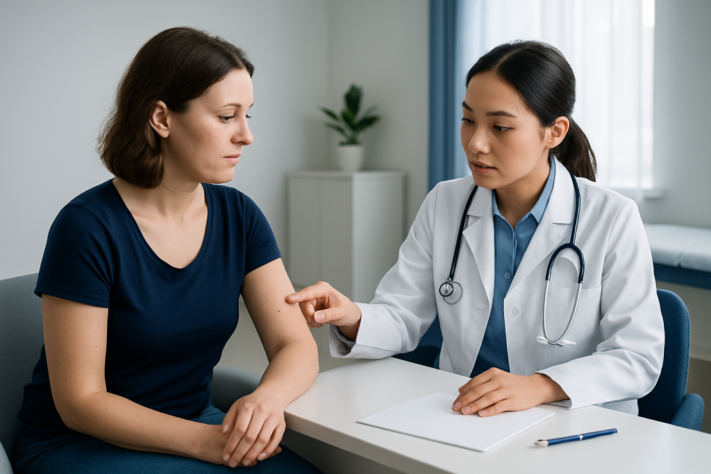
Diagnosis usually begins with a detailed skin examination. The clinician looks at the lesion’s size, color, shape, borders, and evolution, and may compare it with other moles on the body. A careful history is also important, including when the lesion first appeared, whether it has changed, and whether there is any itching, bleeding, or trauma.
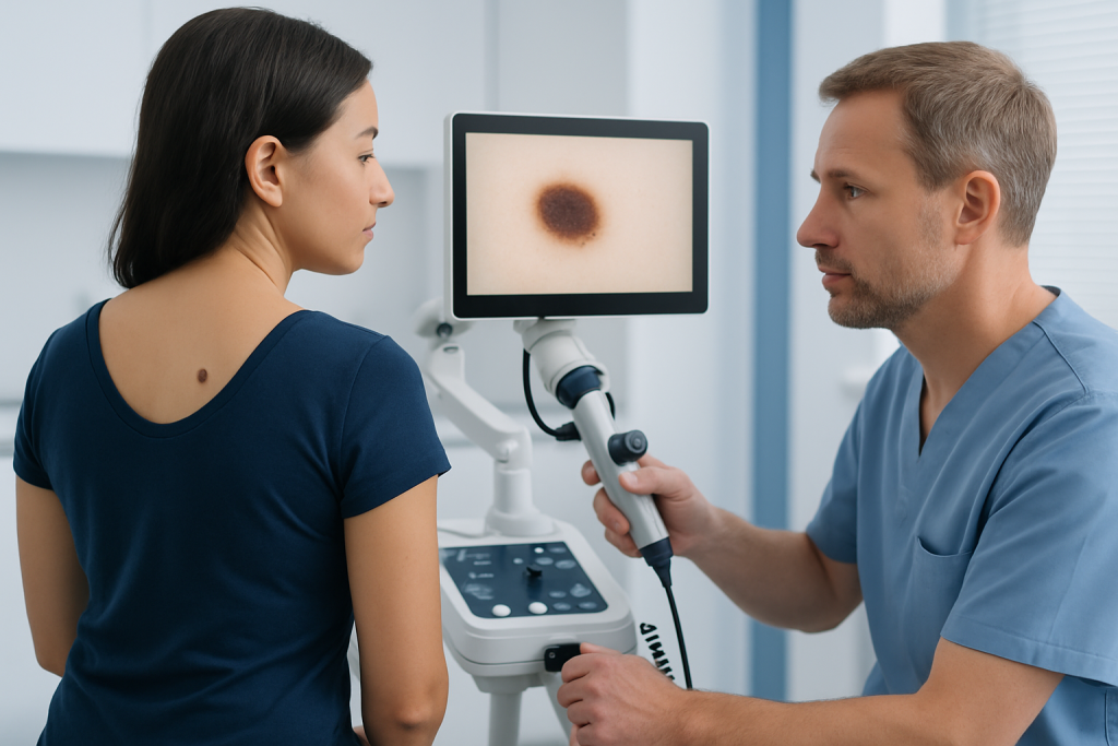
Dermoscopy is a common next step. This handheld tool allows the clinician to see patterns within the mole that are not visible to the naked eye, which can improve confidence in distinguishing benign nevi from lesions that need closer investigation.
If the diagnosis is uncertain, a biopsy may be recommended. That means removing all or part of the lesion so it can be examined under a microscope. For patients arriving from abroad, it is useful to ask how results will be shared, whether follow-up should happen locally or remotely, and whether the care team can help organize the next step before travel ends.
Treatment options
Many nevi do not require treatment. If a mole looks typical and has not changed, observation is often the safest and simplest approach. The goal is not to remove every mole, but to treat the ones that are bothersome, suspicious, or difficult to monitor.
Removal may be considered when a lesion is irritated by clothing, shaving, or repeated friction, when it causes cosmetic concern, or when the clinician wants a complete tissue diagnosis. Methods can include surgical excision or other removal techniques chosen by the dermatologist or surgeon based on the lesion’s size, depth, and location.
When a nevus is removed for medical reasons, the tissue may be sent to pathology to confirm the diagnosis. Patients should know that a simple procedure still needs aftercare: keeping the area clean, following wound instructions, and watching for signs of infection or unusual bleeding. If follow-up will happen after travel, arranging the timing of suture removal and pathology review in advance can prevent unnecessary delays.
Prevention and self-care
Not every nevus can be prevented, especially when genetics play a major role. Still, protecting the skin from ultraviolet exposure is a practical way to lower the chance of developing new lesions and to reduce overall skin damage. Daily sunscreen, protective clothing, shade, and avoiding tanning beds are all sensible habits.
Self-checks are equally important. A monthly look at the skin, using a mirror or help from a family member for hard-to-see areas, can make it easier to notice change early. Paying attention to the “ugly duckling” concept is useful: a mole that looks noticeably different from a person’s other moles deserves a professional opinion.
Helpful self-care habits include:
- Photographing moles that need monitoring
- Not scratching or picking at lesions
- Recording any change in size, color, or sensation
- Wearing sun-protective clothing during long outdoor exposure
- Booking periodic skin exams if there are many moles or a family history of skin cancer
When to see a doctor
A doctor or dermatologist should review any mole that changes quickly, develops uneven colors or borders, bleeds without injury, or becomes persistently itchy or painful. A new lesion in adulthood, especially one that stands out from a person’s other moles, also deserves attention.
People with numerous nevi, atypical moles, prior skin cancer, or a strong family history may benefit from regular professional surveillance even when nothing seems urgently wrong. For international patients, this can be particularly reassuring because a clear plan for baseline photography, biopsy if needed, and follow-up timing can be created during one visit and then continued at home.
Acibadem Health Point’s multidisciplinary specialists and JCI-accredited hospitals diagnose and treat nevi for international patients, with care plans that can be organized around travel, recovery, and follow-up needs.
If a lesion is causing concern, it is reasonable to ask for a dermatology assessment sooner rather than later. Early evaluation often leads to simple reassurance, and when treatment is needed, it allows the patient to proceed with a clear plan.
Frequently asked questions
Are all nevi harmless?
Most nevi are benign and do not turn into anything dangerous. The important part is to watch for change, because a mole that evolves in size, shape, color, or symptoms should be assessed by a clinician.
What is the difference between a mole and a nevus?
In everyday language, the words are often used to mean the same thing. “Nevus” is the medical term, while “mole” is the common term many patients use.
Can a nevus become skin cancer?
Most nevi never become cancer. However, some suspicious lesions can resemble a nevus early on, which is why new or changing moles should be evaluated rather than assumed to be harmless.
How can a person monitor moles at home?
A monthly self-check, good lighting, and comparison photos can help track changes over time. It also helps to note whether a lesion is different from the person’s other moles or begins to itch, bleed, or grow.
Is mole removal always necessary?
No, many moles are best left alone if they are stable and typical. Removal is usually considered for diagnostic reasons, symptoms, repeated irritation, or cosmetic preference after medical discussion.
What happens if a mole needs a biopsy?
A small procedure is done to remove all or part of the lesion so it can be examined under a microscope. The result helps confirm whether the lesion is benign and whether any further treatment is needed.
References
- American Academy of Dermatology
- Mayo Clinic
- National Cancer Institute
- World Health Organization
This article is for general information only and is not a substitute for professional medical advice. Please consult a qualified doctor about your individual situation.