Benign Breast Lumps: When Imaging Alone Is Enough and When Biopsy Follows

Key Takeaways
- A breast lump is evaluated by history, examination, and imaging, often before any biopsy is considered.
- Ultrasound and mammography can sometimes show a benign-appearing lesion with enough confidence to monitor it safely.
- Biopsy is usually recommended when imaging is unclear, the lump has suspicious features, or there is a mismatch between symptoms and scan findings.
- Common benign causes include cysts, fibroadenomas, and hormone-related changes.
- Follow-up matters, especially when a lump changes in size, feels different, or appears with new skin or nipple changes.
Medically reviewed by the Acıbadem clinical team — June 13, 2026
Most breast lumps are not cancer, but they still deserve careful evaluation. In many cases, imaging can show a clearly benign pattern; in others, a biopsy is needed to confirm what the lump is and guide next steps.
Overview
Finding a breast lump can be unsettling, but the word “lump” does not automatically mean cancer. Many breast lumps turn out to be benign changes such as fluid-filled cysts, fibroadenomas, or areas of normal tissue that feel more prominent because of hormonal shifts.
The key question is not only whether a lump is benign, but how confidently that can be shown. Doctors usually begin with a clinical breast exam and targeted imaging. If the scan pattern is clearly reassuring, the lump may be watched over time rather than sampled immediately.
That approach matters for patients who are planning care from another country as well. Some can leave with a clear imaging-based follow-up plan, while others may need a biopsy before travel decisions are made. The safest path depends on the full picture, not on the lump alone.
Symptoms
Benign breast lumps often feel smooth, mobile, or rubbery, though that description is not universal. Some are tender and seem to fluctuate with the menstrual cycle, while others are noticed by chance during self-exam or routine screening.
Common benign patterns include a lump that has been present for a long time without major change, a cyst that feels fuller at certain times, or a fibroadenoma that is firm but well-defined. Some people also notice generalized breast nodularity or thickened tissue rather than a single distinct lump.
- A lump that moves slightly under the skin
- Breast fullness or tenderness that changes with hormones
- A smooth, round or oval-feeling mass
- Mild discomfort without skin dimpling or nipple inversion
Even when symptoms seem mild, any new breast lump should be evaluated. Benign and concerning findings can overlap, and a proper workup is what separates watchful follow-up from more immediate testing.
Causes & Risk Factors

Benign breast lumps can arise for several reasons. Hormonal changes are common contributors, especially during the menstrual years, pregnancy, breastfeeding, and perimenopause. In many people, breast tissue becomes more reactive to hormonal shifts and develops cysts or lumpy texture.
Fibroadenomas are among the most common benign solid breast masses, especially in younger women. Breast cysts, fibrocystic changes, fat necrosis after injury, and intraductal papillomas can also create lumps or localized thickening.
Risk factors vary by condition, but a few themes recur:
- Hormonal fluctuation across the menstrual cycle or reproductive years
- Family history of breast disease, which may affect how carefully a lump is assessed
- Prior breast injury or surgery
- Age, since some benign lesions are more common at certain life stages
Most risk factors do not point to a dangerous problem on their own. They simply help the doctor decide which imaging test is most useful and whether the lump deserves biopsy or short-interval monitoring.
Diagnosis
Diagnosis usually starts with a careful history and breast examination. The clinician considers how long the lump has been present, whether it changes with the cycle, whether it is painful, and whether there are associated changes such as nipple discharge or skin alteration.
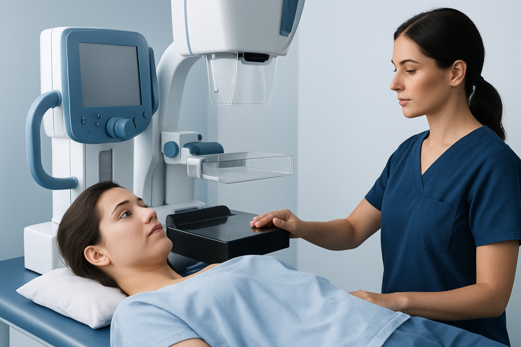
Imaging is the next step for many patients. Ultrasound is especially helpful for distinguishing a fluid-filled cyst from a solid mass, while mammography can reveal calcifications, distortion, or patterns that are not felt on exam. In some situations, MRI may be used to clarify an uncertain finding or to assess more complex breast tissue.
Imaging alone may be enough when the finding has classic benign features and matches the exam. For example, a simple cyst on ultrasound is often managed without biopsy. By contrast, if imaging is indeterminate, the lump is new and persistent, or the scan findings do not fully explain what is felt on exam, biopsy may be recommended to remove uncertainty.
Doctors also pay attention to “concordance,” meaning whether the physical exam and imaging tell the same story. When they do not, further tissue sampling is often the safest choice, even if the images do not look overtly alarming.
Treatment Options
Treatment depends on what the lump is. Many benign breast lumps do not need removal. A simple cyst may be observed, drained if painful, or left alone if it settles on its own. Fibroadenomas that are stable and well characterized may only need periodic imaging follow-up.
Biopsy is usually the next step when imaging cannot confidently classify the lump. Core needle biopsy is commonly used because it provides tissue while remaining minimally invasive. The result can confirm a benign diagnosis, identify a high-risk lesion, or show a problem that needs a different treatment plan.
If the lump is benign but bothersome, treatment may still be appropriate. Some people choose surgical removal because of pain, growth, size, anxiety, or cosmetic concerns. Others prefer surveillance once they understand that the imaging features are reassuring and the follow-up schedule is clear.
For patients travelling internationally, treatment planning often includes a practical timeline: whether biopsy should happen before departure, when follow-up imaging is due, and how pathology results will be shared. A coordinated plan reduces the risk of gaps in care.
Prevention & Self-care
Not every benign breast lump can be prevented, because many arise from normal hormonal or tissue changes. Still, some habits make breast monitoring easier and may reduce unnecessary worry.
Monthly awareness of how the breasts normally feel can help a person notice changes earlier. This is less about performing a rigid self-exam and more about recognizing what is typical for one’s own body. Keeping track of when tenderness or fullness appears in the cycle can also help distinguish a stable pattern from something new.
- Wear a supportive bra if breasts feel tender or heavy
- Note whether a lump changes with menstruation
- Avoid repeated squeezing or checking that can increase soreness
- Bring prior imaging reports when seeking second opinions or care abroad
If imaging follow-up is recommended, keeping the schedule matters. A lump that is watched over time needs the next scan at the planned interval so that any change is caught early and interpreted correctly.
When to See a Doctor
Any new breast lump should be assessed by a qualified clinician, even if it seems likely to be benign. It is especially important to seek evaluation if the lump is hard, fixed, enlarging, or different from the person’s usual breast tissue.
Prompt medical review is also important if there is nipple discharge, especially bloody discharge; skin dimpling, redness, or thickening; nipple pulling inward; or a lump in the underarm area. These findings do not confirm a serious condition, but they do justify a more careful workup.
People should also return sooner if a previously monitored lump changes in size or character, or if a scan and the physical exam do not match. In those situations, doctors may recommend repeat imaging, biopsy, or referral to a breast specialist. Acibadem Health Point’s multidisciplinary specialists and JCI-accredited hospitals can help diagnose and treat this condition for Myomectomy Recovery for International Patients: The Usual Travel Timeline" class="ahp-ilk">international patients when coordinated care is needed.
Clear information, timely imaging, and the right biopsy strategy can turn a frightening discovery into a manageable plan. The goal is not to rush every patient into a procedure, but to choose the least invasive option that still gives a trustworthy answer.
Frequently asked questions
Can imaging alone prove a breast lump is benign?
Sometimes, yes. If ultrasound or mammography shows a classic benign pattern and the scan matches the physical exam, a doctor may recommend observation rather than biopsy. If there is any uncertainty, tissue sampling is usually the safer way to confirm the diagnosis.
What is the difference between a cyst and a solid lump?
A cyst is a fluid-filled sac, while a solid lump is made of tissue. Ultrasound is particularly useful for telling the difference. Simple cysts are often benign and may not need biopsy unless they are painful or atypical.
Does every benign breast lump need to be removed?
No. Many benign lumps can be watched over time if they are stable and well understood on imaging. Removal is usually considered only if the lump grows, causes symptoms, affects appearance, or remains uncertain after testing.
How is a breast biopsy performed?
The most common approach is a core needle biopsy, which uses imaging guidance to take small tissue samples. It is generally done with local anesthesia and is designed to be less invasive than surgery. Your doctor will explain how to prepare and what to expect afterward.
What if the lump hurts but imaging looks benign?
Pain does not automatically mean something dangerous is present. Some benign lumps, especially cysts and hormonally sensitive tissue, can be tender. If the imaging is reassuring, the doctor may suggest symptom relief and follow-up rather than immediate biopsy.
Should someone get a second opinion before biopsy?
A second opinion can be helpful, especially when imaging findings are unclear or care is being coordinated across countries. What matters most is that the exam, imaging, and planned next step all fit together logically. If they do not, another specialist review can be valuable.
References
- American College of Radiology
- American Cancer Society
- National Comprehensive Cancer Network
- Mayo Clinic
- World Health Organization
This article is for general information only and is not a substitute for professional medical advice. Please consult a qualified doctor about your individual situation.
More from the Health Library



Related Specialists

Prof. Dr. A. Taner Usta
Gynecology & Obstetrics
Prof. Dr. Ahmet Tayyar
Gynecology & Obstetrics
Prof. Dr. Ahmet Cem Batukan
Gynecology & Obstetrics
Dr. Sevgi Selen
Gynecology & Obstetrics