Skin Cancer on the Head Under Hair: Warning Signs
Skin cancer on the head under hair may be difficult to notice because the scalp is covered, but it can often be found through regular checks and professional examinations. A new, changing, bleeding, crusted, or non-healing scalp spot should be assessed by a qualified clinician.
Overview: Can Skin Cancer Develop Under Hair?
Yes. Skin cancer on head under hair can develop on any part of the scalp, including areas covered by thick hair. Hair provides some physical shade, but it does not block all ultraviolet (UV) radiation. The scalp may also receive repeated sun exposure along a hair part, at the hairline, through thinning hair, or during outdoor activities.
The most common skin cancers that affect the scalp are basal cell carcinoma, squamous cell carcinoma, and melanoma. Basal cell carcinoma usually grows slowly and rarely spreads, while squamous cell carcinoma may be more likely to grow deeper or spread if left untreated. Melanoma is less common but requires prompt assessment because it can spread to other parts of the body.
Scalp skin cancer can be easy to miss because hair can conceal early changes. A person may first notice a rough area while washing their hair, a recurring scab after a haircut, a spot that bleeds when combing, or a changing mole noticed by a partner, hairdresser, or clinician. Most scalp changes are not cancer, but persistent or changing lesions deserve professional evaluation.
What Scalp Skin Cancer May Look or Feel Like
Skin cancer does not always look the same. Its appearance varies by skin cancer type, skin tone, location, and how long it has been present. Some lesions are painless, so the absence of discomfort does not rule out a problem. A new lesion in adulthood or an existing spot that noticeably changes should be checked.
Possible signs include a pearly, shiny, pink, skin-colored, brown, or black bump; a scaly or rough patch that does not clear; a crusted or bleeding sore; or a firm, growing lump. On darker skin, a concerning lesion may look darker than surrounding skin, bluish, purplish, gray, or simply different from other marks. It may not appear red.
- A sore, scab, or ulcer that does not heal or repeatedly returns
- A rough, tender, thickened, or flaky patch on the scalp
- A mole or pigmented spot that changes in size, shape, color, or sensation
- Bleeding, oozing, itching, pain, or tenderness without a clear cause
- A lesion with irregular borders or several colors
- A spot that looks noticeably different from a person’s other moles, sometimes called the “ugly duckling” sign
For moles and dark spots, clinicians often use the ABCDE guide: asymmetry, irregular border, uneven color, diameter that increases, and evolution or change over time. This guide is useful but not complete. Any unusual or evolving scalp lesion should be examined, even if it does not meet every ABCDE feature.
Why It Develops: Causes and Risk Factors

Most skin cancers result from damage to skin-cell DNA. UV radiation from sunlight is a major cause, and exposure can accumulate over many years. The scalp is particularly vulnerable in people with baldness, thinning hair, a wide part, short hair, or a history of frequent outdoor work or recreation.
Risk is higher in people with fair skin, light-colored eyes, freckles, a tendency to burn, or a history of intense sun exposure and sunburns. However, skin cancer can occur in every skin tone and hair type. In people with darker skin, diagnosis may sometimes be delayed if an unusual lesion is mistaken for a harmless mark or scalp condition.
Other factors include older age, a personal or family history of skin cancer, previous Brain Cancer Radiation Treatment: Options and Outlook" class="ahp-ilk">radiation treatment, long-term immune suppression, certain inherited conditions, and exposure to tanning devices. Chronic wounds, scars, or longstanding inflammation can also rarely be associated with certain squamous cell cancers.
Common scalp conditions such as dandruff, psoriasis, folliculitis, cysts, and seborrheic dermatitis can cause itching, scale, or bumps and are much more common than skin cancer. Still, a lesion that persists despite usual care, changes steadily, or behaves differently from other scalp symptoms should not be assumed to be benign.
How Doctors Examine and Diagnose a Scalp Lesion
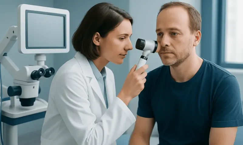
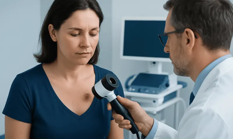
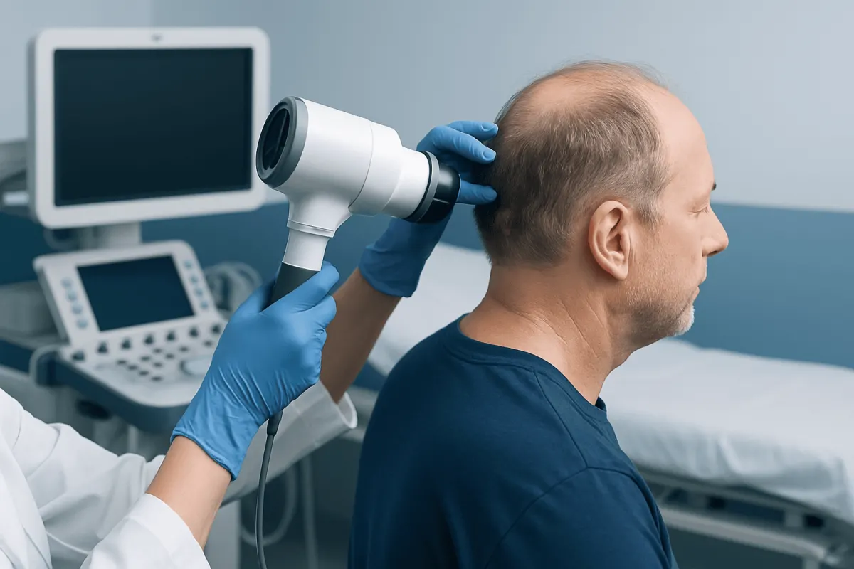
A dermatologist or other qualified clinician starts with a focused history and skin examination. They may ask when the spot was first noticed, whether it has changed, whether it bleeds or hurts, and about sun exposure, previous skin cancers, medicines, and immune health. The clinician examines the entire scalp by separating the hair in sections and may also check the face, ears, neck, and the rest of the skin.
A dermatoscope, a handheld magnifying device with specialized lighting, may help reveal details not visible to the naked eye. Photographs or digital monitoring can sometimes be used for selected lesions, but a suspicious lesion should not be watched indefinitely without a clear clinical plan.
A biopsy is the only way to confirm whether a lesion is cancerous and to identify its exact type. During this minor procedure, a clinician removes all or part of the lesion after numbing the area. A pathologist examines the sample under a microscope. The result guides treatment planning and helps determine whether additional testing is necessary.
If a melanoma or higher-risk squamous cell carcinoma is diagnosed, the care team may assess lymph nodes and consider imaging or other staging tests when clinically appropriate. These tests are not required for every person or every type of scalp skin cancer.
Treatment Options for Skin Cancer on the Scalp
Treatment is individualized according to the cancer type, depth, size, location, microscopic features, overall health, and a person’s preferences. Most scalp skin cancers can be treated effectively, especially when identified early. The goal is to remove or destroy the cancer while preserving function and achieving appropriate wound healing.
Surgery is often the main treatment. Standard surgical excision removes the visible cancer along with a margin of surrounding skin. For cancers in high-risk areas, recurrent tumors, or lesions with poorly defined edges, Mohs micrographic surgery may be considered. This technique removes tissue in stages and examines the edges during the procedure, helping conserve as much healthy tissue as possible.
Depending on the diagnosis, other options may include curettage and electrodessication, cryotherapy for selected precancerous lesions, topical medicines, photodynamic therapy, radiation therapy, or systemic treatments such as immunotherapy or targeted therapy for advanced disease. Treatment for melanoma usually involves surgical removal, with additional therapies considered when the melanoma has a higher risk of recurrence or has spread.
After removal, a small wound may heal naturally, be closed with stitches, or require a skin graft or flap in some cases. The clinician will explain wound care, activity restrictions, scar expectations, and follow-up skin examinations. Ongoing monitoring is important because people who have had one skin cancer have a higher chance of developing another.
Checking the Scalp and Protecting It From Sun Exposure
Regular self-checks can help identify changes between professional skin examinations. A person can use a hand mirror and a larger mirror, or ask a trusted partner, family member, or hair professional to look at areas that are difficult to see. Checking the scalp in sections—along the part, hairline, crown, behind the ears, and back of the neck—can make the process more manageable.
Sun protection is useful throughout the year, especially during prolonged outdoor exposure. A broad-brimmed hat or cap designed to provide UV protection can cover the scalp. For exposed areas such as a part line, hairline, bald scalp, or thinning areas, broad-spectrum sunscreen may be appropriate. Products formulated for the scalp, including lotions, sticks, sprays, and powders, may be easier to use, but should be applied according to their instructions.
Additional practical measures include seeking shade when possible, avoiding tanning beds, wearing protective clothing, and reapplying sunscreen after swimming, sweating, or prolonged time outdoors. Sun protection reduces future UV damage but does not replace clinical assessment of a suspicious spot.
People with a history of skin cancer, many moles, immune suppression, or significant cumulative sun exposure may benefit from regular full-body skin examinations at intervals recommended by their clinician.
When to Seek Medical Care
A person should arrange a medical appointment promptly for a scalp spot that is new and growing, changes in color or shape, repeatedly bleeds, forms a persistent crust, becomes painful, or does not heal within several weeks. It is also sensible to seek assessment for a mole that looks different from others or a rough patch that remains despite appropriate treatment for common scalp irritation.
More urgent evaluation is appropriate if a lesion is rapidly enlarging, severely painful, ulcerated, associated with swollen lymph nodes, or accompanied by unexplained symptoms such as persistent fatigue or weight loss. These symptoms have many possible causes, but timely assessment is important.
It is best not to pick, shave over repeatedly, burn, or use over-the-counter wart removers on an unexplained scalp lesion. These actions can irritate the area and make assessment more difficult. A clinician can determine whether the change is benign, inflammatory, infectious, or cancerous and recommend suitable care.
Acıbadem Health Point’s multidisciplinary specialists and JCI-accredited hospitals diagnose and treat skin cancer for international patients, with care plans based on the confirmed diagnosis and individual clinical needs.
Frequently asked questions
01Can skin cancer grow under thick hair?
Yes. Thick hair may reduce some direct sun exposure, but it does not completely protect the scalp from UV radiation. Skin cancer can also develop for reasons beyond sun exposure, so any changing or persistent scalp lesion should be assessed.
02What does skin cancer on the scalp feel like?
It may feel like a rough patch, firm bump, sore, crusted area, or tender spot. Some lesions itch, bleed, or hurt, while others have no symptoms at all. A persistent change is more important than whether it is painful.
03Is a scab on the scalp always skin cancer?
No. Scabs commonly result from scratching, acne-like folliculitis, eczema, psoriasis, injury, or infection. However, a scab that repeatedly returns in the same place, bleeds easily, or does not heal should be examined by a clinician.
04How can someone check their scalp for skin cancer?
They can use two mirrors to inspect the scalp in sections, including the part line, crown, hairline, behind the ears, and neck. Asking a partner, family member, or hairdresser to report unusual spots can also be helpful. Taking a clear photograph may help document change, but it does not replace a medical examination.
05Can a dermatologist diagnose scalp cancer without a biopsy?
A dermatologist may strongly suspect a diagnosis based on examination and dermoscopy. However, a biopsy is generally needed to confirm skin cancer and identify the specific type. This information helps select the most appropriate treatment.
06Does skin cancer on the scalp require surgery?
Surgery is common because it can remove the cancer and allow tissue testing, but it is not the only option. Depending on the type and stage, treatment may include topical therapy, cryotherapy, radiation, photodynamic therapy, or medicines that work throughout the body. The recommended approach depends on the confirmed diagnosis.
This article is for general information only and is not a substitute for professional medical advice. Please consult a qualified doctor about your individual situation.
Hair transplant in Turkey — costs, graft counts & a free assessment
JCI-accredited · board-certified surgeons · reply within 24h
Add us as a Preferred Source to see more of our trusted health content across Google Search, AI Overviews and Discover.