Skin Cancer on Leg Pictures: Signs, Diagnosis and Care
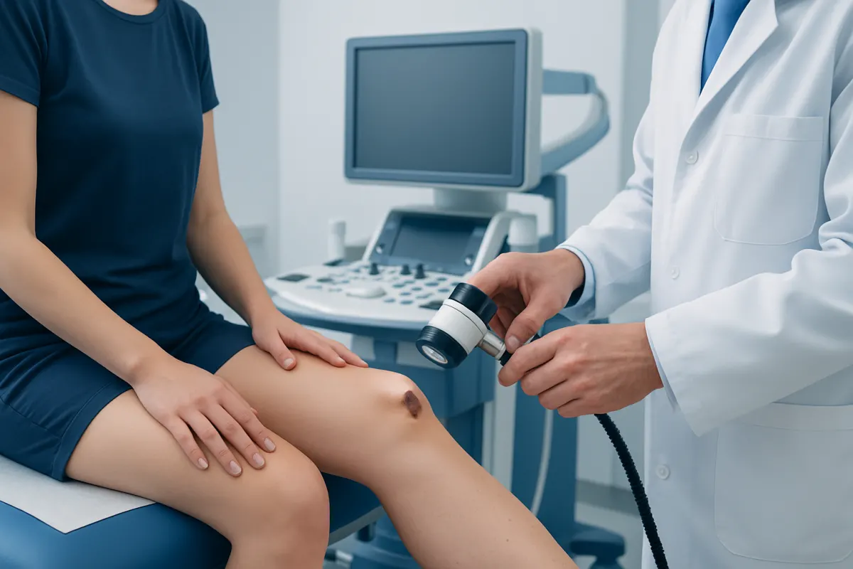
Skin cancer on leg pics may help a person notice concerning changes, such as an evolving mole, a persistent sore or a scaly patch, but photographs cannot diagnose skin cancer. A dermatologist can examine the area and, when needed, perform a biopsy to identify the type of lesion and guide treatment.
What can skin cancer on leg pics show?
Skin cancer on leg pics may show a new dark spot, an existing mole that is changing, a pink or pearly bump, a rough scaly area, or a sore that repeatedly crusts, bleeds or does not heal. These images can be useful for recognising that a lesion needs attention, especially when a person compares photographs taken over time. However, lighting, skin tone, camera quality and the wide range of harmless skin changes mean that a photo cannot reliably confirm or exclude cancer.
The legs are a common location for skin cancers because they receive intermittent or cumulative ultraviolet exposure. Melanoma can develop anywhere on the skin and is often seen on the legs in women, while squamous cell carcinoma and basal cell carcinoma may occur on sun-exposed areas of the lower leg. Any new or changing lesion should be assessed rather than judged by an online image alone.
Useful photographs for a medical appointment include one image showing the general location and another close image taken in good light. A ruler or another size reference can help document change. Photographs should support, not delay, a professional skin examination.
Common appearances and symptoms on the leg
Melanoma often appears as a new or changing pigmented mark. The ABCDE approach can help identify features worth checking: Asymmetry, irregular Borders, varied Colours, increasing Diameter, and Evolution or change over time. Some melanomas are pink, red, skin-coloured or dark blue rather than brown or black, so a changing non-pigmented bump should not be ignored.
Basal cell carcinoma may look like a shiny or translucent bump, a pink patch, a recurring sore, or an area with a slightly raised edge. Squamous cell carcinoma may present as a firm red nodule, a thickened scaly patch, a tender crusted lesion or a non-healing ulcer. These appearances can overlap with eczema, insect bites, infections, warts, scars and other non-cancerous conditions.
- A mole or spot that changes in size, shape, colour or sensation
- A sore that does not heal within several weeks or repeatedly heals and returns
- Persistent bleeding, crusting, itching, tenderness or pain in one area
- A new rapidly growing bump, especially if it is firm, scaly or unusually coloured
- A lesion that looks noticeably different from a person’s other moles, sometimes called the “ugly duckling” sign
Symptoms alone do not establish a diagnosis. Some skin cancers cause no discomfort at all in the early stages, which is why visual change and persistence matter.
How serious is skin cancer on the legs?

How serious skin cancer on the legs is depends mainly on its type, size, depth, location and whether it has spread beyond the skin. Basal cell carcinoma is the most common type and usually grows slowly. It rarely spreads to distant parts of the body, but it can enlarge and damage nearby skin and tissue if left untreated.
Squamous cell carcinoma is also often curable when treated early. Some lesions have a greater likelihood of spreading, particularly when they are large, deeply invasive, recurrent, located in higher-risk areas, or occur in people with a weakened immune system. Melanoma is less common but can spread to lymph nodes or other organs, so early assessment is particularly important.
A diagnosis does not automatically mean advanced disease. Many leg skin cancers are identified while localised and can be removed or treated successfully. A clinician determines outlook after assessing the lesion and, if cancer is confirmed, reviewing pathology findings and any appropriate staging tests. For broader information on this group of conditions, see skin cancer.
Causes and risk factors
Most skin cancers are linked to damage from ultraviolet radiation, including sunlight and artificial tanning devices. Sunburns, especially severe or repeated burns, can increase risk. Ultraviolet exposure also accumulates over time, so people may develop skin cancer even if they do not recall a single major sunburn on their legs.
Other factors include fair skin that burns easily, light eyes or hair, a personal or family history of skin cancer, many moles, atypical moles, older age, immune suppression, certain genetic conditions and previous radiation exposure. People with darker skin can also develop skin cancer; changes on the legs, soles, nails and other less sun-exposed areas should still be evaluated.
Risk factors help explain why assessment may be advisable, but they cannot determine whether one particular mark is cancer. A person with few recognised risks may still develop skin cancer, while many people with risk factors never do.
Diagnosis: why a biopsy matters
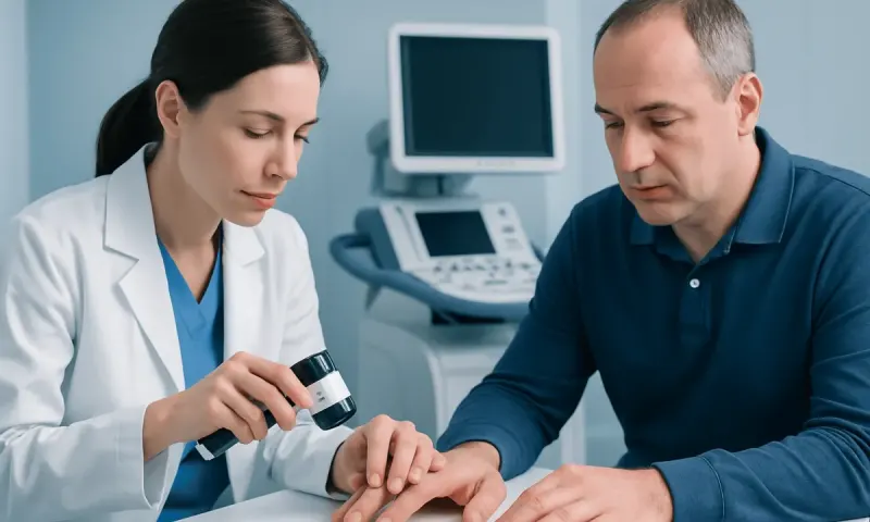
A dermatologist or another qualified clinician will ask when the lesion appeared, whether it has changed, and whether there is a history of sun exposure, prior skin cancer or immune suppression. They will inspect the spot and may examine the wider skin surface for other concerning lesions. A dermatoscope, a handheld magnifying device with specialised lighting, can reveal patterns not visible to the naked eye.
If a lesion is suspicious, the clinician usually takes a biopsy. This involves removing all or part of the area after numbing the skin and sending the tissue to a laboratory. The pathology report identifies the lesion type and may describe important features such as tumour thickness or depth. A biopsy is more definitive than photos, visual inspection or blood tests.
For confirmed melanoma or higher-risk non-melanoma skin cancer, further evaluation may be recommended to determine whether there is any spread. This can include lymph node examination, imaging in selected situations, or a sentinel lymph node biopsy for some melanomas. The care plan is tailored to the diagnosis rather than based on appearance alone.
How deep does basal cell carcinoma grow?
Basal cell carcinoma begins in cells in the lower part of the epidermis, the outer skin layer. It may remain relatively superficial for a long time, but untreated lesions can gradually extend into the dermis, fatty tissue, cartilage, muscle or, rarely, bone. Growth rate and depth vary by subtype, location, size and how long the lesion has been present.
Unlike melanoma, basal cell carcinoma is not usually described by one standard thickness measurement that determines stage. Instead, clinicians assess whether the tumour has high-risk features, such as an infiltrative growth pattern, recurrence after prior treatment, poorly defined borders, larger size or invasion into deeper structures. Most basal cell carcinomas are detected and treated before deep invasion occurs.
A lesion that has persisted, enlarged, ulcerated or repeatedly bled should be examined even if it seems small. Prompt treatment can limit the amount of tissue that needs to be removed and may simplify reconstruction when surgery is necessary.
Modern treatment approaches and everyday outlook
Treatment is based on the cancer type, its site and size, pathology results, a person’s overall health and cosmetic or functional considerations. Surgical removal is commonly used for melanoma and many basal or squamous cell carcinomas. Techniques may include standard excision or Mohs surgery, in which tissue is removed and examined in stages to preserve as much healthy skin as possible in appropriate cases.
Some superficial, low-risk non-melanoma skin cancers may be managed with carefully selected non-surgical options, such as topical medicines, freezing, curettage and electrodessication, photodynamic therapy or radiation therapy. Advanced melanoma or advanced non-melanoma skin cancer may require treatments that work throughout the body, including immunotherapy, targeted therapy or other oncology-directed care. These decisions should be made with a dermatology and oncology team.
Can you live a normal life with skin cancer? Many people do, particularly after successful treatment of an early, localised skin cancer. Follow-up skin examinations, sun protection and awareness of new or changing lesions become part of ongoing care. A diagnosis of melanoma or advanced disease can require closer monitoring and more intensive treatment, but support, rehabilitation and symptom management are available throughout care.
Acıbadem Health Point’s multidisciplinary specialists and JCI-accredited hospitals diagnose and treat skin cancer for international patients, with care planned according to pathology and individual clinical needs.
When to seek medical care
A person should arrange a dermatology appointment promptly for a new, changing or unusual lesion on the leg, especially one that bleeds, crusts, ulcerates, grows quickly or does not heal. A rapidly changing dark spot, a mole with ABCDE features, or a lesion that differs from surrounding moles should not wait for a routine skin check.
More urgent assessment is appropriate if a known skin cancer changes quickly, a wound becomes increasingly painful or infected-looking, or there are new concerning symptoms such as a growing lump in the groin. These symptoms have many possible explanations, but they should be evaluated clinically.
What are the symptoms of terminal skin cancer? Advanced or terminal cancer can affect people differently and cannot be diagnosed from symptoms alone. Possible concerns in widespread disease may include worsening fatigue, unintentional weight loss, persistent pain, reduced appetite, breathlessness, neurological symptoms or declining ability to manage daily activities. Anyone with these symptoms should contact their treating team promptly; palliative care can provide active support for symptoms, comfort and quality of life at any stage of serious illness.
To reduce future risk, use broad-spectrum sun protection, wear protective clothing when outdoors, seek shade when practical and avoid tanning beds. Regular self-checks are helpful, but they do not replace professional review for a suspicious finding.
Frequently asked questions
01Can a photograph diagnose skin cancer on the leg?
No. A photograph can document a lesion and help a person notice changes over time, but it cannot reliably diagnose skin cancer. A clinical examination and, when indicated, a skin biopsy are needed for confirmation.
02What does melanoma on the leg look like?
Melanoma may look like a new or changing mole with asymmetry, irregular borders, several colours or noticeable evolution. It can also be pink, red, skin-coloured or dark blue. Any spot that is changing or looks unlike a person's other moles should be assessed.
03Is a non-healing sore on the lower leg always skin cancer?
No. Poor healing can occur with injuries, infection, circulation problems, diabetes and inflammatory skin conditions. However, a sore that persists, repeatedly bleeds or heals and returns should be examined to rule out skin cancer and other treatable causes.
04Can skin cancer on the leg spread to lymph nodes?
Melanoma and some squamous cell carcinomas can spread to nearby lymph nodes, including nodes in the groin. Basal cell carcinoma very rarely spreads, although it can invade nearby tissue when untreated. The need for lymph node assessment depends on the diagnosed cancer type and its risk features.
05How is skin cancer on the leg treated?
Many lesions are removed surgically, often under local anaesthetic. Depending on the exact cancer and its risk level, other options may include topical treatment, cryotherapy, radiation therapy, Mohs surgery, immunotherapy or targeted therapy. Treatment should follow a confirmed diagnosis and personalised care plan.
06How often should someone check their legs for skin cancer?
A monthly self-check can help a person become familiar with their skin and recognise changes early. It is important to inspect the front and back of the legs, feet, soles, between toes and around the nails. People with previous skin cancer, many moles or other risk factors may need clinician-led checks on an individual schedule.
This article is for general information only and is not a substitute for professional medical advice. Please consult a qualified doctor about your individual situation.
Oncology care in Turkey — second opinion and treatment plan
JCI-accredited · board-certified surgeons · reply within 24h
Add us as a Preferred Source to see more of our trusted health content across Google Search, AI Overviews and Discover.