Pulled Muscle MRI: What the Scan Can Show
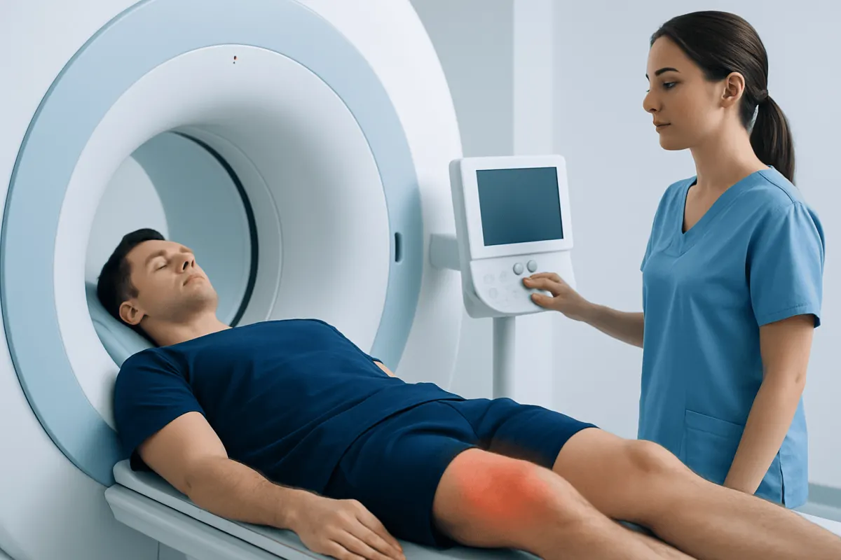
A pulled muscle MRI can provide detailed images of muscles, tendons, and nearby soft tissues when symptoms are severe, unclear, persistent, or suggest a more significant injury. Most uncomplicated muscle strains are diagnosed clinically and improve with conservative care, so MRI is not routinely needed for every pulled muscle.
Pulled Muscle MRI: the answer at a glance
A pulled muscle MRI is a magnetic resonance imaging scan used to examine a suspected muscle strain in detail. It can show swelling, small or large muscle-fiber tears, bleeding within tissue, tendon involvement, and other conditions that may cause similar pain.
Most mild muscle strains do not require an MRI. A clinician can often make the diagnosis from the history, physical examination, and how the injury responds to early care. MRI becomes more helpful when the injury is severe, the diagnosis is uncertain, an athlete or active person needs a precise return-to-activity plan, or symptoms are not improving as expected.
Muscle strains commonly affect the hamstrings, calf, groin, shoulder, chest, and lower back. A pulled back muscle MRI may be considered when back pain follows an injury but has unusual features, persists, causes marked limitation, or needs to be distinguished from a spine, nerve, or other soft-tissue problem.
What an MRI can show after a muscle injury
MRI creates detailed images of soft tissues by using a strong magnetic field and radio waves. Unlike X-rays and CT scans, it does not use ionizing radiation. The images can help a radiologist and treating clinician locate the injured muscle, estimate the extent of tissue damage, and assess nearby tendons, fascia, bones, joints, nerves, and blood vessels.
In a recent injury, MRI may show fluid and inflammation around damaged fibers, known as edema. It can also identify partial or complete tears, a collection of blood called a hematoma, tendon avulsion from bone, or scarring from an older injury. These findings may affect rehabilitation planning, activity restrictions, and whether a surgical opinion is appropriate.
Pulled muscle imaging is not limited to MRI. Ultrasound can be a useful, accessible option for some superficial muscles and can assess tissue movement in real time. However, MRI often provides a broader and more detailed view, particularly for deeper muscles, complex injuries, or uncertainty about the source of pain.
Who may benefit from a pulled muscle MRI

A doctor may recommend MRI when there was a sudden pop, substantial bruising or swelling, visible deformity, pronounced weakness, or inability to use the affected limb normally. It may also be appropriate when clinical examination suggests a high-grade tear or tendon injury, especially around the hamstring, quadriceps, calf, shoulder, or groin.
Imaging can be useful when symptoms do not improve with an appropriate period of rest and rehabilitation, when pain recurs repeatedly, or when the diagnosis remains unclear. For example, pain that seems like a muscle strain may sometimes arise from a stress injury, joint problem, nerve irritation, infection, or another condition requiring different care.
MRI may support return-to-sport decisions in selected cases, but scan findings should not be used alone. Recovery also depends on pain, strength, movement, function, and the demands of work or sport. A sports medicine or orthopedic clinician can coordinate imaging with a tailored rehabilitation plan through orthopedic care.
- Severe pain or major loss of function after an injury
- Concern for a complete muscle or tendon tear
- Persistent pain, weakness, swelling, or bruising
- Repeated strains in the same area
- Symptoms that do not match a straightforward muscle strain
How the procedure works: preparation and step-by-step scanning
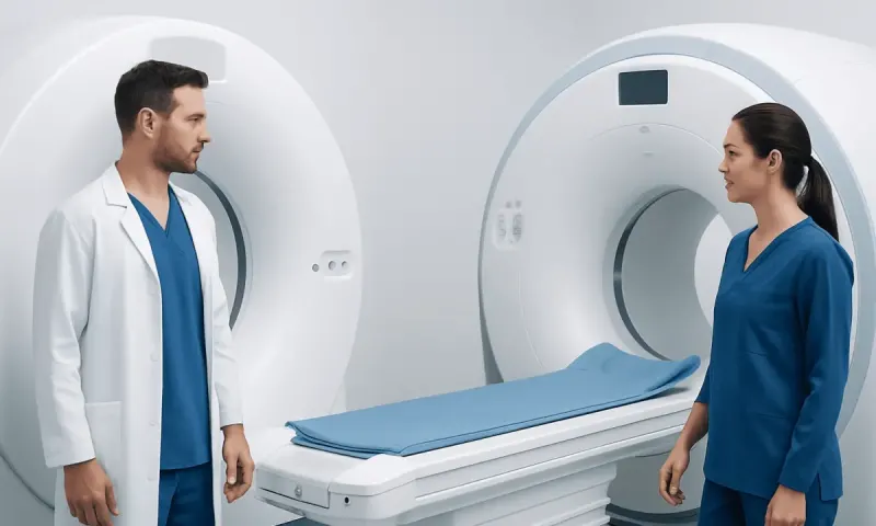
Before the scan, the imaging team reviews medical history, previous surgery, implanted devices, kidney health when contrast may be considered, and possible pregnancy. Many implants are MRI-compatible, but some devices or metal fragments require special assessment. It is important to mention pacemakers, neurostimulators, cochlear implants, aneurysm clips, metalworking exposure, and any implanted hardware.
For most muscle MRI examinations, no special preparation is needed. The patient removes jewelry, watches, hearing aids, and clothing with metal components, then changes into a gown if needed. Contrast dye is not routinely required for an acute uncomplicated strain, although it may be used when clinicians need to evaluate a mass, infection, inflammation, or another less typical concern.
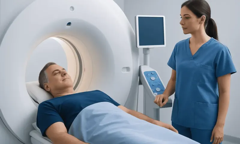
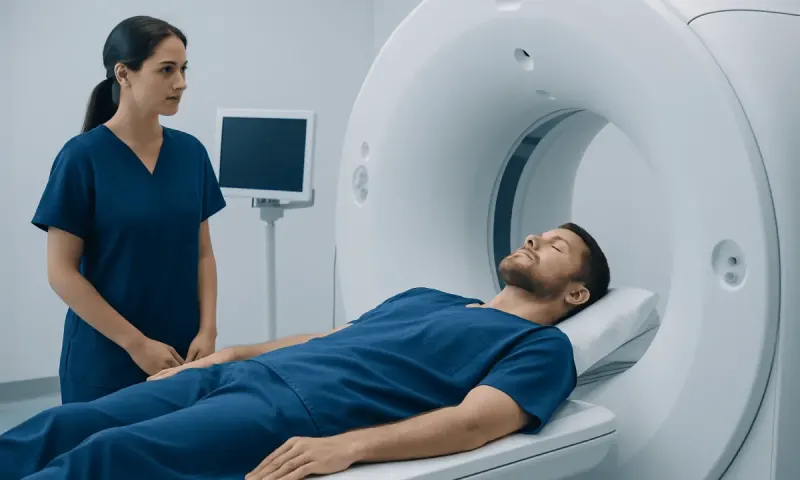
During the scan, the patient lies on a padded table that moves into the MRI scanner. Positioning supports or coils may be placed around the body part being examined. The scanner makes loud tapping or knocking sounds, and ear protection is provided. Staying still is important because movement can blur the images. Depending on the body area and protocol, a muscle MRI commonly takes about 20 to 45 minutes.
People with claustrophobia should tell the care team before the appointment. Options may include coaching, music or communication during the scan, a wider-bore scanner where available, or clinician-prescribed medication when appropriate. The team can usually communicate with the patient throughout the examination.
Results, benefits, and limitations of MRI
A radiologist reviews the images and sends a report to the referring clinician, who interprets the findings alongside the examination and symptoms. The report may describe the injured muscle, the location of a tear, the amount of edema, whether a tendon is involved, and any additional findings. The timing for results varies by facility and clinical urgency.
The main benefit of MRI is detailed soft-tissue assessment without radiation exposure. It can help distinguish a mild strain from a more extensive injury and can identify findings that may change treatment. This can be particularly valuable when a person has persistent symptoms or a potential surgical injury.
However, MRI has limitations. The scan may reveal changes that are unrelated to the current pain, particularly in people with previous injuries or age-related tissue changes. Image findings do not always predict exactly how long recovery will take. MRI also may not be necessary if the diagnosis and treatment plan are already clear from a clinical assessment.
In uncommon situations, imaging may identify an area that needs further investigation. An MRI guided muscle biopsy is not a standard test for a pulled muscle. It is a specialized procedure considered only when imaging raises concern for an unexplained muscle abnormality, such as suspected inflammatory, infectious, or tumor-related disease, and it is planned by an appropriate specialist team.
Treatment and recovery after a muscle strain
Most muscle strains improve with relative rest from painful activity, gradual return to movement, and a rehabilitation plan that restores flexibility, strength, balance, and sport- or work-specific function. In the first days after injury, protecting the area and avoiding activities that worsen pain can be helpful. A clinician may advise cold therapy for comfort and appropriate pain relief based on the person’s health history.
As pain settles, prolonged complete rest is usually avoided unless a clinician recommends it. Progressive exercises are important because returning too quickly can increase the risk of reinjury, while avoiding movement for too long can lead to stiffness and weakness. A physiotherapist can adapt exercises to the muscle involved and the person’s goals through physical therapy and rehabilitation.
Recovery varies widely. Mild strains may improve over days to a few weeks, while more significant partial tears can take longer. Complete tears or tendon avulsions may require specialist review and, in selected cases, surgery. Follow-up decisions should be based on function as well as symptoms rather than an expected calendar date alone.
Acıbadem Health Point’s multidisciplinary specialists and JCI-accredited hospitals assess muscle and tendon injuries for international patients, coordinating diagnostic imaging, orthopedic review, and rehabilitation when needed.
When to seek medical care
Prompt medical assessment is advisable after an injury that causes severe pain, a popping sensation followed by weakness, inability to bear weight or use the affected area, rapidly increasing swelling, extensive bruising, or a visible change in the muscle’s shape. These symptoms can occur with a substantial tear or another injury that should not be managed as a minor strain alone.
Medical advice is also important if pain is worsening rather than improving, symptoms continue beyond the expected early recovery period, or the same muscle is repeatedly injured. A clinician can determine whether examination, ultrasound, MRI, or another test is appropriate and can help guide safe return to activity.
Emergency care is needed for symptoms such as a cold, pale, blue, or numb limb; severe pain out of proportion to the injury; rapidly progressive tight swelling; chest pain; shortness of breath; fainting; or fever with redness and increasing pain. These are not typical features of a simple pulled muscle and require urgent evaluation.
Frequently asked questions
01Do I need an MRI for a pulled muscle?
Usually, no. Many muscle strains can be diagnosed through a medical history and physical examination and improve with conservative treatment. MRI is more likely to be recommended for severe injuries, uncertain diagnoses, persistent symptoms, or suspected tendon or complete muscle tears.
02Can MRI tell how bad a muscle strain is?
MRI can show the location and extent of muscle edema, fiber disruption, bleeding, and tendon involvement. This helps clinicians assess whether the injury appears mild or more significant. The scan is interpreted alongside pain, strength, movement, and function because imaging alone cannot determine recovery time precisely.
03Is a pulled muscle MRI painful?
The MRI scan itself is painless and does not use radiation. A sore area may feel uncomfortable when positioned or held still, and the scanner can be noisy. Patients can tell the imaging team if positioning causes significant discomfort.
04How long does a muscle MRI take?
The scan commonly takes about 20 to 45 minutes, depending on the body area and number of image sequences needed. The total appointment may be longer because of registration, screening, changing clothes, and positioning. Contrast, if needed, can add time.
05Can a pulled back muscle show on MRI?
Yes. MRI can show injury-related changes in back muscles and surrounding soft tissues. However, clinicians may also use it to assess whether symptoms could relate to spinal discs, nerves, bones, or other structures, particularly when pain is severe, persistent, or accompanied by neurological symptoms.
06Can I exercise after a muscle strain MRI?
Having an MRI does not usually create activity restrictions by itself. Activity should instead follow the advice of the clinician managing the injury and depend on pain, strength, and suspected injury severity. It is generally sensible to avoid activities that reproduce pain until a safe progression plan is in place.
This article is for general information only and is not a substitute for professional medical advice. Please consult a qualified doctor about your individual situation.
Joint and spine care in Turkey — expert assessment & treatment
JCI-accredited · board-certified surgeons · reply within 24h
Add us as a Preferred Source to see more of our trusted health content across Google Search, AI Overviews and Discover.