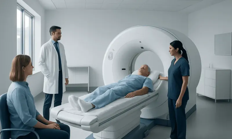
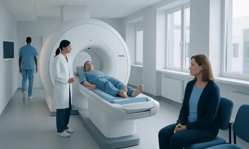
What Can a CT Scan Show That an MRI Cannot?

CT and MRI scans provide different types of information, and neither test is universally better. CT is often the preferred test for fast assessment of acute bleeding, bone fractures, lung conditions, certain calcifications and some abdominal emergencies, while MRI gives greater soft-tissue detail in many other situations.
Overview: what can a CT scan show that an MRI cannot?
What can a CT scan show that an MRI cannot? A CT scan can be better at showing acute internal bleeding, many bone fractures, lung changes, air in the body, some calcium deposits and urinary stones. It is also usually much faster than MRI, which can be important when clinicians need answers quickly after an injury or during a suspected stroke or other emergency.
CT, or computed tomography, uses multiple X-ray images processed by a computer to create detailed cross-sectional views of the body. MRI, or magnetic resonance imaging, uses a powerful magnetic field and radio waves rather than X-rays. The tests are complementary: the most appropriate examination depends on the part of the body being assessed, the clinical question, the person’s medical history and how urgently imaging is needed.
A CT finding is not automatically more important than an MRI finding, and a normal scan does not always rule out every possible condition. Radiologists interpret images in context, and a clinician may recommend another imaging test, blood tests, monitoring or specialist review when symptoms and scan findings do not fully match.
Where CT has practical advantages over MRI
CT is widely used in emergency and acute-care settings because it can be completed in minutes. It is less sensitive to movement than MRI and may be easier for people who are in severe pain, have difficulty lying still, need close monitoring or are uncomfortable in confined spaces. CT can also be performed in many people with implanted medical devices that may make MRI unsuitable, although the device type always needs to be checked.
CT is particularly strong at displaying dense structures. Bone cortex, calcium and metal are usually clearly seen. CT also provides excellent views of the chest because it shows air-filled lungs and the spaces around them well. Contrast dye may be used in either CT or MRI to make blood vessels, inflammation or masses easier to evaluate; the choice of contrast depends on the examination and a person’s kidney function, allergy history and other health factors.
- Acute bleeding: CT can rapidly identify many fresh bleeds in the brain, chest, abdomen or pelvis.
- Bone injury: It can define complex fractures, small bone fragments and some joint injuries.
- Lung and chest findings: It can show pneumonia patterns, lung nodules, collapsed lung and blood clots in the lung arteries when CT angiography is used.
- Calcification and stones: It is highly useful for kidney stones and many types of abnormal calcification.
What can a head CT scan show that an MRI cannot?

In the brain, the question of what can a CT scan show that an MRI cannot is often linked to speed and acute care rather than a complete difference in what is visible. A non-contrast head CT is commonly the first scan used after a significant head injury or sudden neurological symptoms because it can quickly show many cases of fresh bleeding, skull fractures, swelling that shifts brain structures, and pressure-related complications such as hydrocephalus.
CT can show skull bone particularly well. This includes fractures at the skull base, facial bones and around the sinuses, where MRI is not usually the first examination. CT can also identify air inside the skull after trauma or surgery, and it may demonstrate calcified lesions or calcification within an older injury.
However, MRI is often more sensitive for subtle brain injury, small strokes, inflammation, infection, tumors and many causes of seizures or chronic neurological symptoms. For suspected stroke, CT may first be used to exclude bleeding, while CT angiography or CT perfusion and MRI may be selected according to the timing of symptoms and local stroke protocols. Sudden weakness, speech difficulty, facial drooping, severe new headache or loss of consciousness requires urgent medical assessment rather than waiting for an outpatient scan.
Can an MRI see everything a CT scan can?
No. MRI cannot always replace CT, even though it can provide more detailed pictures of many soft tissues. MRI is less useful for some tiny fractures, subtle bone cortex changes, lung air-space detail, acute blood in certain settings, calcified structures and urinary stones. The longer scan time can also make MRI less practical when immediate assessment is needed.
MRI images may be affected by motion, and the examination can take considerably longer than CT. Some people cannot undergo MRI because of particular implanted devices, metal fragments or other safety concerns. Many modern implants are MRI-conditional rather than automatically incompatible, so imaging staff review the exact device and its documentation before scheduling a scan.
That said, MRI may still be chosen after CT if more tissue detail is needed. For example, a CT may identify a fracture or bleeding after trauma, while MRI can later assess associated ligament injury, spinal cord involvement or subtle damage to brain tissue. The two scans are often used at different stages of the same evaluation.
What cancers show up on a CT scan?
CT can detect or help assess many cancers, including cancers affecting the lung, liver, pancreas, kidney, bladder, colon, ovaries and lymph nodes. It can show a mass, organ enlargement, enlarged lymph nodes, fluid around organs, bone destruction and signs that cancer may have spread to the chest, abdomen, pelvis or other areas. CT is also used to guide some biopsies and to monitor how a known cancer responds to treatment.
CT does not diagnose every abnormality as cancer, and not every cancer is visible on a CT scan. Very small tumors, early surface lesions and tumors that blend with nearby tissue may be difficult to detect. A suspicious CT finding may require additional imaging, laboratory testing, endoscopy or a biopsy for a definite diagnosis.
Different cancers require different imaging approaches. Low-dose CT is used in selected people for lung cancer screening based on age and smoking history, while MRI, ultrasound, mammography, PET imaging or other tests may be more suitable in other settings. Cancer evaluation should be directed by a qualified clinician who can select the right tests and explain what the results mean.
What diseases can an MRI detect that a CT scan cannot?
MRI is particularly valuable when a condition affects soft tissue or when subtle changes need to be seen in detail. It may detect early or small strokes, multiple sclerosis plaques, inflammation of the brain or spinal cord, certain brain tumors, pituitary abnormalities, epilepsy-related structural changes and infections that may be less visible on CT. MRI is also often preferred for examining the spinal cord, discs, nerves, ligaments, tendons, cartilage, muscles and bone marrow.
In the abdomen and pelvis, MRI can provide additional information about the liver, bile ducts, pancreas, uterus, ovaries, prostate and pelvic floor. It may help characterize a lesion seen on CT or ultrasound without exposing the person to ionizing radiation. MRI can also assess blood flow and blood vessels in many situations, sometimes without contrast material.
There are important exceptions. CT may still be better for a specific question even when MRI is available. For example, a person with severe flank pain may have CT to look for a urinary stone, and someone with a sudden severe headache may have an urgent head CT to look for bleeding. The best test is determined by the likely diagnosis, not by a general ranking of CT versus MRI.
What is more serious, a CT scan or MRI?
Neither CT nor MRI is “more serious” in terms of what the test itself means. Both are diagnostic tools, and being referred for either scan does not necessarily indicate a severe illness. Clinicians order imaging to investigate symptoms, clarify an earlier result, monitor a known condition or safely rule out urgent problems.
The tests differ in practical considerations. CT uses ionizing radiation, so clinicians aim to use it when the expected benefit outweighs the small potential radiation-related risk. MRI does not use ionizing radiation, but it uses a strong magnetic field and may not be appropriate for everyone. MRI can also be noisy, longer and challenging for people with claustrophobia or severe pain.
Contrast agents can occasionally cause side effects or allergic reactions with either test. People should tell the imaging team about kidney disease, pregnancy, possible pregnancy, previous contrast reactions, asthma, implanted devices and any metal fragments. The imaging team can then determine whether contrast is needed and whether precautions or an alternative test are appropriate.
Preparing for imaging and when to seek medical care
Preparation depends on the type of scan. Some CT or MRI studies require fasting or contrast dye, while others require no special preparation. Patients should bring relevant previous images or reports if available and tell staff about medicines, allergies, kidney problems, pregnancy and implants. They should not stop prescribed medication unless their clinician specifically advises it.
Medical care should be sought urgently for symptoms that could indicate a serious acute condition, such as sudden weakness or numbness on one side, trouble speaking, new confusion, seizure, fainting, a sudden severe headache, chest pain, serious breathing difficulty, major head injury or severe abdominal pain. Emergency clinicians can determine whether CT, MRI or another test is needed.
For ongoing symptoms such as recurrent headaches, persistent pain, unexplained weight loss, new neurological changes or a concerning lump, an appointment with a doctor is appropriate. Acıbadem Health Point’s multidisciplinary specialists and JCI-accredited hospitals diagnose and treat conditions requiring advanced imaging for international patients, with the imaging plan tailored to the individual clinical question.
Frequently asked questions
01Can a CT scan show something an MRI cannot?
Yes. CT may show bone fractures, lung air-space changes, calcifications, urinary stones and acute bleeding more clearly or more quickly in certain situations. MRI can show many of these findings in some cases, but it is not always the preferred or fastest test.
02What will an MRI show that a CT scan will not?
MRI can reveal subtle abnormalities in the brain, spinal cord, nerves, ligaments, cartilage, muscles and bone marrow that may not be visible on CT. It is often more sensitive for early ischemic stroke, multiple sclerosis, soft-tissue injuries and some tumors or inflammatory conditions.
03Why is CT often used first after a head injury?
A head CT is fast and effective for identifying many urgent problems, including fresh bleeding, skull fractures and swelling that can raise pressure inside the skull. MRI may be used later when clinicians need a more detailed assessment of brain tissue or subtle injury.
04Does a CT scan show all cancers?
No. CT can identify many tumors and signs of cancer spread, but it cannot detect every cancer or confirm that every mass is cancerous. Further tests, including biopsy, may be necessary to establish a diagnosis.
05Is CT radiation dangerous?
CT uses radiation, but imaging teams use protocols designed to keep exposure as low as reasonably achievable while obtaining medically useful images. When a CT is clinically justified, its diagnostic benefit generally outweighs the potential risk; patients can discuss concerns and alternatives with their clinician.
06Can a person have both a CT and an MRI?
Yes. In some cases, the tests answer different questions and are used together. For example, CT may assess an acute injury quickly, while MRI may later evaluate soft tissues, nerves or subtle brain changes in greater detail.
This article is for general information only and is not a substitute for professional medical advice. Please consult a qualified doctor about your individual situation.
Joint and spine care in Turkey — expert assessment & treatment
JCI-accredited · board-certified surgeons · reply within 24h
Add us as a Preferred Source to see more of our trusted health content across Google Search, AI Overviews and Discover.