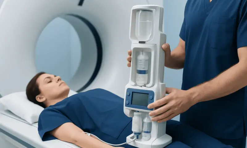
CT Scan, CAT Scan, and MRI: What Is the Difference?
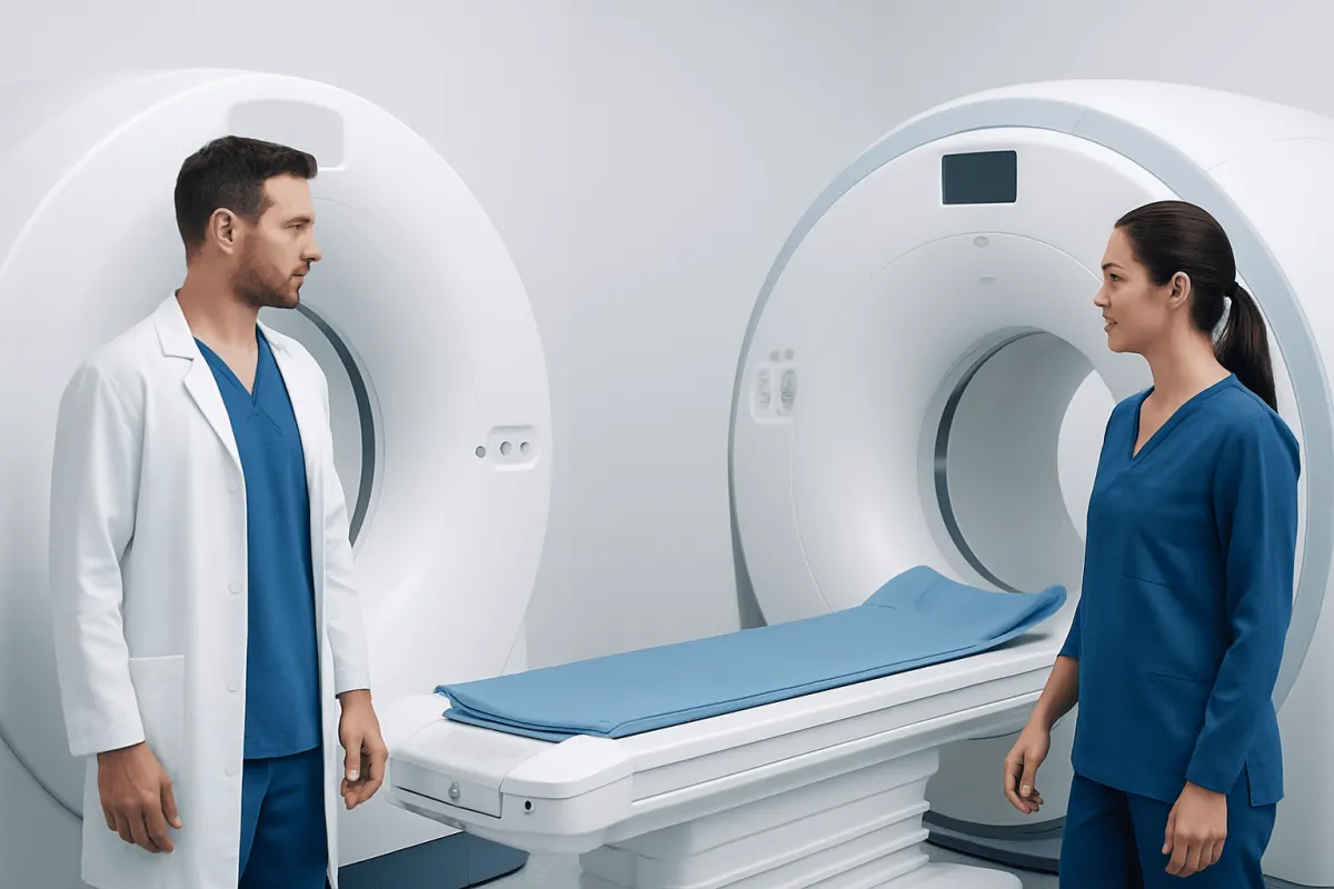
A CT scan and a CAT scan are two names for the same imaging test. MRI is a different scan that uses magnetic fields rather than X-rays, and the best choice depends on the body area, symptoms, urgency, and clinical question.
CT Scan vs CAT Scan vs MRI: Side-by-Side Comparison
In the comparison of CT scan vs CAT scan vs MRI, the first point is simple: a CT scan and a CAT scan are the same examination. CAT stands for computed axial tomography, an older term that is still commonly used. Today, clinicians usually say CT, meaning computed tomography.
CT uses a series of X-rays and computer processing to create detailed cross-sectional images of the body. MRI, or magnetic resonance imaging, uses a strong magnetic field and radio waves to generate images. MRI does not use ionizing radiation, but it commonly takes longer than CT and requires the person to lie still in a narrow scanner.
- CT/CAT scan: Fast, uses X-rays, and is especially helpful for bones, lungs, internal bleeding, trauma, and many urgent abdominal or chest conditions.
- MRI: Uses magnets and radio waves, with high soft-tissue detail; often used for the brain, spinal cord, joints, nerves, pelvic organs, and certain tumors.
- Contrast material: Either test may use contrast to make blood vessels, inflammation, organs, or abnormalities easier to assess. The type of contrast differs between CT and MRI.
- Choice of scan: Depends on the suspected condition, the body part being examined, how quickly information is needed, prior imaging, kidney function, pregnancy status, and medical implants.
How Clinicians Tell CT and MRI Apart
Clinicians do not choose between CT and MRI based only on which test produces a clearer-looking image. They first define the medical question: for example, whether there may be a fracture, stroke, infection, tumor, torn ligament, kidney stone, blood clot, or internal bleeding. Each test highlights different tissues and changes within the body.
CT is usually preferred when speed is essential. It can be performed quickly and is widely used after significant injury, for sudden severe headache when bleeding is a concern, and for rapid evaluation of the chest or abdomen. It is also particularly informative for bone detail, calcium, many lung conditions, and urinary stones.
MRI takes more time but can distinguish soft tissues in greater detail. It can show subtle differences in the brain, spinal cord, bone marrow, muscles, tendons, cartilage, ligaments, and pelvic structures. The radiologist interprets the findings alongside the scan technique, contrast use, symptoms, examination findings, and previous studies.
What Does an MRI Show That a CT Scan Will Not?
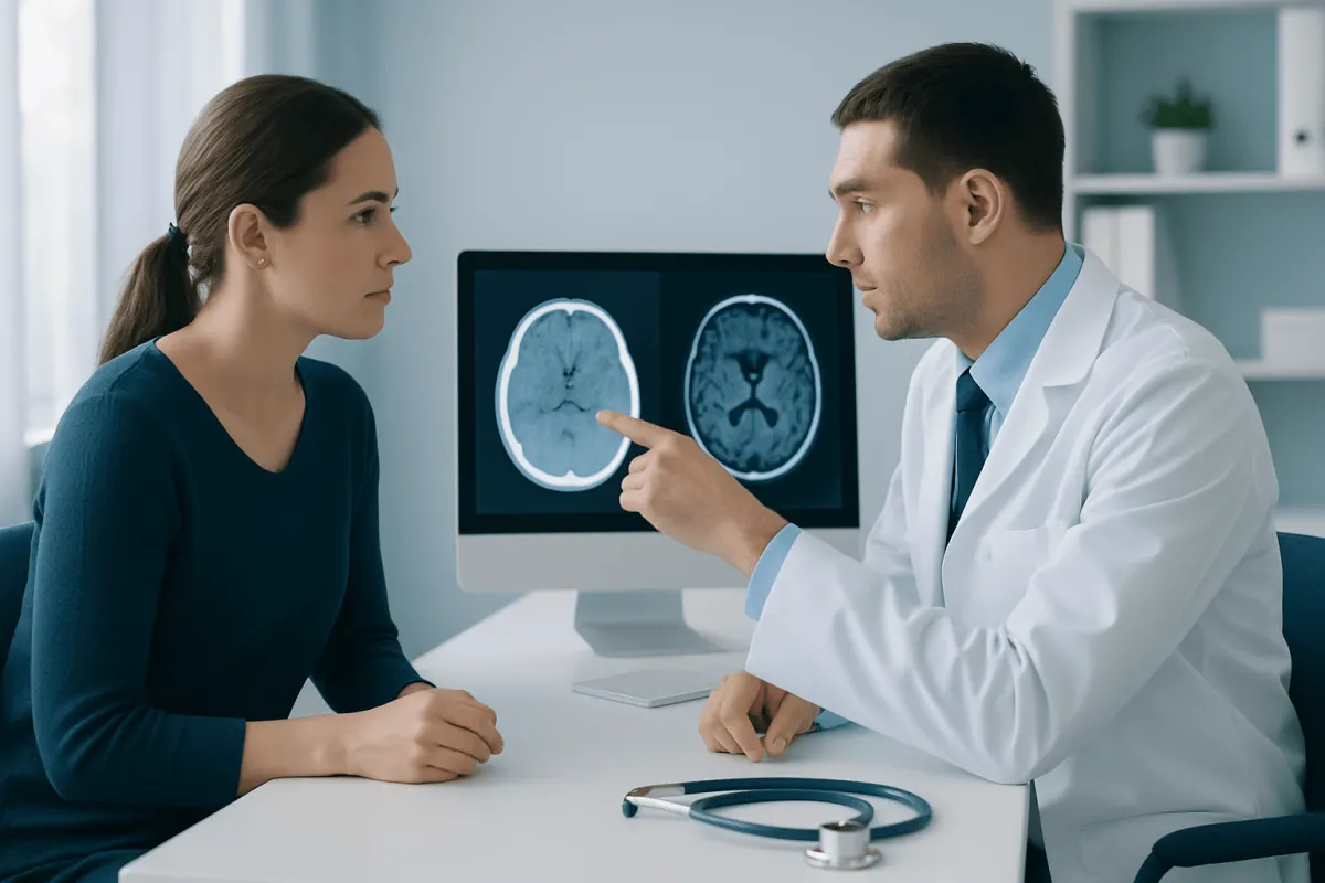
MRI can often show soft-tissue details that may be difficult to see, or less clearly defined, on CT. These include many abnormalities involving the brain and spinal cord, such as inflammation, certain early stroke changes, multiple sclerosis plaques, spinal disc problems, nerve compression, and some tumors. MRI may also better assess ligaments, tendons, cartilage, muscles, bone marrow, and some pelvic organs.
However, MRI does not replace CT in every situation. CT can be more useful for detecting acute bleeding in some emergency settings, evaluating complex fractures, examining the lungs, identifying calcifications, and detecting many kidney stones. In some cases, clinicians use both tests because the information is complementary rather than competing.
An MRI finding must always be interpreted in context. Small changes may be unrelated to symptoms, while a normal MRI does not rule out every condition. A qualified clinician can explain what a result means for the individual and whether further assessment is needed.
How to Differentiate Between CT and MRI Report?
The report header usually identifies the examination clearly. A CT report may be titled “CT head,” “CT chest,” “CT abdomen and pelvis,” or “computed tomography.” An MRI report may say “MRI brain,” “MRI lumbar spine,” or “magnetic resonance imaging.” The report may also note whether intravenous contrast was used.
CT reports often describe density or attenuation, terms related to how X-rays pass through tissue. They may comment on fractures, bleeding, lung nodules, stones, calcification, or organ injury. MRI reports often describe signal intensity and sequences, such as T1, T2, FLAIR, diffusion, or contrast enhancement. These technical terms help radiologists identify tissue characteristics but should not be used alone to judge seriousness.
The most useful parts for patients are generally the “Findings” and “Impression” sections. The impression is a concise summary of the main observations and may include recommendations for follow-up. It is best to review the report with the clinician who ordered the scan, as they can connect the result to symptoms and medical history.
What Gives Better Results, CT Scan or MRI?
Neither CT nor MRI always gives better results. The better test is the one most likely to answer the specific clinical question accurately and safely. For example, CT may provide the best immediate information after a major injury, when there is concern about a lung condition, or when a clinician needs to identify a fracture or kidney stone quickly.
MRI may give better results for many conditions affecting the brain, spinal cord, joints, muscles, ligaments, tendons, and nerves. It is also often used to characterize certain abnormalities seen on ultrasound or CT. For suspected stroke, the preferred initial study can vary according to timing, symptoms, local emergency pathways, and the need to exclude bleeding rapidly.
Radiation exposure is another consideration. CT uses X-rays, so clinicians aim to use it only when it is medically justified and to keep radiation as low as reasonably achievable. MRI does not use radiation, but it may be impractical for people who cannot lie still, have certain implanted devices, or need very rapid imaging. Claustrophobia, kidney health, allergies, and pregnancy may also influence the plan.
What Cancers Are Best Seen on MRI?
MRI is especially valuable for evaluating cancers in areas where soft-tissue contrast is important. It is commonly used in assessment of tumors involving the brain and spinal cord, prostate, liver, uterus, cervix, ovaries, rectum, breast, muscles, bones, and other soft tissues. MRI may help define a tumor’s size, relationship to nearby structures, and response to treatment.
Still, MRI is not the best single test for every cancer or for every stage of cancer care. CT is frequently used to examine the chest, abdomen, and pelvis, including the lungs, and to look for spread of disease. Mammography, ultrasound, PET imaging, endoscopy, laboratory tests, and biopsy may also have important roles depending on the suspected cancer type.
Imaging can identify areas that need further assessment, but it usually cannot confirm cancer on its own. A biopsy, in which a tissue sample is examined by a pathologist, is often needed for a definite diagnosis. For patients undergoing cancer evaluation, imaging choices are typically coordinated by radiology, oncology, surgery, and other relevant specialists.
What to Do Before, During, and After Each Scan
Before a CT or MRI scan, patients should tell the imaging team about pregnancy or possible pregnancy, kidney disease, previous reactions to contrast material, asthma or allergies, diabetes medicines, and all implants or metal in the body. For MRI, this includes pacemakers, cochlear implants, aneurysm clips, insulin pumps, neurostimulators, metal fragments, and previous surgery. Many modern devices are MRI-conditional, but the team must verify the exact device and safety requirements.
Some CT and MRI examinations require fasting for a period beforehand, particularly when contrast is planned, while others require no special preparation. Patients should follow the instructions from their imaging center. Metal objects such as jewelry, watches, hearing aids, removable dental appliances, and credit cards must be removed before MRI because of the magnetic field.
During CT, the scanner usually moves quickly around the body. During MRI, the machine makes loud tapping or knocking sounds, and ear protection is provided. The team can communicate throughout the test, and patients should mention pain, anxiety, breathing difficulty, itching, or any unusual symptoms promptly. After contrast imaging, the team may provide individual guidance about hydration or medicines.
Acıbadem Health Point’s multidisciplinary specialists and JCI-accredited hospitals can coordinate diagnostic imaging and follow-up care for international patients when CT, MRI, or further assessment is needed.
When to Seek Medical Care
People should seek urgent medical care for symptoms that may require rapid assessment, including sudden weakness or numbness on one side of the body, new trouble speaking, sudden severe headache, fainting, seizures, major head injury, chest pain, severe shortness of breath, coughing blood, severe abdominal pain, or significant trauma. Emergency clinicians decide whether CT, MRI, or another test is needed based on the situation.
Medical review is also appropriate for persistent or worsening headaches, unexplained neurological symptoms, ongoing back pain with weakness or bladder or bowel changes, a new lump, unexplained weight loss, persistent bone pain, or symptoms that do not improve as expected. Imaging should be requested by a clinician when it is likely to guide care rather than used as a routine screening test without a clear reason.
Anyone who has received an imaging report with uncertain or concerning wording should arrange a discussion with the ordering clinician. They can clarify whether the finding is urgent, incidental, benign-appearing, or likely to need repeat imaging, specialist review, or further tests.
Frequently asked questions
01Is a CAT scan the same as a CT scan?
Yes. CAT scan and CT scan refer to the same imaging technology. CAT is an older abbreviation for computed axial tomography, while CT, meaning computed tomography, is now the more common term.
02Which is safer, CT or MRI?
Both can be safe when appropriately selected and performed by trained professionals. CT uses ionizing radiation, while MRI does not, but MRI may not be suitable for people with certain implants or metal objects. The safest option depends on the reason for imaging and the person's medical history.
03Can CT and MRI both use contrast dye?
Yes, but the contrast agents are different. CT commonly uses iodine-based contrast, while MRI commonly uses gadolinium-based contrast. The imaging team reviews kidney function, prior contrast reactions, and other relevant health information before using contrast.
04Why would a doctor order an MRI after a CT scan?
A CT may identify an abnormality or exclude an urgent problem quickly, while MRI can provide more detail about soft tissues. MRI may be ordered to clarify a brain, spine, joint, nerve, pelvic, or tumor-related finding. Having both scans does not necessarily mean that a problem is serious.
05Is MRI better than CT for headaches?
The most appropriate scan depends on the type of headache, symptoms, examination findings, and urgency. CT is often used quickly in emergency situations, particularly when bleeding or injury is a concern. MRI may be more informative for certain persistent, recurrent, or neurological headache patterns.
06Can an MRI miss something that a CT scan finds?
Yes. Each scan has strengths and limitations. CT may be better for some fractures, lung abnormalities, calcifications, acute bleeding patterns, and kidney stones, while MRI may better show many soft-tissue and neurological conditions.
This article is for general information only and is not a substitute for professional medical advice. Please consult a qualified doctor about your individual situation.
Joint and spine care in Turkey — expert assessment & treatment
JCI-accredited · board-certified surgeons · reply within 24h
Add us as a Preferred Source to see more of our trusted health content across Google Search, AI Overviews and Discover.