What Does a PET Scan Show in the Body?
A PET scan shows how active tissues and organs are by detecting a small amount of radioactive tracer in the body. It can help doctors evaluate cancer, heart disease, certain brain conditions and inflammation, but its findings always need interpretation alongside medical history and other tests.
Overview: What Does a PET Scan Show?
A positron emission tomography (PET) scan shows how tissues and organs are functioning at a cellular level. It uses a very small amount of a radioactive tracer, most often a glucose-like substance called FDG, to identify areas that are using more or less energy than expected. The scan can help doctors see patterns that may be linked with cancer, inflammation, infection, heart muscle damage or some neurological conditions.
For many examinations, PET is combined with computed tomography (PET-CT) or magnetic resonance imaging (PET-MRI). The PET images show biological activity, while the CT or MRI images show the location and structure of organs and tissues. Together, they can give a more complete picture than either test alone.
A PET scan does not usually provide a diagnosis by itself. Its results are assessed by a radiologist or nuclear medicine physician in the context of symptoms, examination findings, laboratory tests, previous scans and, when needed, tissue biopsy results.
How PET Imaging Works and What It Can Assess
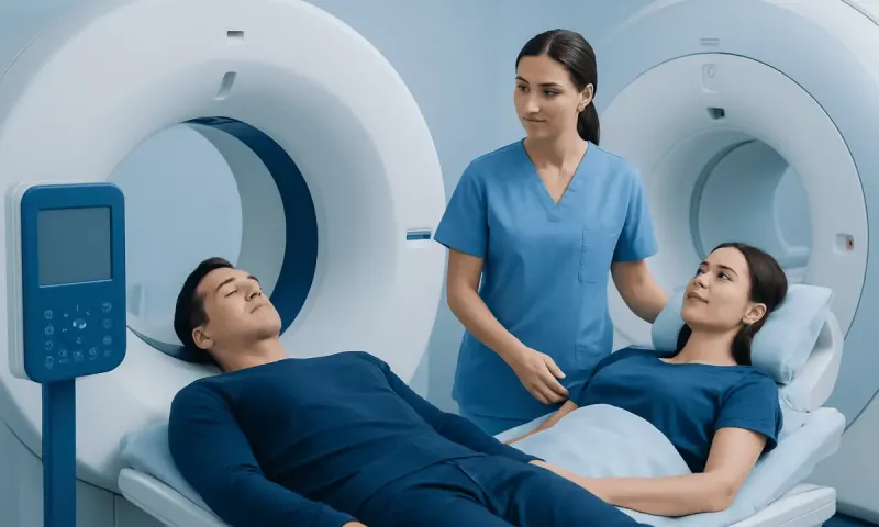
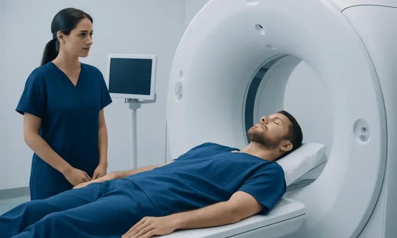
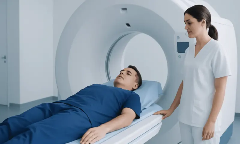
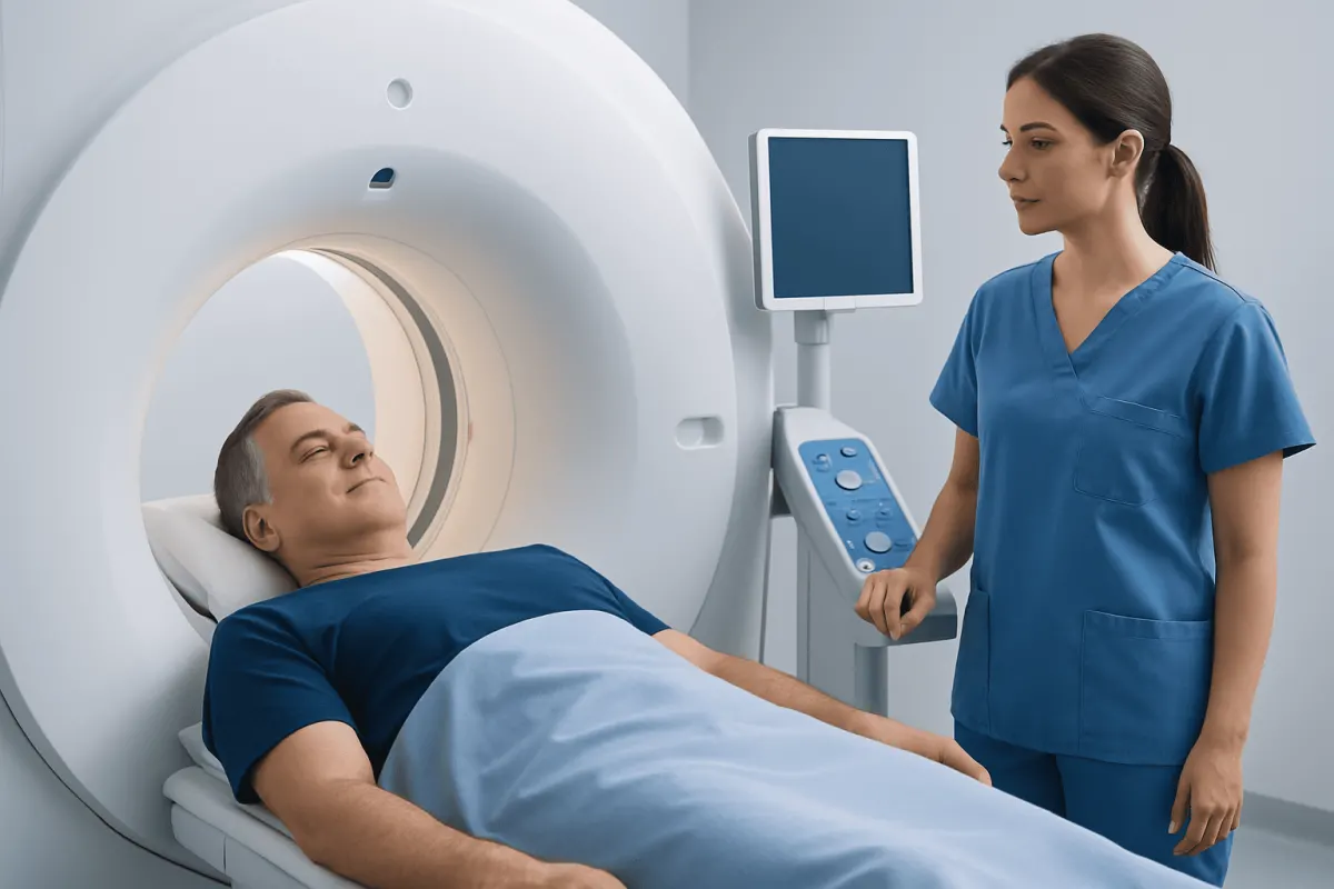
Before the scan, a healthcare professional injects the tracer into a vein. The tracer travels through the bloodstream and collects in tissues according to the process being studied. With FDG, cells that use more glucose may appear as areas of increased uptake, sometimes described as “hot spots.” The patient rests while the tracer distributes, then lies still on a scanning table while images are taken.
What does a PET scan show you? Depending on the tracer and clinical question, it may show the activity of a suspected tumour, whether disease may have spread, how well heart muscle is supplied with blood, or changes in brain metabolism. Other specialized tracers can target particular receptors or proteins, allowing more tailored imaging in selected conditions.
In cancer care, PET can help determine the extent of disease, guide the best site for a biopsy, support treatment planning and monitor response to therapy. It may also help distinguish active disease from scar tissue after treatment, although this distinction is not always certain.
PET Compared With CT and MRI

What does a PET scan show that a CT scan does not? CT creates detailed cross-sectional pictures of anatomy, such as the size, shape and location of a mass, enlarged lymph node or organ abnormality. PET can show whether an area has unusually high or low metabolic activity, which may identify active disease before a clear structural change is visible on CT.
However, a PET scan is not inherently better than CT. CT is often more useful for fine anatomical detail, lung assessment, bone structure, acute injury and many other questions. PET-CT combines the strengths of both tests and is commonly used when doctors need to correlate metabolic activity with a precise body location.
What does a PET scan show that an MRI does not? MRI provides excellent detail of soft tissues, including the brain, spinal cord, joints, liver and pelvic organs. PET adds information about metabolism or specific molecular targets. PET-MRI may be useful in selected cases, but the most suitable scan depends on the part of the body and the reason for testing.
PET Scans for Cancer, Heart and Other Conditions
What does a PET scan show for cancer? Many cancers use glucose more actively than surrounding tissues and therefore take up more FDG tracer. PET may help identify the primary tumour, lymph node involvement or distant spread in some cancer types. It is frequently used in conditions such as lymphoma, lung cancer, colorectal cancer, head and neck cancers and melanoma, among others.
What is the most common diagnosis for a PET scan? There is no single diagnosis made by PET scanning, but cancer evaluation is one of its most common uses. In oncology, PET is often requested to stage a known cancer, assess whether treatment is working, investigate a possible recurrence or clarify an uncertain finding on another test. The scan is also used in some heart and brain assessments.
What does a PET scan show for prostate cancer? Standard FDG PET is not ideal for many prostate cancers. A prostate-specific membrane antigen (PSMA) PET scan may be used in suitable patients to look for prostate cancer that has returned or spread, and to support treatment planning. The choice of scan depends on the person’s PSA level, prior treatment, symptoms and clinical situation.
What does a PET scan show for multiple myeloma? PET-CT can identify metabolically active myeloma lesions in bone or soft tissue and may help assess the extent of disease or response to treatment. It is interpreted alongside blood and urine tests, bone marrow findings and other imaging because no one test answers every question in multiple myeloma.
What does a PET scan show of the heart? Cardiac PET can evaluate blood flow to heart muscle and, in some cases, whether weakened heart muscle is still viable and may benefit from restored blood supply. It may be used when other cardiac tests are inconclusive or when a detailed assessment of coronary circulation is needed.
Will a PET Scan Show All Cancer?
No. A PET scan will not show all cancers, and a normal scan cannot fully rule out cancer. Some tumours are too small to detect, have low metabolic activity, or occur in areas where normal tracer uptake can make interpretation difficult. Certain cancer types also do not absorb a particular tracer strongly.
FDG PET can be less reliable for some slow-growing tumours and for some cancers of the prostate, kidney, liver or brain, depending on the clinical setting. Doctors may choose a different tracer, CT, MRI, ultrasound, endoscopy or another investigation when these methods are more appropriate.
Increased tracer uptake is not specific to cancer. Recent surgery, healing tissue, infection, inflammation, arthritis, active muscles and normal uptake in organs such as the brain, heart and urinary tract can affect the images. For this reason, PET findings should never be interpreted in isolation.
What Should You Not Do Before a PET Scan?
Preparation instructions vary according to the tracer and type of PET examination, so the imaging department’s advice should always take priority. For many FDG PET scans, patients are asked to avoid eating for several hours beforehand and to drink plain water. This helps reduce normal glucose use in the body and can improve image quality.
Patients are often advised not to do strenuous exercise for at least 24 hours before an FDG PET scan. Exercise can cause muscles to take up more tracer and may make images harder to interpret. They should also avoid chewing gum, smoking or using nicotine on the day of the scan if instructed, as these activities can sometimes affect uptake patterns.
People with diabetes need individualized preparation because blood glucose levels can influence FDG imaging. They should not change insulin, diabetes tablets or meals without guidance from the imaging team or their clinician. It is important to tell staff about pregnancy, possible pregnancy, breastfeeding, diabetes, current medications, recent infections and recent procedures.
- Wear comfortable clothing and remove metal items if requested.
- Bring previous scans and relevant medical records when available.
- Plan to remain still during the scan and allow time for tracer uptake beforehand.
- Follow instructions about hydration and using the restroom, which may help reduce tracer in the urinary system.
What Happens If a PET Scan Is Positive?
A positive PET scan means that an area has taken up more tracer than expected, or occasionally less tracer than expected, depending on the study. It is an imaging finding rather than a final diagnosis. The meaning depends on the location, intensity and pattern of uptake, as well as the patient’s symptoms and medical history.
If a PET scan is positive, the doctor may compare it with CT, MRI or earlier PET images; request blood tests; recommend short-term follow-up imaging; or arrange a biopsy if confirmation is needed. A biopsy is often the most reliable way to determine whether a suspicious area is cancerous and, if so, identify its type.
For someone already being treated for cancer, increased uptake may indicate active disease, treatment-related inflammation, infection or healing. The care team will explain what the result means in that individual context and discuss whether any further tests or treatment changes are appropriate.
When to Seek Medical Care
A PET scan is usually arranged by a clinician after symptoms, examination findings or another test suggests that further assessment may be useful. People should contact their doctor promptly if they have persistent unexplained symptoms such as a new or growing lump, unexplained weight loss, ongoing bleeding, persistent pain, worsening shortness of breath or a significant change in bowel, urinary or neurological function.
After a PET scan, most people can return to normal activities unless they receive different instructions. Mild bruising at the injection site can occur. Medical advice should be sought for signs of a significant allergic reaction, such as difficulty breathing, facial swelling or widespread hives, although serious reactions to PET tracers are uncommon.
For people who need coordinated assessment, Acıbadem Health Point’s multidisciplinary specialists and JCI-accredited hospitals diagnose and treat conditions requiring advanced imaging for international patients. The next step after an abnormal scan should be discussed with a qualified doctor who can place the result in the full clinical context.
Frequently asked questions
01How long does a PET scan take?
The scanning portion commonly takes around 20 to 40 minutes, but the full appointment is often longer. Time is needed for tracer injection, a quiet uptake period and preparation, so patients should allow several hours unless their imaging center advises otherwise.
02Is a PET scan painful?
A PET scan is generally not painful apart from the brief needle prick for the tracer injection. Patients need to lie still on a table, which may be uncomfortable for some people, but the scanner itself does not cause pain.
03Does a hot spot on a PET scan mean cancer?
No. A hot spot means an area has increased tracer uptake, but it can occur with cancer, infection, inflammation, healing tissue or normal body activity. A specialist interprets the finding with other scans, medical information and sometimes a biopsy.
04Can a PET scan detect cancer spread?
PET scans can help identify areas that may represent cancer spread in lymph nodes, bones or other organs for certain cancers. Their accuracy varies by cancer type, tumour size, tracer used and body location, so additional tests may still be needed.
05Can you be around other people after a PET scan?
In most cases, patients can be around other people after a PET scan because the tracer amount is small and decreases naturally over time. The imaging team may give temporary precautions, especially around pregnant people or young children, based on the tracer used.
06How soon are PET scan results available?
A nuclear medicine physician or radiologist reviews the images and prepares a report for the referring doctor. Timing varies by facility and clinical urgency, but the referring clinician will explain when and how the results will be discussed.
This article is for general information only and is not a substitute for professional medical advice. Please consult a qualified doctor about your individual situation.
Medical weight-loss options in Turkey — costs & eligibility
JCI-accredited · board-certified surgeons · reply within 24h
Add us as a Preferred Source to see more of our trusted health content across Google Search, AI Overviews and Discover.