Ultrasound vs CT Scan: How the Images Differ
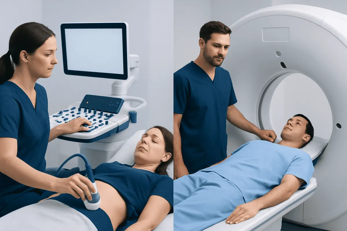
The difference between ultrasound and CT scan is mainly the technology used and the detail each test provides. Ultrasound uses sound waves and has no ionizing radiation, while CT uses X-rays to create detailed cross-sectional images that can better assess many internal injuries, organs and structures.
Overview: Ultrasound and CT at a Glance
The difference between ultrasound and CT scan is that ultrasound creates images with high-frequency sound waves, while computed tomography (CT) combines multiple X-ray images to produce detailed cross-sectional views of the body. Both are widely used diagnostic tools, but they answer different clinical questions and are selected according to the person’s symptoms, medical history and the area being examined.
Ultrasound is portable, does not use ionizing radiation and can provide real-time images. CT is particularly useful when clinicians need a fast, detailed overview of the chest, abdomen, pelvis, head, bones or blood vessels. A doctor may begin with ultrasound and recommend CT if more information is needed.
- Ultrasound: sound waves, real-time imaging, no radiation.
- CT scan: X-rays processed by a computer, detailed cross-sectional images, uses radiation.
- Choice of test: based on the suspected condition, urgency, body part and individual circumstances such as pregnancy or kidney function.
Side-by-Side Comparison: Ultrasound vs CT Scan
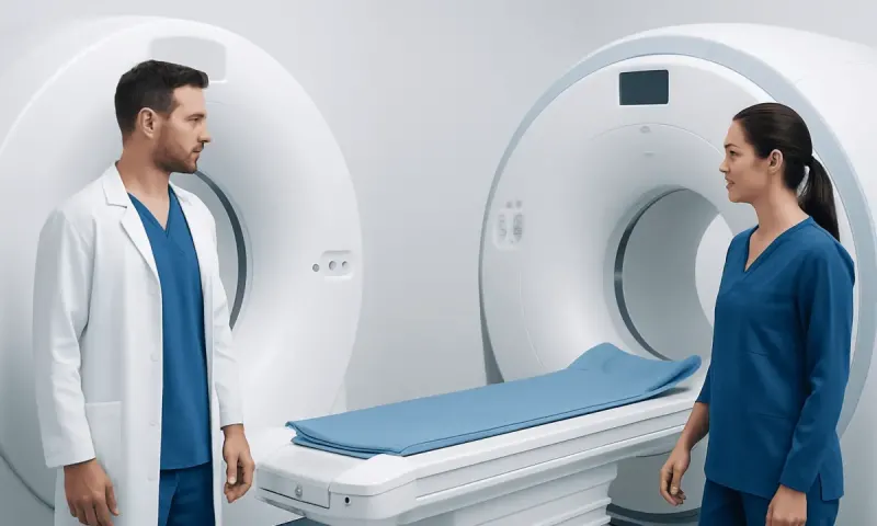
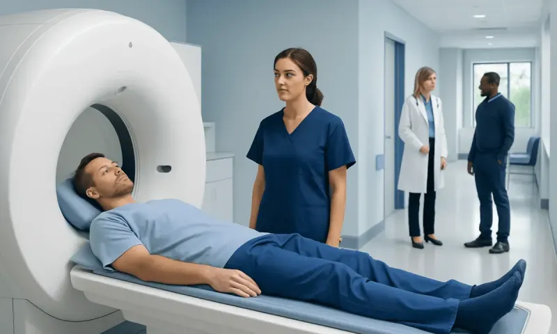
Although both tests are non-surgical imaging examinations, the experience and information they provide are different. Ultrasound generally involves moving a small handheld probe over gel applied to the skin. For CT, the person lies on a table that moves through a donut-shaped scanner while images are taken.
| Feature | Ultrasound | CT scan |
|---|---|---|
| Technology | Sound waves | X-rays and computer processing |
| Radiation | No ionizing radiation | Uses ionizing radiation |
| Best visualized areas | Pregnancy, pelvic organs, gallbladder, thyroid, tendons, superficial lumps and blood flow | Brain, lungs, bones, internal injuries, complex abdominal conditions and many blood vessels |
| Image detail | Excellent for selected soft tissues and moving structures; can be limited by bowel gas or body habitus | Detailed cross-sectional images with a wide field of view |
| Speed | Often quick, but varies by examination and operator | Usually very fast, especially in urgent care |
| Contrast material | Usually not needed for routine studies | May be given by mouth or vein for certain examinations |
Results are interpreted by appropriately trained clinicians, commonly radiologists, alongside the person’s symptoms, physical examination and other test results. An imaging result is only one part of a full clinical assessment.
How Clinicians Tell Which Scan Is Needed

Clinicians do not choose between ultrasound and CT simply because one is newer or more detailed. They first consider the likely cause of symptoms and whether the test can safely and clearly examine the relevant body area. For example, ultrasound may be appropriate for pelvic pain, suspected gallstones, a thyroid nodule or assessment of blood flow in a leg vein.
CT is often preferred when there is concern about a serious injury, bleeding, appendicitis, kidney stones, a complicated infection, lung disease or a condition involving several organs at once. In emergency settings, its speed and broad view can be especially helpful. For symptoms suggesting appendicitis, CT may be considered when ultrasound is inconclusive or when a more complete abdominal assessment is needed.
Age, pregnancy status, previous imaging, kidney health, allergy history and the possible need for contrast material also influence the decision. In children and pregnant people, clinicians generally seek to avoid unnecessary radiation and may use ultrasound first whenever it can answer the clinical question reliably.
What Can a CT Scan Show That an Ultrasound Does Not?
CT can show many structures that ultrasound cannot assess well because sound waves are blocked or distorted by air and bone. CT is particularly useful for examining the lungs, brain, skull, spine, ribs and other bones. It can also provide a broad view of deep abdominal and pelvic structures that may be difficult to see with ultrasound because of bowel gas.
With contrast material when appropriate, CT can identify patterns of inflammation, internal bleeding, abscesses, some tumors, blood vessel narrowing or blockage, and damage after trauma. It is commonly used to evaluate complex injuries because it can show bones, organs and blood collections in the same examination.
This does not mean CT replaces ultrasound. Ultrasound can be more informative for certain problems, including fetal assessment, gallstones, testicular concerns, superficial soft-tissue lumps and real-time blood-flow evaluation. Each modality has strengths that complement the other.
What Is More Accurate, Ultrasound or CT Scan?
Neither ultrasound nor CT is universally more accurate. Accuracy depends on what clinicians are looking for, where it is located, how advanced the condition is and whether the images are technically clear. A test is most accurate when it is matched to the medical question.
For many deep abdominal conditions, injuries and lung or bone problems, CT often gives more complete and detailed information. It may be more reliable when bowel gas, depth of tissue or a person’s body shape limits ultrasound images. CT can also reveal findings outside the area where pain is felt, which may help identify an unexpected cause.
Ultrasound is highly accurate for several targeted uses, such as evaluating the developing fetus, detecting many gallstones, examining the thyroid, checking blood flow with Doppler techniques and assessing some pelvic conditions. It also allows the examiner to see movement in real time and to focus on the exact area of tenderness. If the first study does not fully answer the question, a clinician may recommend additional imaging rather than assuming a normal or unclear result rules out every condition.
What Is the Major Disadvantage of CT Scan?
The major disadvantage of CT scanning is exposure to ionizing radiation. The amount varies according to the body part scanned and the type of examination, and imaging teams use protocols designed to keep radiation as low as reasonably achievable while still obtaining useful images. A single medically necessary CT may be appropriate when its potential diagnostic benefit outweighs the risk.
Some CT examinations use iodinated contrast material to make blood vessels and organs easier to see. Contrast reactions are uncommon but possible, and people should tell their care team about prior contrast reactions, asthma, kidney disease, thyroid conditions, diabetes medicines, pregnancy or other relevant health concerns. The care team can decide whether contrast is suitable and whether precautions or another test are preferable.
CT also may detect small incidental findings that are unrelated to symptoms. These findings can sometimes require further review or follow-up imaging. A clinician can explain whether a reported finding needs attention and how it relates to the person’s health.
What Cannot Be Detected by Ultrasound?
Ultrasound cannot reliably detect every medical problem. It is limited when sound waves must pass through air-filled bowel or lungs, dense bone, or very deep tissues. As a result, ultrasound is generally not the main test for examining most brain conditions in adults, detailed lung disease, many bone injuries or structures hidden behind bowel gas.
Some small, deep or early abnormalities may be difficult to identify, particularly if the imaging window is limited. For example, ultrasound may not clearly show the appendix in every person, may not fully assess the pancreas, and may provide incomplete views of deep abdominal organs in some circumstances. An unclear ultrasound does not necessarily mean there is no medical problem.
When ultrasound has limitations, the next step may be CT, MRI, laboratory testing or clinical observation, depending on the situation. For suspected urinary stones, clinicians may use kidney stone evaluation and treatment pathways that consider symptoms, ultrasound findings and CT when needed.
Preparing for Imaging and When to Seek Medical Care
Preparation depends on the examination. Some abdominal ultrasounds require fasting for a period beforehand, while pelvic ultrasound may require a comfortably full bladder. CT preparation may include avoiding food for a short time if contrast is planned. The imaging department will provide tailored instructions, and patients should bring information about medications, allergies, prior scans and relevant health conditions.
Medical care should be sought urgently for severe or worsening abdominal pain, sudden weakness or difficulty speaking, chest pain, severe shortness of breath, fainting, significant head injury, heavy bleeding, high fever with severe illness, or new symptoms after major trauma. These symptoms do not always require CT, but they need prompt clinical assessment so the appropriate tests can be arranged.
For non-urgent symptoms that persist, recur or interfere with daily life, a qualified doctor can determine whether imaging is needed and which test is most suitable. Acıbadem Health Point’s multidisciplinary specialists and JCI-accredited hospitals diagnose and treat a broad range of conditions for international patients, with imaging choices guided by clinical need.
Frequently asked questions
01Is an ultrasound safer than a CT scan?
Ultrasound does not use ionizing radiation, which is an important advantage when it can provide the needed information. CT uses X-rays, but it may be the more appropriate test when detailed and rapid imaging is necessary. A clinician weighs the expected benefit of imaging against any potential risks.
02Can ultrasound replace a CT scan?
Sometimes ultrasound can provide all the information needed, particularly for pregnancy, gallbladder problems, pelvic organs and blood-flow assessment. However, it cannot replace CT for every situation because CT better evaluates many deep structures, lungs, bones and urgent injuries. The tests are complementary rather than interchangeable.
03Why might a doctor order CT after an ultrasound?
A doctor may order CT if ultrasound images are unclear, if the area cannot be adequately visualized, or if symptoms suggest a condition that CT assesses more effectively. CT may also help confirm a suspected diagnosis, show its extent or identify alternative causes of symptoms. This does not necessarily mean that the ultrasound result was abnormal.
04Does a CT scan hurt?
A CT scan is painless and usually takes only a short time. The person needs to lie still while the table moves through the scanner. If contrast is injected, some people briefly notice a warm sensation or metallic taste, which usually passes quickly.
05Can ultrasound detect cancer?
Ultrasound can identify masses or changes in tissue that need further assessment, and it can help guide biopsies. However, ultrasound alone often cannot determine whether a mass is cancerous. Further imaging, follow-up and sometimes biopsy may be needed for a definite diagnosis.
06Should pregnant people have a CT scan?
CT is usually avoided during pregnancy unless it is medically necessary and the expected benefit is important. Ultrasound is commonly used first because it does not use radiation. If CT is needed for urgent diagnosis, the medical team can use appropriate precautions and discuss the reason for the examination.
This article is for general information only and is not a substitute for professional medical advice. Please consult a qualified doctor about your individual situation.
Explore treatments in Turkey — costs, top hospitals & a free quote
JCI-accredited · board-certified surgeons · reply within 24h
Add us as a Preferred Source to see more of our trusted health content across Google Search, AI Overviews and Discover.