Melanoma Cancer Signs, Diagnosis, Outlook, and Treatment
A melanoma cancer sign is most often a new, changing, unusual-looking, or non-healing spot on the skin. Melanoma can be highly treatable when found early, so a prompt assessment of suspicious skin changes is important.
Melanoma cancer signs: an overview
A melanoma cancer sign may be a new mole, a mole that changes in color, shape, size, or sensation, or a skin spot that looks noticeably different from others. Melanoma is a type of skin cancer that begins in melanocytes, the cells that produce skin pigment. It can develop on skin that receives frequent sun exposure, but it may also occur on the scalp, soles of the feet, palms, under nails, genitals, or in the eye.
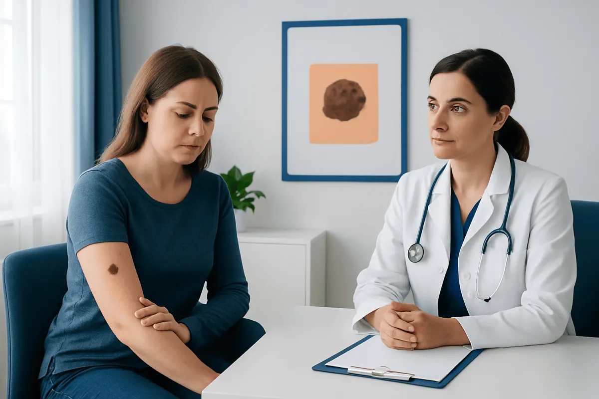
Many skin marks are harmless, and a suspicious-looking spot does not automatically mean melanoma. However, melanoma can grow and spread if it is not identified and treated. A dermatologist or qualified clinician can examine the skin and, when appropriate, remove a small sample or the whole lesion for laboratory testing. Information about melanoma may help patients understand the condition and the role of early evaluation.
Melanoma is not always dark brown or black. It may be pink, red, purple, blue-gray, skin-colored, or without pigment. This is particularly relevant for people with darker skin tones, in whom melanoma may appear on the palms, soles, nails, or mucosal surfaces rather than on sun-exposed areas.
How to recognize a possible melanoma cancer sign
The ABCDE approach is a practical way to notice changes in moles and other skin lesions. A stands for asymmetry, when one half does not resemble the other. B stands for an irregular, blurred, notched, or uneven border. C refers to varied color, such as different shades of brown, black, tan, red, white, or blue within one lesion. D means diameter, especially a spot larger than about 6 millimeters, although melanomas can be smaller. E means evolving: any change over weeks or months in appearance, size, texture, bleeding, or symptoms.
Another useful observation is the “ugly duckling” sign. This refers to a mole or spot that stands out because it looks different from a person’s other moles. A lesion that itches, hurts, crusts, bleeds, or does not heal should also be assessed, particularly if these changes persist or recur.
Not every melanoma starts in an existing mole. A significant number arise as new lesions on previously normal skin. Regularly checking the full skin surface, including the back, scalp, between the toes, and under the nails, can make changes easier to notice. A partner, family member, or clinician may help examine hard-to-see areas.
Causes and factors linked with melanoma risk
Melanoma develops when pigment-producing cells acquire genetic changes that allow abnormal growth. Ultraviolet, or UV, radiation from sunlight and tanning devices is an important preventable contributor. Intense intermittent sun exposure and sunburns, particularly blistering sunburns, may increase risk. Tanning beds expose skin to concentrated UV radiation and are not a safe alternative to sunlight.
Risk is also influenced by personal and family factors. People may have a higher likelihood of melanoma if they have many moles, unusual-looking moles, fair skin that burns easily, light eyes or hair, a previous melanoma or other skin cancer, a close relative with melanoma, or a weakened immune system. Melanoma can nevertheless affect people of every skin tone, age, and background.
Having one or more risk factors does not mean that melanoma will occur, and some people diagnosed with melanoma have no clear risk factor. A clinician can recommend an appropriate schedule for professional skin examinations based on individual history, skin type, and the number and appearance of moles.
How melanoma is diagnosed and staged
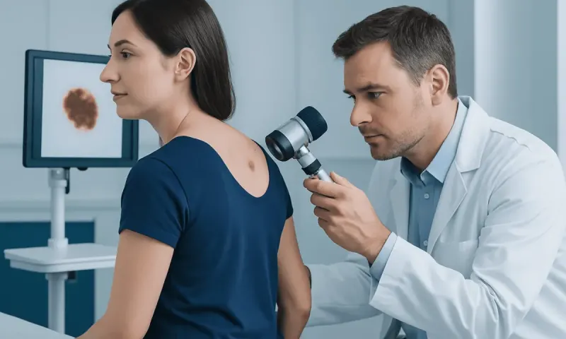
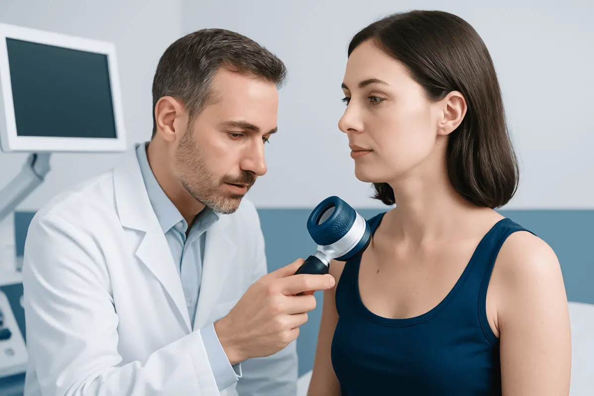
Diagnosis usually begins with a skin examination and a discussion of when the lesion appeared and how it has changed. A dermatologist may use dermoscopy, a handheld device that magnifies and illuminates skin structures. Dermoscopy can help identify concerning features, but it does not replace a biopsy when melanoma is suspected.
A biopsy is the only way to diagnose melanoma with certainty. Whenever possible, the clinician removes the entire suspicious lesion with a narrow margin of normal-appearing skin. A pathologist examines the tissue under a microscope and reports features that influence treatment planning, including Breslow thickness, ulceration, and the rate of cell division in some cases.
After diagnosis, the care team determines the stage. Early-stage melanoma is confined to the skin. More advanced stages may involve nearby lymph nodes or distant organs. Depending on the tumor’s features and stage, further assessment may include a sentinel lymph node biopsy, ultrasound, computed tomography, magnetic resonance imaging, or other scans. Imaging is not needed for every person with early melanoma.
What are the next steps after being diagnosed with melanoma?
After a melanoma diagnosis, the next step is usually to review the pathology report with a dermatologist, surgical oncologist, or multidisciplinary cancer team. The discussion should cover the melanoma’s thickness, whether ulceration is present, the location of the lesion, whether the biopsy fully removed it, and whether lymph node assessment is recommended. These details help clarify the stage and the treatment plan.
For many patients, the next treatment is a wider local excision. This is a procedure to remove additional normal-appearing skin around the biopsy site, lowering the chance that melanoma cells remain locally. The size of the margin depends on the melanoma thickness and body location. Reconstruction, such as a skin graft or flap, may occasionally be needed when a larger area must be removed.
Patients may also be referred for sentinel lymph node biopsy when the melanoma has features associated with a meaningful risk of spread to nearby lymph nodes. If melanoma is found in lymph nodes or elsewhere, the team may discuss systemic treatment, additional imaging, and structured follow-up. It can be helpful to bring a support person, keep copies of pathology reports, and write down questions before appointments.
Is there a cure for melanoma?
Many early melanomas can be cured with complete surgical removal. The outlook is generally more favorable when melanoma is diagnosed before it has spread beyond the skin. Individual prognosis depends on the stage and on pathology findings, including tumor thickness and ulceration, rather than on appearance alone.
Melanoma that has spread to lymph nodes or distant sites can be more complex, but treatment options have improved substantially. Immunotherapy helps the immune system recognize and attack cancer cells. Targeted therapies may be appropriate for melanomas with particular gene changes, commonly involving the BRAF gene. Radiation therapy, surgery for selected sites of disease, and clinical trials may also have a role in some circumstances.
Even after successful treatment, follow-up remains important because melanoma can occasionally return and because a person who has had melanoma may develop another skin cancer. Follow-up plans vary by stage and individual circumstances. They commonly include regular skin and lymph node examinations, self-skin checks, and advice about sun protection.
Can stage 1 melanoma make you tired?
Stage 1 melanoma is limited to the skin and often causes no symptoms beyond the visible lesion. Tiredness is not typically caused directly by stage 1 melanoma. Fatigue is common and can have many explanations, including stress after diagnosis, poor sleep, anemia, infection, thyroid conditions, medications, depression, and other medical concerns.
Feeling tired after receiving a Prostate Cancer Diagnosis: Tests and Next Steps" class="ahp-ilk">cancer diagnosis is understandable, and it should not be dismissed. A person should tell their clinician if fatigue is new, persistent, worsening, or affecting everyday activities. The healthcare team can review symptoms, medical history, sleep, emotional well-being, and, when appropriate, arrange tests to look for treatable causes.
Fatigue alone cannot determine melanoma stage or whether melanoma has spread. Staging relies on pathology findings, lymph node evaluation, and imaging only when clinically indicated. Patients should avoid trying to interpret general symptoms without discussing them with their care team.
Treatment, follow-up, and when to seek medical care
Treatment is individualized according to melanoma stage, location, pathology results, general health, and personal preferences. Surgery is central to treating localized melanoma. For higher-risk or advanced melanoma, care may involve medical oncology and treatments such as immunotherapy or targeted therapy. These treatments are selected only after careful staging and, for targeted therapy, appropriate tumor testing.
People should seek medical care promptly for a new or changing mole, a spot that bleeds or fails to heal, a dark streak under a nail that is widening, or a lesion that looks unlike their other moles. A person already diagnosed with melanoma should contact their care team about new or enlarging lumps, persistent unexplained symptoms, treatment side effects, or concerns between scheduled visits. Urgent medical assessment is appropriate for severe symptoms or signs of an acute illness.
Sun protection supports skin health before and after treatment. Useful measures include seeking shade when UV exposure is strong, wearing protective clothing and a broad-brimmed hat, using broad-spectrum sunscreen as directed, and avoiding tanning beds. Acıbadem Health Point’s multidisciplinary specialists and JCI-accredited hospitals diagnose and treat melanoma for international patients, with care plans based on pathology, stage, and individual clinical needs.
Frequently asked questions
01What is the first melanoma cancer sign to notice?
The first noticeable sign is often a new or changing mole or skin spot. Changes in asymmetry, border, color, size, or evolution over time should be assessed by a qualified clinician. Some melanomas are pink, red, or skin-colored rather than dark.
02What happens if you are diagnosed with melanoma?
The diagnosis is confirmed through a biopsy examined by a pathologist. The care team then reviews the tumor features and stage, and may recommend a wider excision of the area, lymph node assessment, or further tests depending on the individual situation. Treatment and follow-up are tailored to the melanoma stage.
03Can melanoma be diagnosed from a photograph?
Photographs can document a skin change and may be useful during a consultation, but they cannot confirm melanoma. A clinician needs to examine the lesion, and a biopsy is required for a definite diagnosis. Anyone concerned about a changing lesion should arrange an in-person assessment.
04Is a melanoma always a black mole?
No. Melanoma may be brown, black, tan, pink, red, blue-gray, purple, or skin-colored. It can also occur in less visible locations, including under a nail, on the soles or palms, and on the scalp. Any changing or unusual lesion deserves medical attention regardless of color.
05Can stage 1 melanoma make you tired?
Stage 1 melanoma does not usually directly cause fatigue because it is confined to the skin. Tiredness may result from stress, sleep disruption, another health condition, or other factors. Persistent or concerning fatigue should be discussed with a clinician.
06How often should someone check their skin after melanoma treatment?
Many clinicians advise regular self-skin checks, often monthly, as part of ongoing surveillance. The schedule for professional examinations depends on melanoma stage, treatment history, and individual risk factors. A treating dermatologist or oncology team can provide a personalized follow-up plan.
This article is for general information only and is not a substitute for professional medical advice. Please consult a qualified doctor about your individual situation.
Hair transplant in Turkey — costs, graft counts & a free assessment
JCI-accredited · board-certified surgeons · reply within 24h
Add us as a Preferred Source to see more of our trusted health content across Google Search, AI Overviews and Discover.