Image of Melanoma Skin Cancer: Signs and Next Steps
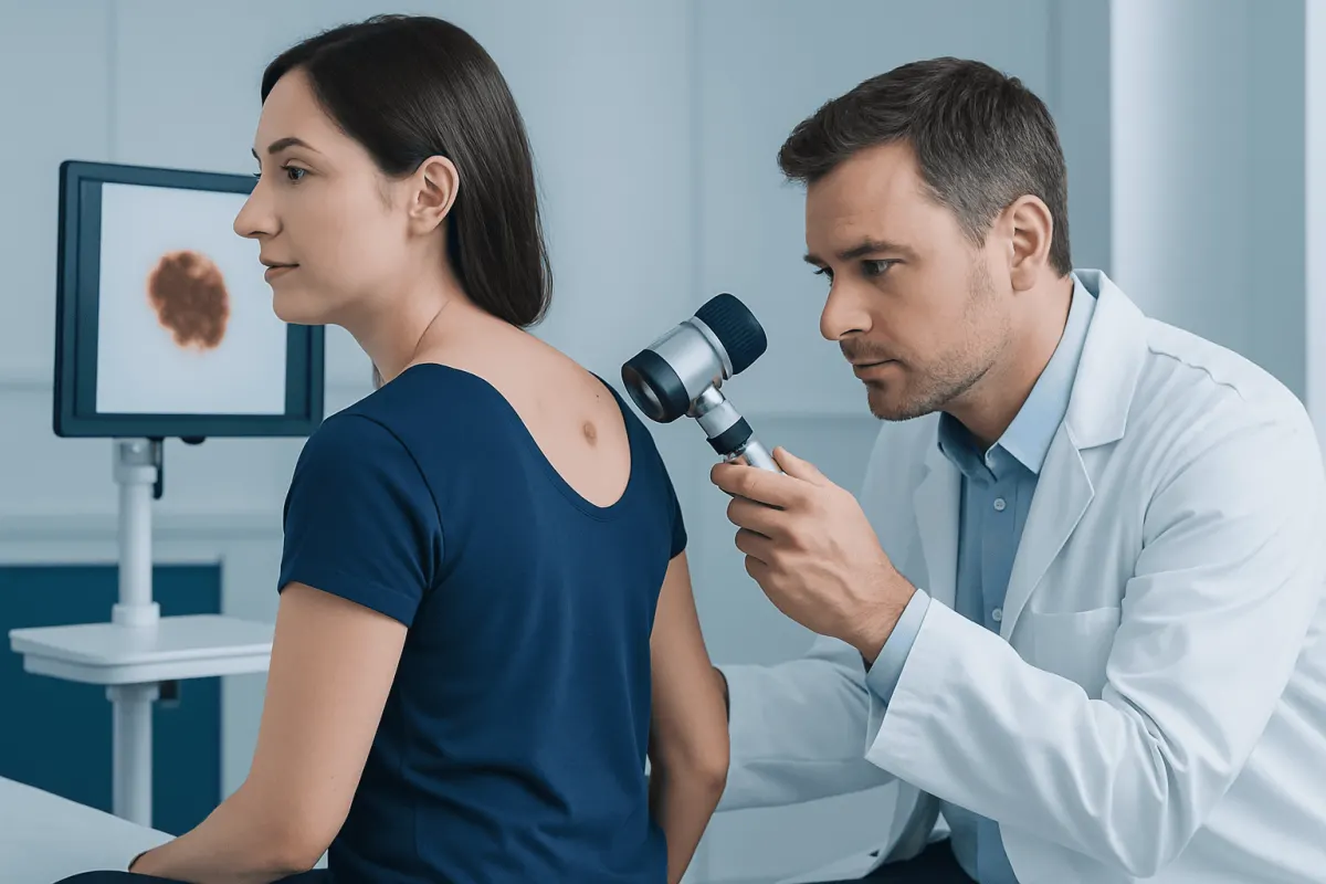
An image of melanoma skin cancer can help people recognize potentially concerning changes, such as an asymmetric mole, irregular border, varied color, or a spot that is evolving. Images cannot confirm melanoma, however; a prompt clinical skin examination and, when needed, biopsy are essential for diagnosis.
What an Image of Melanoma Skin Cancer Can Show
An image of melanoma skin cancer often shows a mole or skin spot that appears different from surrounding marks. It may be uneven in shape, have a ragged or blurred edge, contain several colors, grow over time, or look unlike the person’s other moles. Melanoma can occur on skin that is regularly exposed to the sun, but it may also develop on areas with little sun exposure.
Photographs are useful for awareness and for monitoring change, but they cannot diagnose melanoma. Benign moles, seborrheic keratoses, blood spots, and other harmless skin changes can sometimes resemble concerning lesions. Conversely, some melanomas do not fit the typical appearance shown in educational images.
Melanoma may be brown, black, tan, pink, red, purple, blue, skin-colored, or a mixture of shades. In people with darker skin tones, it may be more likely to arise on the palms, soles, under or around the nails, or on mucosal surfaces. Any new, changing, bleeding, crusting, or persistently itchy spot should be assessed by a qualified clinician.
Visual Clues: The ABCDE and Ugly Duckling Checks
Clinicians commonly use the ABCDE checklist to describe visual changes that may be associated with melanoma. It is an awareness tool rather than a diagnostic test. A lesion with one or more of these features should be discussed with a dermatologist, particularly if it is new or changing.
- A — Asymmetry: one half does not closely match the other.
- B — Border: edges are irregular, scalloped, notched, faded, or poorly defined.
- C — Color: the spot contains different shades or has an unusual color.
- D — Diameter: it is wider than about 6 millimeters, although melanomas may be smaller.
- E — Evolving: there is a change in size, shape, color, surface, sensation, or bleeding.
The “ugly duckling” approach is also helpful. Most people’s ordinary moles tend to share a similar pattern. A spot that looks noticeably different from the rest—darker, larger, more raised, or otherwise distinct—may merit examination even if it does not meet every ABCDE feature.
Some melanomas, especially nodular melanoma, can grow as a firm raised bump that is dark, pink, red, or skin-colored. A new lump that enlarges over weeks or months, bleeds easily, or does not heal should not be ignored.
Where Melanoma Can Develop and Who May Be at Risk
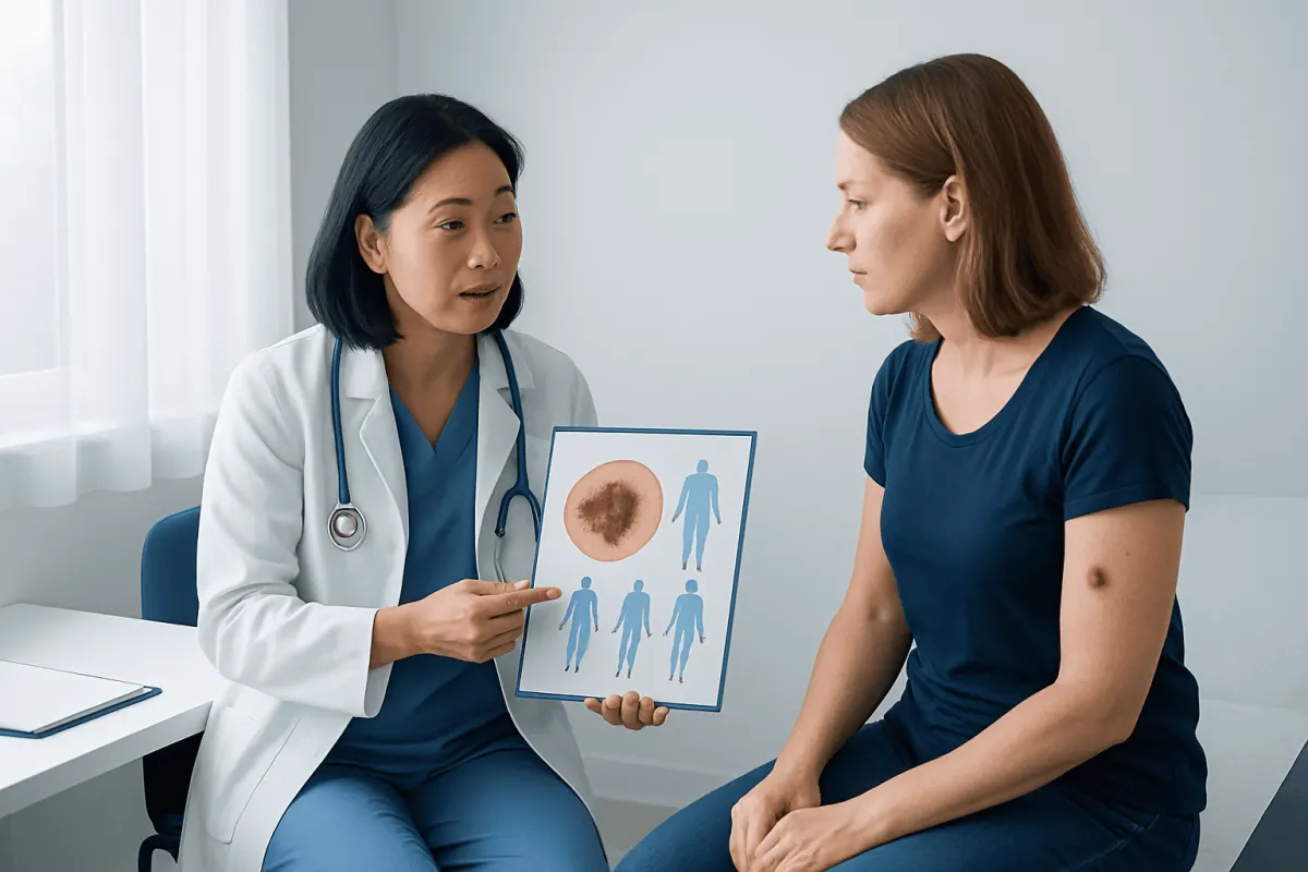
Melanoma begins in melanocytes, cells that produce pigment in the skin. It can develop anywhere melanocytes are present. Common locations include the back, chest, legs, face, neck, and arms. In men, it is often found on the trunk; in women, it is commonly found on the legs, although melanoma can occur in any location in any person.
Risk is influenced by a combination of personal, family, genetic, and environmental factors. Ultraviolet radiation from sunlight and tanning devices is an important preventable risk factor. A history of severe sunburns, especially during childhood, can increase risk. Having many moles, atypical moles, fair skin that burns easily, light eyes or hair, a weakened immune system, or a personal or family history of melanoma can also raise risk.
People with darker skin can develop melanoma and should not assume they are protected. Acral lentiginous melanoma, for example, may appear on the palms, soles, or nail units. A dark streak in a nail, pigment extending onto surrounding skin, or a changing spot on the foot should be medically reviewed rather than attributed automatically to injury or friction.
How Doctors Diagnose a Suspicious Skin Lesion
A dermatologist begins with a medical history and careful examination of the skin. They may ask when the lesion was first noticed, whether it has changed, and whether there is a personal or family history of skin cancer. A full-body skin examination can identify additional spots that may need monitoring.
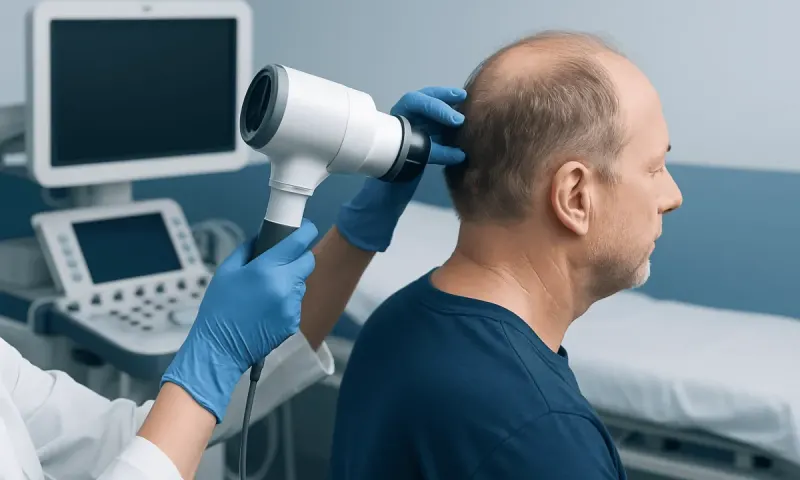
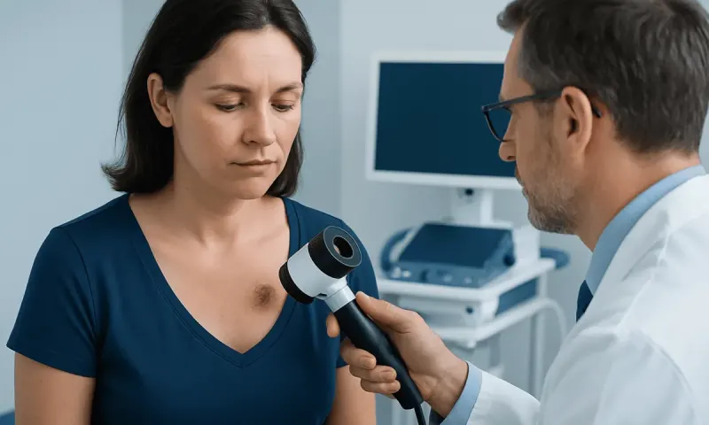
Dermatoscopy, also called dermoscopy, uses a handheld magnifying device with light to examine structures beneath the skin surface. This can help a trained clinician assess a lesion more accurately, but it does not replace biopsy when melanoma is suspected. Clinical photographs or digital mole monitoring may be used to document lesions over time in selected patients.
A biopsy is required to confirm melanoma. Usually, the clinician removes all or part of the suspicious lesion under local anesthetic and sends the tissue to a pathology laboratory. The pathology report describes the melanoma type and features that guide treatment planning, such as thickness and whether the skin surface is ulcerated.
If melanoma is diagnosed, additional assessment may be recommended depending on its features. This can include examination of nearby lymph nodes, sentinel lymph node biopsy, imaging in selected situations, and staging discussions. More detail about this condition is available in melanoma information.
Melanoma Staging and Outlook
Melanoma is staged according to factors including the thickness of the primary tumor, ulceration, involvement of nearby lymph nodes, and whether cancer has spread to more distant parts of the body. Staging helps the care team estimate risk, discuss follow-up needs, and select appropriate treatment options.
Many melanomas are found while still limited to the skin. In these cases, surgery can often remove the cancer completely. A thinner melanoma generally has a more favorable outlook than a thicker lesion, which is one reason why evaluation of a changing mole should not be delayed.
When melanoma has reached lymph nodes or distant organs, treatment has become increasingly individualized. Outlook varies widely and depends on the extent and biology of the cancer, overall health, response to treatment, and other clinical factors. A specialist can explain what staging information means for the individual rather than relying on images or general comparisons online.
Modern Treatment Approaches for Melanoma
Surgical excision is the main treatment for localized melanoma. The surgeon removes the melanoma together with a margin of normal-looking skin around it. Depending on the depth and location of the tumor, a sentinel lymph node biopsy may be discussed to check whether microscopic cancer cells have reached the nearest lymph nodes.
For melanoma with a higher risk of recurrence or disease that has spread, treatment may involve immunotherapy, targeted therapy for tumors with certain gene changes, radiation therapy in selected circumstances, surgery for carefully chosen sites of disease, or combinations of these approaches. The care plan is based on the stage, pathology findings, molecular testing, previous treatment, and the person’s general health.
Immunotherapy helps the immune system recognize and attack cancer cells. Targeted medicines act on particular changes in cancer cells, such as certain BRAF mutations, when testing shows that they are present. These treatments can have side effects, so patients need regular review and prompt advice about new symptoms during care.
Melanoma treatment is best planned by a multidisciplinary team that may include dermatology, Cancer Surgery Is Used and What Recovery Involves" class="ahp-ilk">surgical oncology, medical oncology, pathology, radiology, and supportive-care specialists. Acıbadem Health Point’s multidisciplinary specialists and JCI-accredited hospitals evaluate and treat melanoma for international patients, with treatment plans tailored to clinical findings. Medical oncology care may be part of treatment for melanoma that requires systemic therapy.
Skin Checks, Sun Protection, and Self-Care
Regular self-examination can help people notice change early. A monthly check in good lighting may include the scalp, face, ears, neck, torso, underarms, arms, hands, nails, buttocks, legs, feet, soles, and spaces between the toes. A mirror or help from another person can be useful for difficult-to-see areas such as the back and scalp.
Taking well-lit photographs can help document a spot, especially when a clinician advises monitoring. Photos should supplement—not delay—a medical appointment if a lesion is changing or concerning. It is not recommended to remove, cut, freeze, or apply unproven products to a suspicious mole at home.
Sun protection reduces ultraviolet exposure. Helpful habits include seeking shade when practical, wearing protective clothing and a wide-brimmed hat, using broad-spectrum sunscreen as directed, and avoiding tanning beds. Sunscreen is important but should be used alongside other protection measures rather than as a reason to spend prolonged periods in strong sunlight.
People with a prior melanoma, numerous atypical moles, or a strong family history may need scheduled professional skin examinations. The interval is individualized by the treating clinician based on the person’s risk profile and previous findings.
When to Seek Medical Care
A person should arrange a medical appointment promptly for a new mole or spot that is growing, changing in color or shape, bleeding, crusting, painful, itchy, or not healing. The same applies to a lesion that looks unlike the person’s other moles or to a new dark line or changing pigment around a fingernail or toenail.
There is no need to assume that every unusual mark is melanoma. Many skin lesions are benign, and a professional assessment can provide clarity. However, early evaluation is important because visual inspection alone cannot reliably distinguish harmless lesions from skin cancer.
People who have been diagnosed with melanoma should attend recommended follow-up visits and contact their care team if they notice new suspicious skin lesions, enlarged lymph nodes, unexplained persistent symptoms, or treatment side effects. Follow-up also provides an opportunity to discuss emotional concerns, sun safety, and practical skin-monitoring habits.
Frequently asked questions
01Can an image of melanoma skin cancer confirm that a mole is cancerous?
No. Images can show warning features, but many benign lesions can resemble melanoma and some melanomas look atypical. A dermatologist’s examination and a biopsy, if needed, are required to confirm the diagnosis.
02What does melanoma usually look like?
Melanoma may appear as a new or changing mole with asymmetry, an irregular border, multiple colors, or growth over time. It can also be a pink, red, dark, or skin-colored raised bump, so color alone is not reliable.
03Can melanoma occur without a pre-existing mole?
Yes. Melanoma can arise in an existing mole or begin as a new spot on previously normal-looking skin. This is why new skin lesions, as well as changes in old moles, should be monitored.
04How is melanoma diagnosed?
A clinician examines the skin and may use dermatoscopy to assess a suspicious lesion. If melanoma is suspected, a biopsy is performed and the tissue is examined by a pathologist.
05Is melanoma always dark brown or black?
No. Melanoma may be brown or black, but it can also be pink, red, purple, blue, skin-colored, or mixed in color. In darker skin tones, it may occur on the palms, soles, or nail areas.
06What should a person do if a mole changes?
They should make an appointment with a dermatologist or another qualified healthcare professional, especially if the change is rapid or includes bleeding, itching, pain, crusting, or growth. Taking a photograph may help document the change, but it should not replace medical assessment.
This article is for general information only and is not a substitute for professional medical advice. Please consult a qualified doctor about your individual situation.
Oncology care in Turkey — second opinion and treatment plan
JCI-accredited · board-certified surgeons · reply within 24h
Add us as a Preferred Source to see more of our trusted health content across Google Search, AI Overviews and Discover.