Temporal Bone

Key Takeaways
- The temporal bone sits on the side of the skull and houses important structures for hearing and balance.
- Injury, infection, tumors, and developmental conditions can affect the temporal bone.
- Symptoms may include ear pain, hearing changes, dizziness, swelling, or facial weakness.
- Diagnosis often involves physical examination plus imaging such as CT or MRI.
- Treatment depends on the cause and may include medication, monitoring, surgery, or rehabilitation.
The temporal bone is a dense, complex part of the skull that protects the ear structures and helps support hearing, balance, and jaw movement. Understanding its anatomy and common problems can make symptoms easier to recognize and discuss with a specialist.
Overview
The temporal bone is the sturdy side wall of the skull that sits above and around the ear. It is not a single simple block of bone; instead, it has several parts and contains many small openings, channels, and ridges that carry nerves, blood vessels, and the delicate structures involved in hearing and balance.
Because so much is packed into such a small region, even a minor problem in the temporal bone can affect daily life in noticeable ways. A person may first notice ear pressure, hearing changes, ringing, dizziness, jaw discomfort, or pain behind the ear. In an international-patient setting, these symptoms often lead to questions about whether the issue is in the ear itself, the jaw joint, the skull base, or a deeper structure that needs imaging.
For a clearer picture, it helps to think of the temporal bone as the protective shell and support framework for several functions at once: sound transmission, inner-ear balance, facial nerve pathways, and the joint that allows the jaw to move. That combination is why doctors from ENT and related specialties often evaluate temporal bone problems together rather than in isolation.
Symptoms
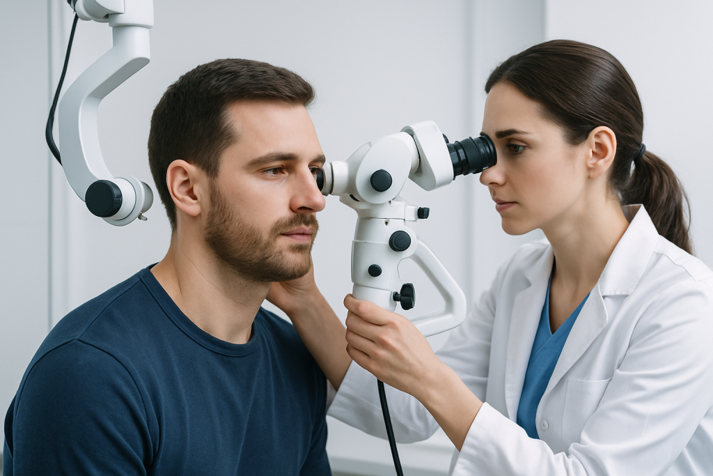
Symptoms linked to the temporal bone vary widely depending on the cause and the exact structure involved. Some people have only mild discomfort, while others develop signs that point to a more urgent injury or infection. The pattern of symptoms often guides the next step in evaluation.
Common symptoms may include:
- Ear pain or fullness
- Hearing loss or muffled hearing
- Ringing in the ear (tinnitus)
- Dizziness or imbalance
- Swelling or tenderness behind the ear
- Jaw pain or difficulty opening the mouth
- Facial weakness or numbness
- Drainage from the ear
Some symptoms develop suddenly, such as after a fall, car accident, or sports injury. Others appear gradually, as with chronic infection, benign growths, or degenerative changes near the ear. New facial weakness, severe dizziness, or a change in hearing after trauma deserves prompt medical attention.
Causes & Risk Factors

There is no single disease of the temporal bone. Problems arise from many different causes, and the source may be in the bone itself, the ear structures inside it, or nearby tissues. In practice, doctors look at the whole region because symptoms can overlap.
Common causes and risk factors include:
- Trauma: Falls, blows to the head, and road accidents can fracture the temporal bone.
- Ear infection: Chronic middle-ear infection or mastoid infection may spread into nearby bone.
- Cholesteatoma: An abnormal skin-lined growth in the middle ear can erode surrounding bone.
- Tumors or growths: Benign and malignant lesions may involve the skull base or ear canal.
- Congenital conditions: Some people are born with structural differences affecting the ear or skull.
- Inflammatory or bone disorders: Less common conditions may alter bone structure or density.
Risk can be higher in people with repeated ear infections, prior head injury, weakened bone health, or symptoms that have been present but not fully evaluated. In adults traveling for care, the history of previous scans or operations can be especially important, because earlier findings help specialists determine whether a change is new or long-standing.
Diagnosis
Diagnosis starts with a careful history and physical examination. A doctor usually asks when the symptoms began, whether there was any trauma, if hearing has changed, and whether dizziness, discharge, or facial weakness is present. An ear examination may show signs of infection, fluid, eardrum changes, or structural abnormalities.
Imaging is often central to assessing the temporal bone because much of it cannot be understood fully by exam alone. A CT scan is commonly used to show fine bony detail, which is helpful for fractures, erosion, or anatomy around the ear. MRI may be chosen when soft tissue, nerve involvement, inflammation, or tumor is a concern. Hearing tests and balance testing can also help define how the condition is affecting function.
In more complex cases, diagnosis may involve several specialists, such as an ENT surgeon, radiologist, neurologist, or neurosurgeon. For patients arriving from another country, bringing prior images, reports, and a list of symptoms in chronological order can make the visit more efficient and help the team compare old and new findings accurately.
Treatment Options
Treatment depends entirely on the cause. Some temporal bone problems are watched over time, while others require medication, urgent drainage, or surgery. The goal is not only to treat the bone itself but also to protect hearing, facial nerve function, and balance whenever possible.
Examples of treatment approaches include:
- Observation and follow-up: Small, stable findings may be monitored with repeat exams or scans.
- Medication: Antibiotics, anti-inflammatory medicines, or other targeted drugs may be used when infection or inflammation is present.
- Surgical treatment: Fractures with complications, cholesteatoma, tumors, or severe chronic infection may need an operation.
- Hearing support: Hearing aids or other assistive devices may help if hearing loss persists.
- Rehabilitation: Balance therapy or facial nerve rehabilitation may be recommended in selected cases.
After treatment, follow-up matters. The temporal bone area is closely linked to functions that affect communication, walking stability, and facial movement, so recovery is often tracked through symptom checks, hearing assessment, and sometimes repeat imaging. For international patients, planning follow-up before travel home can reduce gaps in care and clarify which warning signs should prompt a local review.
Prevention & Self-care
Not every temporal bone condition can be prevented, but a few practical habits can lower risk and make recovery smoother. Protecting the head during sports, using seat belts, and seeking care for persistent ear infections are all sensible steps. People with a history of ear disease should avoid delaying evaluation when symptoms change.
Self-care is most useful when it supports, rather than replaces, medical treatment. Rest, hydration, and avoiding forceful nose-blowing or unnecessary ear cleaning may help in some ear-related conditions. If a clinician has advised activity limits after trauma or surgery, those instructions should be followed closely because pressure changes and strain can affect healing.
It is also helpful to keep a symptom diary. Note whether hearing loss is one-sided or both-sided, whether dizziness occurs with movement, and whether pain is improving or worsening. This kind of detail can help the care team distinguish between a healing pattern and a condition that needs reassessment.
When to See a Doctor
A doctor should be consulted if ear pain, hearing loss, dizziness, or swelling around the ear lasts more than a short time, keeps returning, or interferes with daily activities. Sudden symptoms after head injury should be evaluated promptly, even if they seem mild at first.
Urgent assessment is especially important if there is facial weakness, severe headache after trauma, clear fluid or blood from the ear, high fever, confusion, or a major change in balance. These signs do not always mean a serious condition, but they do deserve timely review so the cause can be identified and treated appropriately.
Patients who are considering treatment abroad often benefit from a coordinated ENT assessment with imaging review before making travel plans. At Acibadem Health Point, multidisciplinary specialists and JCI-accredited hospitals evaluate and treat temporal bone conditions for international patients, helping align diagnosis, treatment, and follow-up in one organized pathway.
Frequently asked questions
What is the temporal bone?
The temporal bone is the part of the skull located at the side and base of the head near the ear. It protects structures involved in hearing, balance, and facial nerve function. Because it is so anatomically dense, problems there can cause several different symptoms.
Can a temporal bone problem affect hearing?
Yes. The temporal bone contains the outer, middle, and inner ear structures, so changes in this area can influence hearing. Hearing loss may be temporary or long-lasting depending on the cause and how quickly it is treated.
How is a temporal bone fracture diagnosed?
Doctors usually suspect a fracture after head injury and confirm it with imaging, most often a CT scan. An ear exam and hearing assessment may also be performed to check for related damage. Additional tests are sometimes needed if facial nerve or balance symptoms are present.
Is temporal bone surgery always necessary?
No. Some temporal bone conditions are monitored, especially if they are small or stable. Surgery is considered when there is fracture complication, persistent infection, cholesteatoma, a concerning growth, or damage that is unlikely to improve on its own.
Can dizziness come from the temporal bone?
Yes. The temporal bone houses the inner-ear structures that help control balance, so dizziness or vertigo can be linked to problems in this area. A clinician will usually look for ear-related, neurologic, and trauma-related causes before deciding on treatment.
What should a patient bring to an overseas specialist visit?
It helps to bring previous scan images, written reports, a list of symptoms with dates, and any past ear or head injury records. This makes it easier for the specialist to compare old and new findings and plan care efficiently. If possible, bringing hearing test results is also useful.
References
- Merck Manual Professional Edition
- American Academy of Otolaryngology–Head and Neck Surgery
- National Institute on Deafness and Other Communication Disorders
- Radiological Society of North America
- Mayo Clinic
This article is for general information only and is not a substitute for professional medical advice. Please consult a qualified doctor about your individual situation.