

Mrcp

Key Takeaways
- MRCP uses magnetic resonance imaging to visualise the biliary and pancreatic ducts in detail.
- It is commonly recommended when doctors need to look for gallstones, blockages, inflammation, or structural changes.
- The test does not use ionising radiation, which makes it a useful imaging option for many patients.
- Preparation is usually straightforward, but patients should tell the care team about implants, claustrophobia, pregnancy, and kidney problems.
- MRCP helps guide treatment decisions, but the results are interpreted alongside symptoms, blood tests, and other imaging findings.
MRCP is a noninvasive MRI-based test that creates detailed images of the bile ducts, gallbladder, pancreas, and nearby structures. It is often used to help doctors investigate pain, jaundice, gallstones, and other problems without needing an endoscopic procedure first.
Overview
MRCP stands for magnetic resonance cholangiopancreatography. It is a special type of MRI scan designed to show the bile ducts, gallbladder, pancreas, and the channels that carry digestive fluids. Instead of opening the body with surgery or using a camera through the mouth, the scan creates detailed images from outside the body.
For many patients, MRCP becomes part of the story after a symptom such as upper abdominal pain, jaundice, repeated pancreatitis, or unexplained liver test changes. Doctors often use it when they need a clear look at the ducts without moving immediately to an endoscopic procedure. That can make the diagnostic process less invasive while still providing important detail.
Because MRCP is based on MRI, it uses magnets and radio waves rather than ionising radiation. The scan is therefore often valued as a safe, informative way to assess the biliary system and the pancreatic ductal anatomy. It is especially helpful when the clinical picture needs a closer look before a treatment plan is chosen.
Why doctors order an MRCP
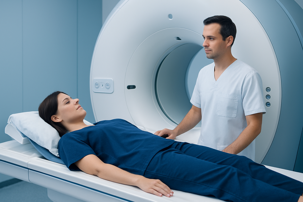
An MRCP is typically ordered when symptoms or test results suggest a problem in the bile ducts or pancreas. Doctors may request it to look for blockages, narrowing, stones, inflammation, congenital duct differences, or complications after surgery. It is also used when ultrasound or CT findings need a more detailed follow-up image.
Common reasons include:
- Possible gallstones in the bile duct
- Unexplained jaundice or dark urine
- Recurrent attacks of pancreatitis
- Suspected bile duct narrowing or stricture
- Evaluation of cysts or duct abnormalities
- Follow-up after certain biliary or pancreatic procedures
In international-patient care, MRCP can be particularly helpful because it gives a focused answer before travel plans, procedures, or follow-up appointments are scheduled. It can help a doctor decide whether a patient needs medication, endoscopic treatment, surgery, or simple observation.
Symptoms and conditions that may lead to MRCP

MRCP is not a treatment in itself; it is a diagnostic tool used when the body is sending signals that the biliary or pancreatic system may not be working normally. A person might be sent for the scan after noticing persistent discomfort in the upper right or upper middle abdomen, especially if pain appears after meals or comes with nausea.
Other signs that may prompt the test include yellowing of the skin or eyes, itching, pale stools, fever with abdominal pain, repeated vomiting, or abnormal blood tests showing liver or pancreatic irritation. Sometimes the scan is ordered even when symptoms are mild, because early imaging can help prevent delays in diagnosis.
Conditions commonly investigated with MRCP include gallstones, choledocholithiasis, pancreatitis, bile duct strictures, biliary obstruction, choledochal cysts, and some post-operative complications. The scan may also be useful in assessing how a known condition is progressing over time.
Causes and risk factors
The findings on an MRCP depend on the underlying condition rather than the scan itself. One of the most common reasons for abnormalities is gallstone disease, where stones form in the gallbladder and may move into the bile ducts. In other cases, inflammation can narrow the ducts or disrupt the normal flow of digestive fluids.
Risk factors vary with the condition being investigated. A higher chance of biliary problems may be associated with a history of gallstones, certain dietary and metabolic factors, rapid weight changes, pregnancy, or previous biliary surgery. Pancreatic issues can be linked to alcohol use, smoking, autoimmune disease, genetic conditions, or repeated episodes of acute pancreatitis.
Some patients have ductal changes from birth or develop scarring after infection, surgery, or long-standing inflammation. Others may have no clear risk factor, which is one reason imaging is useful: it can show a structural cause even when the symptoms alone do not reveal the answer.
How the diagnosis is made
MRCP is usually one part of a broader diagnostic process. A doctor starts with the patient’s symptoms, medical history, examination, and laboratory tests such as liver function tests, bilirubin, and pancreatic enzymes. Ultrasound, CT, or plain MRI may already have been performed before MRCP is recommended.
During the scan, the patient lies still inside the MRI machine while the system collects highly detailed images. Some centres may ask the patient to avoid eating for a few hours beforehand so the bile ducts and gallbladder are easier to evaluate. In some cases, contrast material may be used, but MRCP often relies on special MRI sequences that highlight fluid-filled ducts without the need for contrast.
The images are then reviewed by radiologists and the referring doctor. Findings may show a stone, narrowing, duct dilation, inflammation, a cyst, or a normal result. A normal MRCP can still be clinically useful, because it may help rule out serious ductal obstruction and direct attention to other possible causes of symptoms.
Treatment options after an MRCP
MRCP does not determine treatment on its own; it helps doctors choose the next step. If the scan shows a bile duct stone, a gastroenterologist may recommend endoscopic removal. If there is an obstruction from inflammation, scarring, or a mass, treatment may involve endoscopy, surgery, medication, or careful monitoring depending on the cause.
When the scan shows gallstones in the gallbladder without duct involvement, treatment might range from observation to gallbladder removal if symptoms are recurring. In pancreatitis-related conditions, treatment focuses on the trigger, hydration, pain control, nutrition, and long-term prevention of another attack. A clear image often saves time by reducing trial-and-error decisions.
For patients travelling for care, MRCP can help coordinate the right specialist visit before a procedure is planned. That is particularly valuable when several departments need to work together, such as gastroenterology, radiology, surgery, and anaesthesia. Acibadem Health Point’s multidisciplinary specialists and JCI-accredited hospitals diagnose and treat this condition for international patients, with follow-up plans tailored to each case.
How to prepare and what to expect
Preparation is generally simple, but the care team will give specific instructions. Patients should tell the doctor about any metal implants, pacemakers, cochlear implants, aneurysm clips, pregnancy, kidney disease, or prior reactions to contrast material. Because MRI uses strong magnets, these details are important for safety.
Before the scan, patients may be asked to remove jewellery, watches, cards, and any metal-containing items. Comfortable clothing without metal fasteners is often recommended. Some patients feel anxious in enclosed spaces, so it can help to mention claustrophobia early; the team may offer reassurance, communication during the scan, or practical steps to improve comfort.
The scan itself is painless, but it can be noisy and requires stillness for clear images. Patients can usually return to normal activities soon afterwards unless their doctor gives different instructions. If contrast was used, additional advice may be given based on kidney function or the individual care plan.
Prevention, self-care, and follow-up
MRCP cannot prevent illness, but good self-care can reduce the chance of recurring biliary or pancreatic problems in some people. Balanced eating, maintaining a healthy weight, avoiding rapid weight loss, limiting alcohol, and not smoking are all sensible measures that support digestive health. Patients with known gallstone disease or pancreatitis should follow their doctor’s advice closely, even when symptoms settle.
Follow-up matters because imaging findings are most helpful when they lead to a clear plan. Patients should ask how the results fit with blood tests and other scans, whether another test is needed, and what symptoms would mean the condition is changing. When care is coordinated across countries, keeping copies of reports and images can make later consultations smoother.
In some situations, the next step may involve repeat imaging rather than immediate treatment, especially if findings are small, unclear, or not causing symptoms. A calm, structured follow-up plan is often the safest approach.
When to see a doctor
Medical advice should be sought promptly if a person develops yellowing of the skin or eyes, severe upper abdominal pain, fever with abdominal pain, persistent vomiting, or symptoms that suggest a blocked bile duct or pancreatitis. These signs do not always mean something serious, but they deserve timely assessment.
Anyone with ongoing digestive symptoms, unexplained abnormal liver tests, or a known history of gallstones or pancreatitis should discuss whether MRCP or another imaging test is appropriate. A doctor can decide which scan best answers the clinical question and whether urgent evaluation is needed.
When patients are travelling for care, it is wise to arrange a consultation before the trip if possible so the imaging plan, fasting instructions, and follow-up pathway are clear. A well-organised review helps avoid delays and makes the experience more predictable from first appointment to return home.
Frequently asked questions
What does MRCP stand for?
MRCP stands for magnetic resonance cholangiopancreatography. It is a special MRI technique used to look closely at the bile ducts, gallbladder, and pancreatic ducts. Doctors use it to help find the cause of symptoms or abnormal test results.
Is MRCP the same as MRI?
MRCP uses MRI technology, but it is tailored to show fluid-filled ducts in the biliary and pancreatic system. That makes it different from a standard MRI of the abdomen. It is a focused type of imaging rather than a separate test in a completely different category.
Does MRCP use radiation?
No, MRCP does not use ionising radiation. It relies on magnets and radio waves instead. That is one reason it is often chosen when detailed imaging is needed without X-rays or CT radiation.
Do patients need to fast before an MRCP?
Sometimes, yes. Some centres ask patients not to eat for several hours before the scan so the gallbladder and ducts are easier to evaluate. The exact instructions depend on the hospital and the reason for the test.
Can MRCP show gallstones?
Yes, MRCP can often show stones in the bile ducts and may also give information about gallstones in the gallbladder. It is especially useful when doctors suspect a stone is blocking the duct or causing inflammation. The result is interpreted together with symptoms and blood tests.
Is MRCP painful or unsafe?
MRCP is generally painless and noninvasive. Most people find the main challenge is lying still in the scanner, which can be noisy and a little uncomfortable for some patients. The care team can explain each step and help with any concerns before the scan.
References
- National Institute of Diabetes and Digestive and Kidney Diseases
- Radiological Society of North America
- American College of Radiology
- Mayo Clinic
- NHS
This article is for general information only and is not a substitute for professional medical advice. Please consult a qualified doctor about your individual situation.
More from the Health Library



Related Specialists

Dr. Serdar Özdemir
Internal Medicine
Dr. Neda Zabandan
Emergency Service
Prof. Dr. Salih Marangoz
Orthopedic Surgery & Traumatology
Assoc. Prof. Dr. Recep Kurnaz
Orthopedic Surgery & Traumatology