Cranium

Key Takeaways
- The cranium forms the protective case around the brain and works with the face, jaw, and spine to support head function.
- Symptoms such as persistent headache, swelling, deformity, dizziness, or vision changes after head trauma deserve medical assessment.
- Cranial problems may arise from injury, congenital differences, infection, bone disease, or conditions affecting nearby nerves and tissues.
- Diagnosis often includes a physical exam and imaging studies such as CT or MRI when indicated.
- Treatment depends on the cause and may range from observation and pain control to specialist surgery or rehabilitation.
The cranium is the bony framework that shields the brain and supports key structures of the head and face. Understanding its anatomy, common symptoms related to injury or disease, and when to seek care can help patients make informed decisions about evaluation and treatment.
Overview
The cranium is the bony part of the skull that encloses and protects the brain. It is made up of several bones that fit together in a strong, structured way, creating a stable shield for one of the body’s most sensitive organs.
Beyond protection, the cranium provides attachment points for muscles and helps define the shape of the head and face. In everyday language, people often use “skull” and “cranium” interchangeably, but the cranium specifically refers to the braincase rather than the entire skull, which also includes the jaw and facial bones.
For patients considering care abroad, understanding cranial anatomy can make consultations feel less overwhelming. Whether the concern is a recent injury, a long-standing shape difference, or a neurologic symptom that may be linked to pressure or structural change, the first step is usually a careful evaluation by an appropriate specialist.
Symptoms
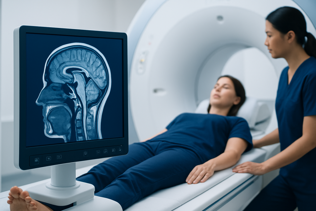
Many cranium-related concerns are noticed only after an injury or during a routine examination. Symptoms depend on the cause, and some problems are visible while others are felt more than seen.
Common signs that may warrant attention include persistent or severe headache, tenderness over the skull, swelling, a palpable bump or indentation, or pain that worsens when the area is touched. After a head injury, dizziness, nausea, confusion, memory gaps, sleepiness, or changes in vision may suggest that the brain or surrounding structures need urgent assessment.
Other symptoms can be more subtle. Some people notice jaw discomfort, asymmetry of the head, hearing changes, facial numbness, or scalp sensitivity. In infants and young children, a healthcare professional may also assess head shape, growth patterns, and the soft spots between skull bones because these can offer clues about cranial development.
- Headache that is new, persistent, or unusually severe
- Visible deformity, swelling, or bruising after trauma
- Confusion, drowsiness, vomiting, or fainting after a head impact
- Facial weakness, numbness, or sensory changes
- Changes in head shape in childhood or over time
Causes & Risk Factors
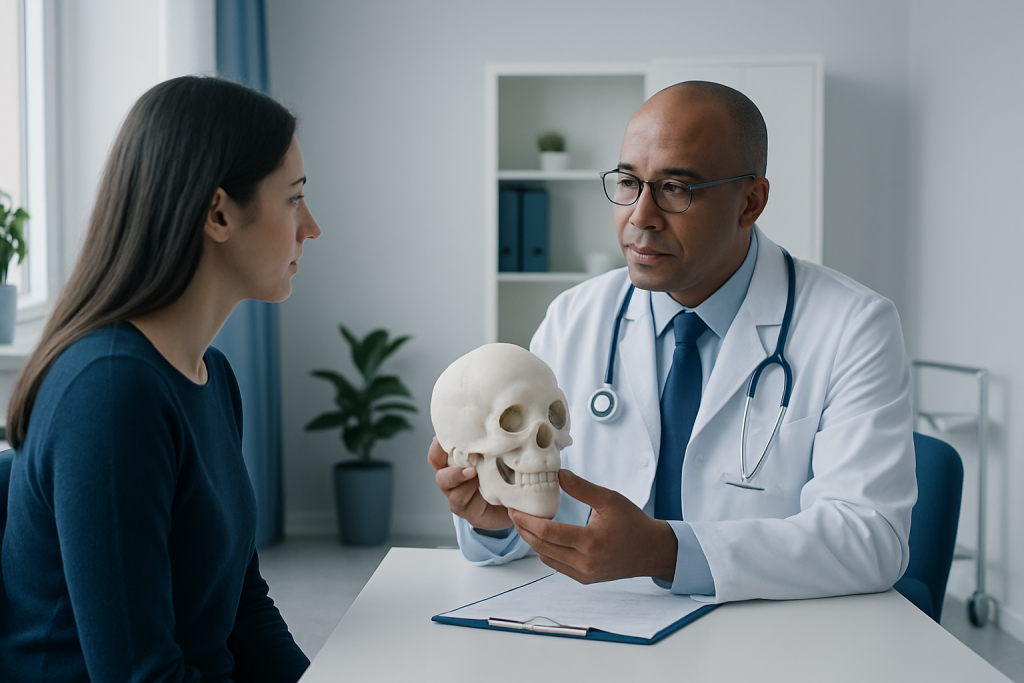
Cranial concerns can begin in many different ways. The most familiar cause is trauma, such as a fall, sports injury, traffic accident, or blunt force to the head. Some injuries affect only the scalp or outer bone, while others may involve fractures or deeper complications.
Not all cranial problems are caused by a sudden event. Congenital conditions, in which the bones of the skull develop differently before birth, can change head shape or affect how the brain grows. Bone disorders, infections, tumors, and inflammatory conditions may also alter the cranium or nearby tissues.
Risk is often higher in infants and children because the skull is still developing, and in older adults because falls are more likely and bones may be more fragile. Certain sports, contact activities, seizure disorders, and the use of medications that affect bone strength or blood clotting can also influence risk. A doctor will usually look at the full picture rather than a single factor when deciding what evaluation is needed.
Diagnosis
Diagnosis starts with a detailed medical history and a physical examination. The clinician will ask how symptoms began, whether there was an injury, and whether there are related neurologic signs such as numbness, vision changes, or balance problems.
Imaging is often the next step when a fracture, structural abnormality, or deeper problem is suspected. A CT scan is commonly used to assess bone detail quickly, especially after trauma. An MRI may be more helpful when the concern involves soft tissue, brain structures, or certain complex conditions. In some situations, X-rays, laboratory tests, or referral to a specialist such as a neurologist, neurosurgeon, ENT specialist, or craniofacial surgeon may be appropriate.
For patients traveling from another country, it helps to bring any previous scans, operative notes, medication lists, and a timeline of symptoms. Clear records can shorten the diagnostic process and help the care team compare old and new findings accurately.
Treatment Options
Treatment depends entirely on the cause. Minor injuries without fracture or neurologic concern may be managed with observation, rest, cold compresses, and general pain relief recommended by a clinician. The goal is to watch for delayed symptoms while supporting recovery.
More significant fractures, deformities, infections, or growth-related conditions may require specialized care. Some patients need surgery to repair bone, relieve pressure, correct shape, or treat complications. Others benefit from medications, close monitoring, or rehabilitation if the injury has affected balance, cognition, jaw movement, or vision.
When treatment is planned overseas, it is reasonable to ask how follow-up will work after returning home. A good team will explain wound care, activity limits, warning signs, and whether local follow-up imaging or specialist review is expected. Recovery plans are usually individualized, because the cranium protects highly delicate structures and even small changes can matter.
Common treatment approaches may include:
- Observation and symptom monitoring
- Pain management and rest
- Antibiotics or other medications when infection is present
- Surgical repair for fractures or structural problems
- Rehabilitation for related neurologic or functional issues
Prevention & Self-care
Not every cranial condition can be prevented, but everyday precautions can lower the chance of injury. Seat belts, child safety seats, helmets for cycling or contact sports, and fall-prevention measures at home all help reduce risk. For people with medical conditions that affect balance or bone strength, regular review with a clinician is especially useful.
After a minor head injury, self-care should be calm and practical rather than improvised. Rest, hydration, and avoiding alcohol or strenuous activity may help recovery, but any worsening symptoms should be checked promptly. It is also wise to avoid returning to sports, work at height, or driving until a clinician says it is safe.
Patients managing a chronic cranial or craniofacial condition often do best with consistent follow-up. Keeping imaging results, notes from appointments, and questions in one place can make care smoother, especially if treatment is shared between a home physician and a specialist center abroad.
When to See a Doctor
Medical assessment is important after any head injury that causes loss of consciousness, repeated vomiting, confusion, severe headache, seizure, weakness, or noticeable behavior change. These symptoms do not always mean something serious has happened, but they should be evaluated promptly.
Even without a recent injury, a doctor should review persistent skull pain, a new lump or indentation, unexplained head shape changes, facial asymmetry, or symptoms that suggest pressure on nearby nerves or structures. In children, concerns about head growth, development, or fontanelle changes should be brought to a pediatric specialist.
If care is being arranged internationally, a coordinated clinic can help match the patient with the right specialty and imaging pathway. Acibadem Health Point’s multidisciplinary specialists and JCI-accredited hospitals diagnose and treat cranial conditions for international patients while helping them understand each step of the plan. Urgent symptoms, however, should always be treated as emergencies wherever the patient is located.
Living With Cranial Conditions
Many cranial conditions are manageable once the cause is understood. The experience is often less about the bone alone and more about how the bone, brain, nerves, and surrounding tissues interact. That is why care may involve several specialists working together rather than a single appointment.
Patients recovering from surgery or injury may need time for pain control, wound care, activity adjustment, and follow-up imaging. Others may live with long-term structural differences that are monitored over time rather than immediately treated. In both situations, the aim is to preserve function, ease symptoms, and protect neurologic health.
Questions are worth bringing to the care team: What changes should prompt concern? How soon can travel resume? Will there be scar care, physical therapy, or additional imaging? Clear answers make it easier to recover with confidence, especially when the patient is balancing treatment with international travel and family logistics.
Frequently asked questions
What is the cranium?
The cranium is the bony portion of the skull that surrounds and protects the brain. It is made of several bones that join together to form a strong protective case. The jaw and facial bones are part of the skull, but not the cranium itself.
Can a cranium problem be serious?
It can be, depending on the cause. A minor bump may only affect the scalp or outer bone, while a fracture, infection, or condition that changes pressure inside the head may need urgent care. The safest approach is to have new or concerning symptoms assessed by a doctor.
What symptoms suggest a possible skull fracture?
Signs may include swelling, tenderness, a dent or step in the bone, bruising around the eyes or behind the ear, or neurologic symptoms after an injury. Confusion, vomiting, drowsiness, or loss of consciousness after a head hit should be treated seriously. Imaging is often needed to confirm whether a fracture is present.
How is a cranial condition diagnosed?
Doctors usually begin with a history and physical examination, then use imaging if needed. CT scans are often helpful for bone detail, while MRI can show soft tissue and brain structures more clearly. The exact tests depend on the suspected cause and symptoms.
Does every cranial injury need surgery?
No. Many minor injuries are managed with observation and symptom control. Surgery is usually considered when there is a significant fracture, pressure on important structures, infection, or another problem that cannot be treated safely in a less invasive way.
What should a patient do before traveling for cranial treatment?
It helps to gather previous scans, medical reports, medication lists, and a clear symptom timeline. Patients should also ask how follow-up will be handled after returning home. This makes it easier for the receiving team to plan treatment and aftercare responsibly.
References
- World Health Organization
- National Institute of Neurological Disorders and Stroke
- Mayo Clinic
- Cleveland Clinic
- MedlinePlus
This article is for general information only and is not a substitute for professional medical advice. Please consult a qualified doctor about your individual situation.
More from the Health Library



Related Specialists

Dr. Zeynep Karaca
Pediatrics
Dr. Bayarmaa Khishigsuren
Medical Oncology
Dr. Edip Yılmaz
Orthopedic Surgery & Traumatology
Dr. Cevat Rifat Cündübey
Gynecology & Obstetrics