Bone Island

Key Takeaways
- A bone island is typically a benign, incidental finding on X-ray, CT, or MRI.
- It often causes no symptoms and is discovered during imaging for another reason.
- Doctors may compare scans over time to confirm that the lesion is stable.
- Treatment is usually not needed unless the diagnosis is uncertain or symptoms suggest another cause.
- If pain is persistent, changing, or associated with other symptoms, medical review is important.
Medically reviewed by the Acıbadem clinical team — July 13, 2026
A bone island, also called enostosis, is a small, harmless area of dense bone that is often found incidentally on imaging. Most people never feel it, and it usually needs only observation rather than treatment.
Overview
A bone island, known medically as an enostosis, is a small focus of compact bone that sits within the softer cancellous bone. On imaging, it often appears as a sharply defined bright spot and is usually found by chance while looking into an unrelated concern such as injury, joint pain, or back discomfort.
For most people, the key point is reassuring: a bone island is generally benign. It is not the same as bone cancer, and it does not usually spread or require active treatment. In many cases, the finding simply becomes part of a patient’s medical record so that future scans can be compared with it.
For international patients who are arranging care from another country, this kind of finding can be especially confusing because it may be mentioned briefly on a report without much context. A careful explanation from an orthopedics or radiology team can help clarify whether the lesion is a classic bone island or whether any additional follow-up is needed.
Symptoms
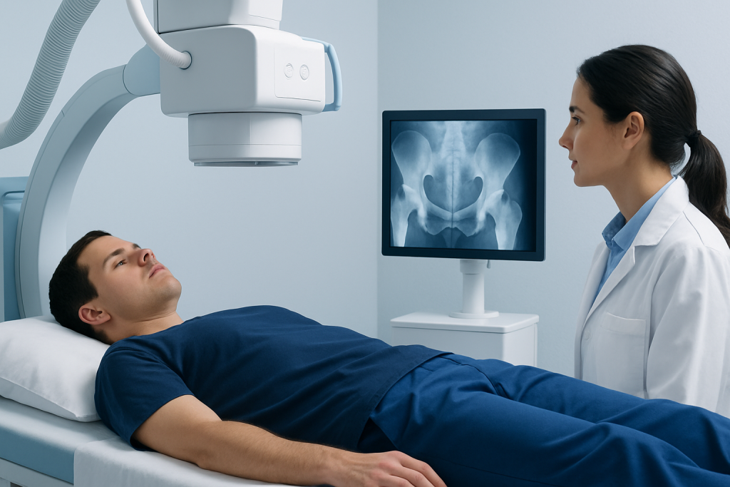
Most bone islands cause no symptoms at all. Many people never know they have one unless it shows up on an X-ray, CT scan, or MRI ordered for a different reason. When a bone island is found in this way, it is often the imaging report rather than the lesion itself that brings the patient into a conversation with a doctor.
Because bone islands are usually silent, pain in the same area should not automatically be blamed on the lesion. Joint strain, tendon irritation, arthritis, old injuries, or spine-related problems are often more likely explanations. Doctors look carefully at the pattern of pain, the location of the finding, and the imaging features before deciding whether the lesion is truly incidental.
In rare situations, a bone island may be noticed because of nearby discomfort, but even then it is not always the direct cause. Any new or persistent pain deserves proper assessment so that the underlying reason is identified accurately.
Causes & Risk Factors

The exact reason bone islands form is not always clear. They are thought to represent a small cluster of dense bone tissue that develops during bone growth or remodeling. In most cases, they are simply a normal variant rather than a sign of disease.
Bone islands can appear in adults of any age, and they are often seen in the pelvis, femur, ribs, spine, or other bones. Having one does not usually point to lifestyle habits, diet, or a specific injury. Most patients do not recall anything that triggered it, because there often is no trigger to identify.
There are no strong everyday “risk factors” in the usual sense, but doctors pay closer attention when imaging features are atypical or when a patient has a history of cancer. That is because some other bone conditions can look similar on scans, and careful interpretation matters more than the label alone.
- Usually discovered incidentally during imaging
- Can occur in several bones throughout the body
- Often unrelated to symptoms or trauma
- Need closer review when imaging features are unusual
Diagnosis
Diagnosis begins with the imaging study that first shows the lesion. On X-ray or CT, a bone island often has a very dense, well-defined appearance with fine radiating edges that blend into the surrounding bone. Radiologists use these visual features, together with the patient’s history, to decide whether the lesion fits the typical pattern.
Sometimes no further testing is needed. If the appearance is classic and the patient has no warning signs, the doctor may simply document the finding and recommend routine follow-up only if future scans are obtained for other reasons. Stability over time is one of the strongest clues that a bone island is harmless.
If the imaging appearance is not completely typical, a doctor may suggest repeat imaging, comparison with older studies, or additional assessment. This is especially important when the patient has pain that does not match the location of the lesion, or when there is any uncertainty about whether the spot could represent another type of bone change.
Treatment Options
Most bone islands do not need treatment. Once the diagnosis is clear, the usual approach is observation. The goal is not to remove the lesion, but to confirm that it behaves like a stable, benign finding and to avoid unnecessary procedures.
If a patient has pain in the same area, treatment is directed at the true cause of the pain rather than the bone island itself. That may mean care for a muscle strain, arthritis, a disc problem, or another orthopedic condition. In that sense, management is often about solving the broader picture rather than focusing narrowly on the incidental scan finding.
Surgery is rarely considered for a bone island. It would generally only enter the discussion if the diagnosis were unclear or if another condition needed to be ruled out. For patients traveling internationally, obtaining and carrying prior imaging reports can be very helpful, because comparison with earlier studies often prevents unnecessary repeat testing.
Prevention & Self-care
There is no known way to prevent a bone island, and most people do not need to change their routine because of one. Since it is usually an incidental bone finding, the more practical focus is on keeping records organized and understanding what the radiology report means.
Self-care is mainly about general bone and joint health. Staying active within comfort limits, maintaining good nutrition, and following a doctor’s advice for any coexisting orthopedic problem can support overall musculoskeletal health. If pain is the reason the imaging was done, it helps to track when the pain occurs, what makes it better or worse, and whether it changes over time.
Patients who are receiving care across borders may find it useful to keep digital copies of prior scans, written reports, and clinic notes. That makes it easier for a new specialist to compare findings and decide whether the lesion has remained unchanged, which is often the most important detail.
- Keep copies of imaging reports and previous scans
- Follow up if new symptoms develop in the same area
- Support bone health with regular movement and balanced nutrition
- Do not assume all pain near a bone island comes from the lesion
When to See a Doctor
A doctor should review the finding if the imaging report is unclear, if the lesion has an unusual appearance, or if there is no older scan available for comparison. This is especially true when the patient has a history of cancer or when the radiologist recommends follow-up. A clear explanation from an orthopedic, radiology, or musculoskeletal specialist can help separate a routine incidental finding from something that needs more attention.
Medical review is also important if pain is persistent, worsening, or accompanied by swelling, reduced function, fever, unexplained weight loss, or night pain. These symptoms do not mean a bone island is dangerous, but they do suggest that another problem may be present and should be evaluated properly.
For international patients who need a coordinated opinion, Acibadem Health Point’s multidisciplinary specialists and JCI-accredited hospitals can help diagnose and treat bone-related findings with a careful, step-by-step approach. The aim is to provide clarity, appropriate imaging review, and a plan that fits the patient’s overall situation.
Living With a Bone Island
Learning that a bone island is present can feel unsettling at first, mostly because it sounds like a structural problem that should be doing something. In practice, it is often one of the most harmless findings seen in musculoskeletal imaging. Once the diagnosis is explained, many patients are relieved to learn that no special treatment is needed.
Living with this finding usually means staying aware, not worried. If future scans are done for other reasons, the lesion may be mentioned again, and comparison with the original report will help confirm that it has remained stable. That stability is often what closes the loop and keeps the finding in perspective.
If questions remain after the first explanation, it is reasonable to ask for a second review of the images or report. A thoughtful discussion can make the difference between an incidental note on a scan and a clear, confident understanding of what it means for daily life.
Frequently asked questions
Is a bone island cancer?
No. A bone island is usually a benign, noncancerous area of dense bone. Doctors still review the imaging carefully to make sure the appearance is typical and to rule out other causes when needed.
Can a bone island cause pain?
Most bone islands do not cause pain. If pain is present, doctors look for other more common reasons such as arthritis, muscle strain, spine problems, or a separate bone condition.
Does a bone island need surgery?
Usually not. Most bone islands are simply observed, and surgery is rarely considered unless the diagnosis is uncertain or another problem must be investigated.
How is a bone island found?
It is often found incidentally on X-ray, CT, or MRI done for another reason. The radiologist looks at the shape, density, and stability of the lesion to decide whether it fits a bone island.
Should a bone island be monitored over time?
Sometimes, yes. If the imaging appearance is classic, no special follow-up may be needed, but comparison with future scans can help confirm that it remains stable.
Can bone islands appear in more than one place?
Yes, they can sometimes be multiple. Even then, they are often benign, but a doctor may review the images carefully to be certain that the overall pattern is consistent with enostoses.
References
- Radiopaedia
- American Academy of Orthopaedic Surgeons
- Merck Manual Professional Edition
- Mayo Clinic
This article is for general information only and is not a substitute for professional medical advice. Please consult a qualified doctor about your individual situation.