Atypical Mole Nevus: Symptoms, Causes and Treatment

Key Takeaways
- An atypical mole nevus usually looks irregular compared with a common mole, but it is not automatically cancerous.
- The main concern is change over time, especially new asymmetry, color variation, border irregularity, or itching and bleeding.
- A dermatologist may use skin examination, dermoscopy, and sometimes a biopsy to make the diagnosis.
- People with many atypical moles, fair skin, sunburn history, or a family history of melanoma may need closer monitoring.
- Sun protection and regular self-checks help detect meaningful changes early.
- Any mole that looks different from a person's other moles or changes quickly should be reviewed by a clinician.
Medically reviewed by the Acıbadem clinical team — August 19, 2026
An atypical mole nevus is a noncancerous skin growth that looks different from an ordinary mole in size, shape, color, or border. Most are harmless, but they deserve careful observation because they can resemble melanoma or signal a higher skin cancer risk in some people.
Overview
An atypical mole nevus, also called a dysplastic nevus, is a mole that does not fit the usual pattern of an ordinary brown spot on the skin. It may be larger, flatter, more irregular in shape, or show several shades of color. Many people notice these moles during routine grooming or when a clinician points them out during a skin exam.
Most atypical moles are benign. The reason they matter is not that they are dangerous by default, but that they can sometimes look similar to early melanoma or appear in people who have a higher overall risk of skin cancer. For international patients deciding whether to travel for a dermatology opinion, the key question is often not simply “What is this mole?” but “Does this mole need removal, biopsy, or monitoring?”
Understanding the difference between a common mole and an atypical one helps people respond calmly and appropriately. A thoughtful skin evaluation can clarify whether the spot is stable, whether it should be watched over time, or whether it deserves a closer look right away.
Symptoms and appearance
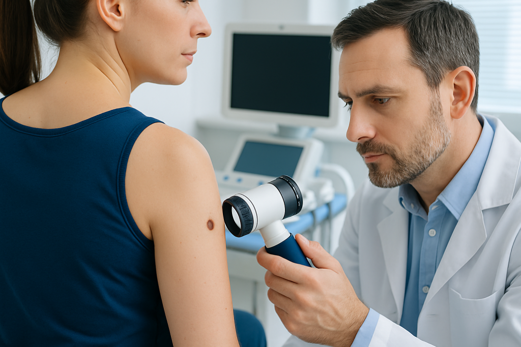
At first glance, an atypical mole may seem only slightly unusual. The changes are often subtle enough that a person may only notice them when comparing one spot with neighboring moles. Dermatologists commonly look for differences in symmetry, border, color, and size.
Typical features can include an oval or irregular outline, a mix of tan, brown, pink, or darker areas within one lesion, and edges that seem uneven rather than smooth. Some atypical moles are flat, while others are slightly raised. They can appear on the back, chest, arms, legs, or anywhere on the body, including areas that are not often exposed to the sun.
Possible warning signs that deserve medical review include:
- Changes in size, shape, or color
- New itching, tenderness, crusting, or bleeding
- A mole that looks very different from a person’s other moles
- A new spot that appears in adulthood and continues changing
It is helpful to remember that symptoms do not confirm cancer on their own. They simply tell a clinician that the spot should be examined carefully, especially if the mole is evolving over time.
Causes and risk factors

An atypical mole develops from pigment-producing cells in the skin called melanocytes. In many people, the exact reason a particular mole becomes atypical is not fully known. Genetics, natural skin characteristics, and sun exposure all seem to play a role.
Risk is higher in people who have many moles, especially if several are atypical. A personal or family history of melanoma is also important, as is a history of sunburns, frequent tanning bed use, or intense cumulative ultraviolet exposure. Fair skin, light eyes, and a tendency to freckle may also be associated with greater skin cancer susceptibility.
Some families have a pattern of many unusual moles and melanoma across generations. In those cases, a dermatologist may recommend more structured surveillance for the person and sometimes for close relatives as well. For people traveling from abroad, it is worth gathering any previous skin photos, pathology reports, and family history details before the appointment, because those records often help the specialist interpret the pattern accurately.
Diagnosis
Diagnosis usually begins with a full skin examination. A dermatologist may compare the suspicious mole with the person’s other moles and ask when it first appeared, whether it has changed, and whether there is any personal or family history of melanoma. This conversation matters because growth patterns often reveal more than a single snapshot does.
A tool called dermoscopy may be used to magnify the lesion and show pigment structures not visible to the naked eye. Dermoscopy can help the clinician decide whether the mole looks stable, whether it should be photographed for follow-up, or whether a biopsy is the safest next step.
If the appearance is uncertain, a biopsy may be recommended. In many cases, the entire mole is removed and sent to pathology so a specialist can examine the cells under a microscope. Pathology is important because atypical moles and early melanoma can sometimes overlap in appearance, and clear tissue analysis provides the most reliable answer.
When the evaluation is part of an international care journey, patients often benefit from an organized plan: skin mapping, comparison with prior photographs, discussion of pathology, and a practical follow-up schedule that can continue with a local physician after returning home.
Treatment options
Not every atypical mole needs removal. If a dermatologist believes the lesion is clearly benign and unchanged, observation may be all that is needed. Monitoring can include clinic visits, photographs, and a plan for the patient to watch for changes at home.
If a mole is clinically concerning, symptomatic, or difficult to classify, surgical removal is often recommended. The procedure is usually straightforward and done under local anesthesia. The goal is to remove the lesion completely when appropriate and to obtain tissue for diagnosis, especially when there is any concern about melanoma.
When pathology confirms an atypical mole, the next steps depend on the degree of atypia, whether margins are clear, and the person’s overall risk profile. Some people only need routine skin checks, while others may need closer surveillance because they have many atypical lesions or a strong family history. The treatment plan is therefore individualized rather than one-size-fits-all.
It can be reassuring for patients to know that treatment is usually focused on clarity and prevention, not aggressive intervention. The most important decision is often the right balance between safe removal, careful monitoring, and avoiding unnecessary procedures.
Prevention and self-care
People cannot always prevent atypical moles from forming, but they can reduce avoidable skin damage and make changes easier to spot. Daily sun protection matters because ultraviolet exposure contributes to skin injury over time. Protective clothing, shade, and broad-spectrum sunscreen are all reasonable parts of routine care.
Regular self-examination is also valuable. A monthly check in good lighting helps people notice whether a mole is evolving or whether a new lesion appears. Many clinicians advise using the “ugly duckling” idea: a spot that looks unlike a person’s other moles should be taken seriously, even if it is small.
Helpful self-care habits include:
- Checking the front, back, scalp, soles, and between the toes
- Using mirrors or asking a partner to help examine hard-to-see areas
- Taking clear photos of monitored spots with dates
- Avoiding tanning beds and intentional sunburn
For travelers, self-care should also include a simple follow-up plan before leaving the country of treatment. Patients may be advised to know which spots were removed, when pathology should be reviewed, and when the next skin exam is due. This makes continuity easier once they are back home.
When to see a doctor
A mole should be examined if it is changing, bleeding, itching, crusting, painful, or growing quickly. It should also be reviewed if it looks distinctly different from surrounding moles or if the person is unsure whether it has always looked that way. These concerns do not mean cancer is present, but they do justify a professional opinion.
People with multiple atypical moles, a history of melanoma, or a close relative with melanoma should consider regular dermatology follow-up even when nothing seems urgent. In higher-risk patients, skin checks are often more useful than waiting for a dramatic change.
If a lesion was previously labeled atypical and a new change appears, it is reasonable to return sooner rather than later. Dermatology assessment is particularly important when a mole changes after sun exposure, after repeated irritation from clothing, or after the person notices that photographs no longer match the current appearance. Acibadem Health Point’s multidisciplinary specialists and JCI-accredited hospitals can help international patients diagnose and treat this condition with coordinated care.
Any rapidly evolving lesion, especially one with irregular colors or bleeding, should be assessed by a qualified doctor promptly. Early review keeps decisions simple and gives the best chance of identifying a harmless mole accurately while also protecting against delayed melanoma diagnosis.
Living with an atypical mole diagnosis
Hearing that a mole is “atypical” can sound unsettling, but in practice it often means the skin needs informed follow-up rather than alarm. Many people live with one or more atypical moles for years without major problems, as long as they remain under observation and know what changes to watch for.
The main goal is consistency. A person who knows which spots are being watched, who keeps follow-up appointments, and who checks the skin regularly is in a strong position to catch meaningful changes early. That is true whether care is being delivered locally or during an international consultation.
For people who travel for care, it helps to leave with a written plan that includes pathology results, the location of any removed lesions, and the recommended timing for the next exam. Good dermatologic care continues after the trip ends, and a clear handoff makes that transition smoother.
Frequently asked questions
What is the difference between an atypical mole and melanoma?
An atypical mole is a mole that looks unusual but is not necessarily cancerous. Melanoma is a skin cancer, and it can sometimes resemble an atypical mole early on, which is why evaluation matters. A dermatologist may use dermoscopy or biopsy to tell them apart.
Do atypical moles always need to be removed?
No, not always. If a dermatologist believes a mole is stable and not suspicious, it may be monitored instead of removed. Removal is more likely when the mole changes, causes symptoms, or cannot be clearly classified.
Can an atypical mole turn into cancer?
Most atypical moles do not become cancer. However, people who have atypical moles may have a higher overall risk of melanoma, especially if they have many such moles or a family history of the disease. That is why regular skin checks are useful.
How can someone monitor an atypical mole at home?
A monthly skin check in good lighting is a practical start. Taking dated photographs can help compare changes over time, and any mole that grows, darkens, bleeds, or becomes itchy should be examined by a clinician.
Is a biopsy painful?
A skin biopsy is usually done with local anesthesia, so discomfort is generally limited. The procedure is typically brief, and the sample is then sent for pathology to provide a more reliable diagnosis.
Who should consider regular dermatology follow-up?
People with many atypical moles, a personal or family history of melanoma, or frequent sun damage should consider ongoing dermatology review. A doctor can suggest how often skin checks should occur based on the person's individual risk.
References
- American Academy of Dermatology
- National Cancer Institute
- Mayo Clinic
- World Health Organization
- American Cancer Society
This article is for general information only and is not a substitute for professional medical advice. Please consult a qualified doctor about your individual situation.
Explore treatments in Turkey — costs, top hospitals & a free quote
JCI-accredited · board-certified surgeons · reply within 24h
Add us as a Preferred Source to see more of our trusted health content across Google Search, AI Overviews and Discover.