Skin Cancer on the Ear: Signs That Need Assessment

Skin cancer on ear most often develops on the sun-exposed outer ear and may look like a sore that does not heal, a crusted or bleeding area, a pearly bump, or a changing pigmented spot. Early medical assessment is important because cancers on the ear can extend into nearby skin, cartilage, or surrounding structures if left untreated.
Overview: What Is Skin Cancer on the Ear?
Skin cancer on ear refers to abnormal growth of skin cells on the visible outer ear, including the rim, earlobe, bowl-shaped central area, and skin behind the ear. It may appear as a persistent scaly patch, a sore that repeatedly crusts or bleeds, a shiny bump, or a mole that changes in shape or color. Because the ear receives frequent sun exposure and is often missed when sunscreen is applied, it is a common location for skin cancer.
The most common forms are basal cell carcinoma and squamous cell carcinoma. These cancers usually grow locally and can often be treated effectively, particularly when found early. Melanoma is less common but can be more serious because it has a greater potential to spread, so a new or changing dark spot on the ear should be assessed without delay.
Not every bump, flaky patch, or itchy area on the ear is cancer. Eczema, psoriasis, infections, pressure-related irritation, and harmless growths can cause similar changes. However, a lesion that persists, grows, changes, or does not heal should be examined by a qualified clinician rather than treated repeatedly at home.
How Ear Skin Cancer May Look and Feel
Ear skin cancer does not always cause pain. A person may first notice a rough area that feels different when washing their hair, wearing glasses, or putting on earrings. Some lesions are tender, itchy, or sensitive to touch, while others have no symptoms at all.
Possible signs include a sore that remains open or returns after healing, a spot that bleeds with minimal contact, a raised pink or pearly growth, or a crusted, thickened patch. Squamous cell carcinoma may look like a persistent red, scaly plaque or firm nodule. Basal cell carcinoma may appear as a shiny, skin-colored or pink bump with a rolled edge, sometimes with visible tiny blood vessels.
Melanoma may develop as a brown, black, blue, red, or mixed-color spot. Changes that deserve attention include asymmetry, an irregular border, varied color, increasing size, or evolution in appearance or sensation. Melanoma can also be amelanotic, meaning it has little or no dark pigment and may resemble a pink or red bump. Checking both the front and back of each ear is helpful, ideally using a mirror or asking another person to look.
- A sore or scab that does not heal within several weeks
- Repeated bleeding, oozing, or ulceration
- A growing lump, thickened patch, or horn-like scale
- A changing mole or new dark spot
- Persistent pain, tenderness, numbness, or itching in one area
Why It Develops: Causes and Risk Factors

Ultraviolet radiation from sunlight is the leading preventable cause of most skin cancers on the ear. Damage accumulates over years, so risk can be higher in people who spend substantial time outdoors for work, sport, travel, or recreation. Tanning beds also expose the skin to harmful ultraviolet radiation and increase skin cancer risk.
The outer ear projects from the head and receives direct sun, especially its upper rim and outer surface. Short hair, thinning hair, baldness, and hairstyles that leave the ears uncovered can increase exposure. The ears may also be missed during routine sunscreen application, even among people who protect the face and neck carefully.
Other factors can raise risk, including fair skin that burns easily, a history of significant sunburns, older age, a personal or family history of skin cancer, and a weakened immune system. People taking medicines that suppress immunity after an organ transplant, or because of certain medical conditions, may develop skin cancers more frequently and may need closer dermatology follow-up. Risk factors guide prevention and screening, but anyone can develop skin cancer, regardless of skin tone.
How Doctors Diagnose a Suspicious Ear Lesion
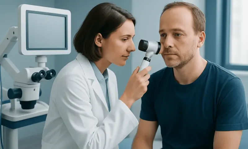
A dermatologist or other trained clinician begins by asking when the skin change began, whether it has grown or bled, and whether there is a history of sun exposure, previous skin cancers, immune suppression, or family history. The clinician examines the entire ear, the skin behind it, the scalp, face, and neck, and may also check nearby lymph nodes when appropriate.
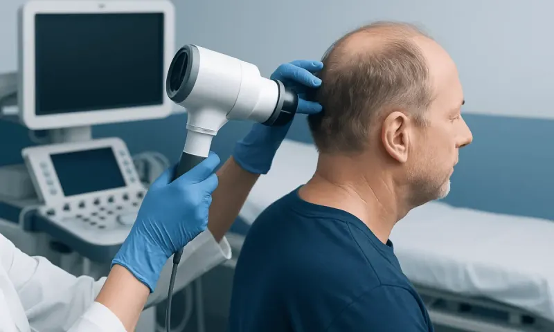
Dermatoscopy, an examination using a handheld magnifying device and light, can help identify patterns not visible to the naked eye. It is useful for assessing pigmented lesions as well as many non-pigmented growths, but it cannot always confirm a diagnosis on its own.
A biopsy is the only way to determine whether a suspicious lesion is cancer and, if so, what type it is. Under local anesthetic, the clinician removes part or all of the lesion and sends it to a laboratory for examination. The pathology report helps determine the diagnosis, depth, growth pattern, and treatment approach. Imaging tests are not needed for most early skin cancers, but may be considered if there is concern that a larger or aggressive cancer has reached deeper tissues or lymph nodes.
Treatment Options for Skin Cancer on the Ear
Treatment depends on the type of cancer, its size and exact location, whether it has returned after previous treatment, its depth, and the person’s general health. The aim is to remove or control the cancer while preserving the ear’s function and appearance as much as possible. A dermatologist, surgical specialist, pathologist, and sometimes radiation or medical oncology team may contribute to care.
Surgical excision removes the cancer along with a margin of normal-looking skin. Mohs micrographic surgery is often considered for cancers on the ear because it examines the edges of removed tissue during the procedure, helping preserve as much healthy tissue as possible while ensuring the tumor is fully removed. Depending on the size and site, the wound may heal naturally, be closed with stitches, or require a skin graft or reconstructive procedure.
Radiation therapy may be an option for selected people who cannot have surgery or for particular cancers with a higher risk of recurrence. For certain superficial, low-risk lesions, clinicians may discuss treatments such as cryotherapy, curettage and cautery, photodynamic therapy, or prescription creams; these are not appropriate for every lesion and are generally not used when invasive cancer is suspected. Advanced squamous cell carcinoma or melanoma may require additional surgery, lymph node assessment, imaging, and systemic treatments such as immunotherapy or targeted therapy.
Follow-up is important after treatment because a person who has had one skin cancer has an increased chance of developing another. The follow-up schedule is individualized according to the cancer type, treatment received, and overall risk profile.
Protecting the Ears and Supporting Skin Health
Sun protection is the most practical way to lower the risk of future ultraviolet-related skin cancers. A broad-spectrum sunscreen with an SPF of 30 or higher can be applied to the front, top, back, and earlobes before going outdoors. It should be reapplied according to the product instructions, especially after swimming, sweating, or prolonged outdoor activity.
A wide-brimmed hat provides more reliable ear coverage than a baseball cap. Shade, sunglasses, and protective clothing add useful protection, particularly during the middle of the day when ultraviolet levels are often strongest. Avoiding tanning beds is also important because there is no safe ultraviolet tanning exposure.
Regular self-checks can help a person become familiar with their skin. They can inspect the ears and surrounding scalp monthly using a handheld mirror and a larger mirror, or ask a trusted person to help. Photographs may be useful for monitoring a spot over time, but they should not replace medical review of a suspicious change. At Acıbadem Health Point, multidisciplinary specialists in JCI-accredited hospitals assess and treat skin cancers for international patients, with care planned around the individual diagnosis.
When to Seek Medical Care
A person should arrange a medical appointment promptly for a new or changing lesion on the ear, especially one that bleeds, ulcerates, forms a recurrent scab, grows, or has not healed after several weeks. A clinician should also assess a mole that changes in color, border, size, or shape, or a spot that looks noticeably different from the person’s other moles.
More urgent assessment is appropriate for a rapidly enlarging lesion, a painful or numb area, a dark spot with marked recent change, or swelling of nearby lymph nodes in front of, behind, or below the ear. These signs do not always mean cancer, but timely evaluation allows an accurate diagnosis and appropriate treatment.
People with a previous skin cancer, a transplant, long-term immune-suppressing treatment, or many actinic keratoses may benefit from regular professional skin examinations. A clinician can recommend an interval that reflects the individual’s risks and previous results.
Frequently asked questions
01What does skin cancer on the ear look like?
It can look like a non-healing sore, a crusted or scaly patch, a shiny pink bump, a bleeding spot, or a changing mole. Skin cancer does not always hurt, so painless changes also deserve attention. A biopsy is needed to confirm the diagnosis.
02Is a crusty spot on the ear always skin cancer?
No. Dry skin, eczema, psoriasis, actinic keratosis, infection, and irritation can all cause crusting. However, a crust that keeps returning, bleeds, grows, or does not heal should be assessed by a clinician.
03Which type of skin cancer is most common on the ear?
Basal cell carcinoma and squamous cell carcinoma are the most common skin cancers on the outer ear. Both are strongly associated with cumulative ultraviolet exposure. Melanoma is less common, but any suspicious pigmented or changing lesion requires prompt assessment.
04Can skin cancer on the ear spread?
Some skin cancers can spread, although many basal cell carcinomas remain local when treated early. Squamous cell carcinoma and melanoma have a greater potential to spread than basal cell carcinoma, particularly when they are larger, deeper, recurrent, or left untreated. The pathology results help the care team assess risk and plan follow-up.
05How is ear skin cancer removed?
Many ear skin cancers are removed surgically, either with standard excision or Mohs micrographic surgery. The choice depends on the cancer type, size, location, and whether preserving nearby healthy tissue is especially important. Other options, including radiation therapy, may be suitable in selected circumstances.
06Can sunscreen prevent skin cancer on the ear?
Sunscreen helps reduce ultraviolet damage and lowers skin cancer risk when used alongside other protective measures. It should be applied to all surfaces of the outer ears and combined with a wide-brimmed hat, shade, and avoidance of tanning beds. It cannot eliminate risk completely, so regular skin awareness remains important.
This article is for general information only and is not a substitute for professional medical advice. Please consult a qualified doctor about your individual situation.
Hair transplant in Turkey — costs, graft counts & a free assessment
JCI-accredited · board-certified surgeons · reply within 24h
Add us as a Preferred Source to see more of our trusted health content across Google Search, AI Overviews and Discover.