Skin Cancer on Neck: Diagnosis, Outlook, and Modern Treatment Approaches
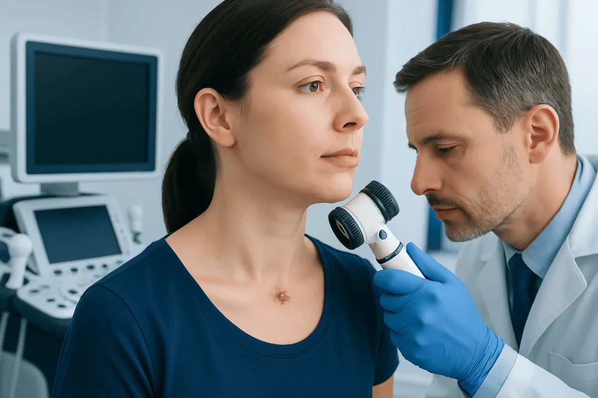
Skin cancer on neck can appear as a persistent scaly patch, pearly bump, changing mole, sore, or area that bleeds and does not heal. A prompt dermatology assessment and biopsy can identify the type of cancer and guide treatment that aims to remove it while preserving healthy tissue.
Overview: Skin Cancer on the Neck
Skin cancer on neck refers to abnormal growth of skin cells on the front, sides, or back of the neck. It is often linked with ultraviolet (UV) exposure over many years, as the neck may receive regular sun exposure but be missed when sunscreen is applied. Most skin cancers found in this area are basal cell carcinoma (BCC) or squamous cell carcinoma (SCC); melanoma is less common but can be more serious.
Diagnosis usually involves a close skin examination followed by a biopsy of the suspicious area. When identified early, many neck skin cancers can be treated effectively with a procedure selected according to the cancer type, size, depth, exact location, and a person’s general health.
The neck is a clinically important site because it is visible, regularly exposed to UV radiation, and close to structures such as nerves, blood vessels, and the ear or jawline. Care is therefore planned to achieve reliable cancer control while considering healing, scar placement, and function.
What Does Early Skin Cancer Look Like on the Neck?
Early skin cancer does not always look dramatic or cause discomfort. It may resemble a minor irritation, dry skin, acne-like bump, wart, or small cut. The most useful clue is persistence: a spot that remains for several weeks, keeps returning, or gradually changes deserves medical attention.
Basal cell carcinoma may look like a shiny or pearly bump, a pink patch, a waxy scar-like area, or a sore that repeatedly crusts or bleeds. Squamous cell carcinoma may present as a rough, scaly, thickened red patch, a tender crusted bump, or a non-healing sore. Melanoma may appear as a new or changing pigmented spot, including one that is uneven in shape or color.
- A sore that does not heal or heals and returns
- A spot that bleeds, crusts, itches, or becomes painful without a clear reason
- A growing pearly, pink, red, brown, or black bump
- A scaly patch that persists despite basic skin care
- A mole or dark mark that changes in size, shape, color, or sensation
The ABCDE guide can help people notice possible melanoma: asymmetry, irregular border, varied color, increasing diameter, and evolution or change. This guide cannot diagnose cancer, however, and some melanomas do not follow every ABCDE feature.
Causes and Risk Factors
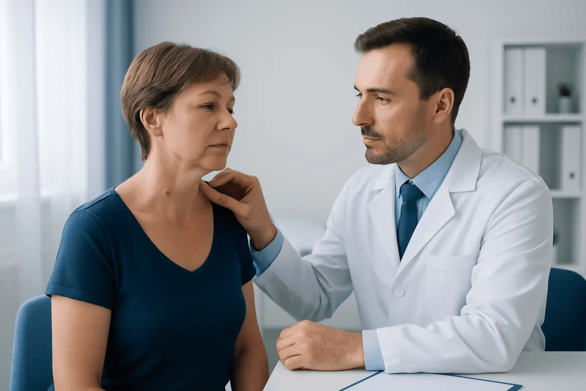
The principal preventable risk factor for neck skin cancer is UV radiation from sunlight and artificial tanning devices. UV exposure damages DNA in skin cells. Over time, accumulated damage can allow abnormal cells to grow. Sunburns, especially blistering burns earlier in life, can further increase long-term risk.
Risk is higher in people with fair skin, light eyes or hair, a tendency to freckle or burn, many moles, or a personal or family history of skin cancer. However, skin cancer can occur in every skin tone. In darker skin, it may be diagnosed later if a changing lesion is mistaken for a benign condition, so persistent changes should never be ignored.
Other factors include older age, outdoor work or recreation, previous radiation treatment, immune suppression after an organ transplant or for certain medical conditions, and exposure to arsenic or some industrial chemicals. People who have had one non-melanoma skin cancer also have a greater chance of developing another, making regular skin checks important.
Some rough sun-damaged patches, called actinic keratoses, can develop on chronically exposed skin and may occasionally progress to SCC. A clinician can evaluate these lesions and recommend monitoring or treatment when appropriate.
Diagnosis and Assessing the Outlook
A dermatologist or other qualified clinician will ask about the lesion’s history, prior sun exposure, immune health, and personal or family history of skin cancer. The skin examination may include dermoscopy, a handheld device that magnifies skin structures and can help distinguish concerning changes from common benign lesions.
If cancer is suspected, a biopsy is needed. A small sample or the entire lesion may be removed under local anesthetic and examined by a pathologist. The pathology report identifies the cancer type and may describe features such as thickness, subtype, depth, and whether cancer cells are seen at the sample edges.
For most BCCs and early SCCs on the neck, the outlook after complete treatment is very favorable. BCC rarely spreads to distant organs, but it can grow into nearby tissue if left untreated. SCC has a greater potential to spread than BCC, particularly when it is large, deep, recurrent, rapidly growing, or occurs in a person with immune suppression.
If melanoma is diagnosed, its thickness and other pathology findings guide staging and treatment planning. The clinician may examine nearby lymph nodes or order further tests when the cancer’s features suggest this is necessary. A diagnosis of skin cancer should always be discussed in the context of the individual pathology report.
How Deep Does Basal Cell Carcinoma Grow?
Basal cell carcinoma does not have one fixed depth. Some BCCs remain very superficial, affecting mainly the upper layers of skin, while others gradually grow downward into the dermis and deeper structures. Depth depends on the subtype, how long the lesion has been present, its location, and individual biological behavior.
On the neck, certain BCCs may grow wider beneath the visible skin surface. Infiltrative, morpheaform, micronodular, and recurrent BCCs can have less clearly defined borders than a typical well-circumscribed lesion. This is one reason a clinician may recommend a treatment method that checks margins carefully during removal.
Although BCC rarely metastasizes, untreated tumors can cause local tissue damage. Over a long period, they may involve deeper skin, fat, muscle, cartilage, or nearby structures. Early assessment usually allows a simpler treatment and may limit the amount of tissue that needs to be removed.
Treatment Options and Modern Approaches
Treatment is individualized. The care team considers the confirmed cancer type, pathology features, lesion size, location on the neck, whether it is a first or recurrent cancer, and the person’s overall health. Surgical excision is a common option: the tumor and a surrounding margin of normal-appearing skin are removed, and the specimen is assessed in a laboratory.
Mohs micrographic surgery is often considered for selected cancers where preserving normal tissue and confirming complete removal are especially important. During Mohs surgery, thin layers are removed and examined in stages until no cancer is seen at the margins. It can be useful for recurrent tumors, tumors with indistinct edges, or tumors in anatomically sensitive areas.
What is the new procedure instead of Mohs surgery? There is no single newer procedure that replaces Mohs surgery for every person. For carefully selected low-risk BCC or SCC, options may include standard excision, curettage and electrodessication, cryotherapy, topical medicines, photodynamic therapy, or radiation therapy. A newer margin-assessment technique called staged excision, sometimes supported by advanced pathology methods, may be used in certain settings, but it is not automatically equivalent to Mohs surgery.
Advanced skin cancers that cannot be managed with local treatment alone may require a multidisciplinary plan involving surgery, radiation, and medicines such as immunotherapy or targeted therapy. Acıbadem Health Point’s multidisciplinary specialists and JCI-accredited hospitals can assess and treat skin cancer for international patients, with treatment decisions based on pathology and individual clinical needs.
What Happens If You Don't Remove the Basal Cell?
Basal cell carcinoma usually grows slowly, but it does not typically disappear on its own. Without treatment, it may gradually enlarge, break down to form a recurring sore, bleed, crust, or become infected. Its slow course can make it easy to delay assessment, even though early treatment is generally less extensive.
Most BCCs remain local rather than spreading to distant sites. However, a neglected lesion can invade nearby tissues and may become more difficult to remove. On the neck, progressive local growth can affect skin and underlying tissue, potentially leading to a larger wound, more complex reconstruction, or additional treatment.
Anyone who has been told they may have BCC should discuss timely management with a dermatologist or skin cancer specialist. If treatment needs to be delayed for personal or medical reasons, the clinician can explain the expected risks, warning signs, and an appropriate monitoring plan.
Prevention, Self-Care, and When to Seek Medical Care
Sun protection lowers the risk of future UV damage and is important after any skin Prostate Cancer Diagnosis: Tests and Next Steps" class="ahp-ilk">cancer diagnosis. Practical measures include seeking shade when UV levels are high, wearing a broad-brimmed hat and neck-covering clothing, using broad-spectrum sunscreen with SPF 30 or higher on exposed skin, and avoiding indoor tanning. Sunscreen should be applied generously and reapplied according to the product instructions, especially after sweating or swimming.
Regular self-checks can help identify new or changing spots. Using a mirror or asking a trusted person to look at the back and sides of the neck may be helpful. People with a previous skin cancer, many moles, immune suppression, or substantial sun exposure may benefit from clinician-directed full-body skin examinations at intervals tailored to their risk.
When to seek medical care: A person should arrange a medical assessment for a new or changing neck lesion, a sore that has not healed within several weeks, unexplained bleeding or persistent crusting, or a mole with evolving color, shape, size, or symptoms. More urgent evaluation is appropriate for a rapidly enlarging lesion, a painful ulcer, or a new dark lesion that changes quickly.
Not every neck spot is cancer, and many are benign. Still, visual inspection alone cannot reliably identify every suspicious lesion. A timely examination and biopsy when indicated provide clarity and allow treatment to be planned safely.
Frequently asked questions
01Is skin cancer on the neck common?
Yes. The neck is frequently exposed to sunlight and may receive less routine sunscreen coverage than the face. Basal cell carcinoma and squamous cell carcinoma are common forms of skin cancer in sun-exposed areas, including the neck.
02Can skin cancer on the neck be cured?
Many early basal cell and squamous cell cancers can be completely removed or controlled with appropriate treatment. The outlook depends on the cancer type, size, depth, location, pathology findings, and whether it has spread. Ongoing follow-up remains important because future skin cancers can develop.
03Does a neck skin cancer biopsy spread cancer?
No. A properly performed skin biopsy does not cause skin cancer to spread. A biopsy is an important diagnostic step because it confirms the diagnosis and helps clinicians choose the most suitable treatment.
04What is the new procedure instead of Mohs surgery?
There is no one new procedure that replaces Mohs surgery in all cases. Depending on the cancer’s risk features, alternatives may include standard surgical excision, radiation therapy, topical treatments, cryotherapy, curettage and electrodessication, or photodynamic therapy. The best approach should be selected after biopsy results are available.
05How deep does basal cell carcinoma grow?
Basal cell carcinoma can range from a superficial lesion near the skin surface to a tumor extending into deeper layers of skin and, when neglected, nearby tissues. Its depth cannot be judged reliably from appearance alone. A biopsy and clinical assessment help determine the appropriate treatment.
06What happens if you don't remove the basal cell?
Basal cell carcinoma commonly continues to grow slowly if it is not treated. It can bleed, ulcerate, become locally destructive, and require a more extensive procedure later. Although spread to distant organs is uncommon, timely treatment is recommended.
07What does early skin cancer look like on the neck?
It may look like a persistent scaly patch, shiny bump, recurring sore, crusted area, changing mole, or spot that bleeds easily. Early lesions can be subtle and painless. Any spot that persists or changes should be checked by a qualified healthcare professional.
This article is for general information only and is not a substitute for professional medical advice. Please consult a qualified doctor about your individual situation.
Oncology care in Turkey — second opinion and treatment plan
JCI-accredited · board-certified surgeons · reply within 24h
Add us as a Preferred Source to see more of our trusted health content across Google Search, AI Overviews and Discover.