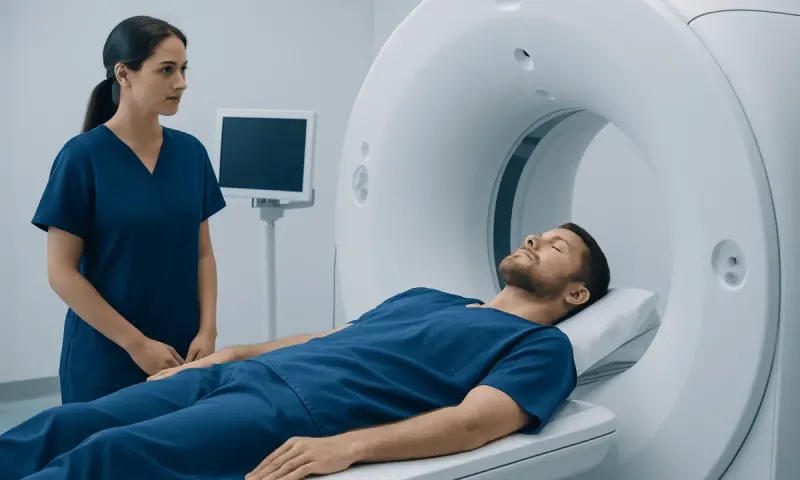
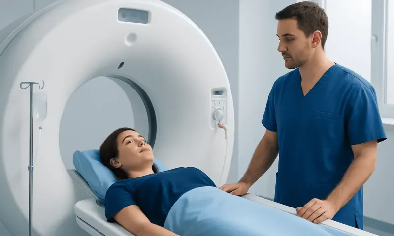
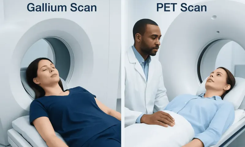
PET Scan vs fMRI: What Does Each Test Show?
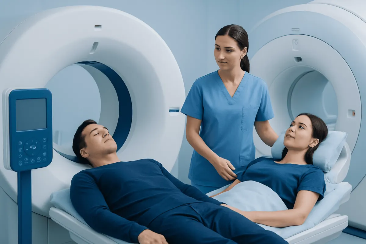
In a PET scan vs fMRI comparison, PET imaging shows how tissues use a small amount of radioactive tracer, while functional MRI (fMRI) maps changes in blood flow associated with brain activity. Neither test is universally better: clinicians choose the examination based on the question being asked, such as cancer assessment, brain structure, or brain function during a task.
PET Scan vs fMRI at a Glance
When comparing a PET scan vs fMRI, the central difference is that PET measures biologic activity using a radiotracer, whereas fMRI measures changes in blood oxygenation that occur when parts of the brain become more active. A standard MRI is different again: it produces detailed images of body structure, including the brain, spinal cord, joints, and soft tissues.
The terms can be confusing because fMRI is a type of MRI, not a type of PET scan. PET and MRI may sometimes be used together in one visit with hybrid PET/MRI technology, but they provide different kinds of information. The right test is selected by a clinician based on symptoms, medical history, previous imaging, and the specific diagnostic question.
| Feature | PET scan | fMRI | Standard MRI |
|---|---|---|---|
| Main information | Metabolism, receptor activity, or other tracer uptake | Task-related brain activity through blood-oxygen changes | Detailed anatomy and tissue structure |
| Typical body areas | Whole body, brain, heart, and other organs | Brain | Brain and many other body areas |
| Radiation | Yes, from the radiotracer | No ionizing radiation | No ionizing radiation |
| Common reasons for use | Cancer staging, treatment monitoring, some neurologic or cardiac assessments | Brain mapping before selected surgery and research | Injury, tumors, inflammation, stroke, and structural abnormalities |
| During the scan | Usually rest quietly after tracer injection | May perform simple tasks, such as moving a hand or answering prompts | Usually remain still; contrast may sometimes be used |
How Clinicians Tell PET, MRI, and fMRI Apart
Clinicians first decide whether they need an anatomic image, a measure of biologic activity, or both. MRI is especially useful when fine structural detail matters, such as assessing the brain, spinal cord, ligaments, muscles, liver, pelvis, or soft tissues. It uses a powerful magnetic field and radio waves rather than X-rays or radioactive tracers.
PET imaging begins with administration of a small amount of radiotracer. Different tracers are designed to highlight different processes. A commonly used tracer reflects glucose use, which can be increased in many cancers but may also rise with infection or inflammation. PET is frequently combined with CT, and sometimes MRI, so metabolic findings can be located precisely in the body.
Functional MRI, often called functional MRI or fMRI, is usually performed when clinicians need to identify brain regions involved in movement, language, sensation, memory-related tasks, or other functions. It does not directly measure nerve-cell firing. Instead, it detects a blood-oxygen-level-dependent signal, which is an indirect marker of local brain activity. This distinction is particularly important in a pet scan vs fmri brain comparison.
What Happens During Each Examination

For a PET scan, preparation depends on the tracer and reason for testing. People may be asked to avoid eating for a period before some examinations, particularly those involving glucose-based tracers. After the injection, there is often a quiet waiting period to allow the tracer to distribute through the body. The scan itself requires lying still on a moving table.
For MRI and fMRI, screening for implanted devices, metal fragments, or certain types of surgical hardware is essential because the scanner uses a strong magnetic field. Some people may receive MRI contrast, generally a gadolinium-based agent, when it can improve the clarity of the examination. The care team checks kidney health and allergy history when relevant.
During an fMRI scan, the person may be asked to complete simple tasks, such as tapping fingers, naming pictures, listening to words, or resting with eyes open or closed. The images are most useful when the tasks are performed as instructed and movement is minimized. MRI scanners can be noisy and enclosed, so patients should tell the imaging team if they have anxiety, claustrophobia, pain, or difficulty lying flat.
- PET involves a small radiation exposure from the tracer.
- MRI and fMRI do not use ionizing radiation.
- All three tests are interpreted alongside symptoms, examination findings, and other test results.
What Is More Accurate, an MRI or a PET Scan?
Neither MRI nor PET is always more accurate. Accuracy depends on what is being investigated. MRI is often more informative for detailed anatomy, including small structural changes in the brain, spine, joints, muscles, and soft tissues. PET may be more informative when the question concerns metabolic activity, the spread of some cancers, treatment response, or selected brain and heart conditions.
For example, a small area of abnormal tissue may be clearly visible on MRI because it changes the structure of an organ, while a PET scan may show whether that area is metabolically active. Conversely, PET may identify abnormal activity before an obvious structural change is visible on a conventional scan. Findings still require careful interpretation because active inflammation, healing tissue, and infection can sometimes resemble cancer on certain PET studies.
In cancer care, clinicians may use imaging alongside biopsy results, laboratory tests, and physical examination. PET results do not by themselves establish a cancer diagnosis, and a normal scan does not always rule out disease. For patients undergoing cancer treatment, the imaging plan is individualized according to cancer type, stage, and the treatment goal.
What Can an MRI Show That a PET Scan Cannot?
MRI can show highly detailed physical anatomy that PET cannot show as clearly on its own. This includes the shape and internal structure of the brain, spinal cord, nerves, discs, tendons, ligaments, cartilage, bone marrow, and many soft tissues. MRI is also valuable for identifying features such as bleeding at certain stages, swelling, scarring, fluid collections, compression of nerves, and many types of tissue injury.
In the brain, MRI can reveal structural changes associated with stroke, multiple sclerosis, developmental differences, trauma, tumors, and some causes of seizures or headaches. A PET scan may add different information by showing altered metabolism, but it does not replace the structural detail of MRI. In some situations, PET/CT or PET/MRI brings functional and structural information together.
The pet scan vs mri vs fmri question is therefore not a choice between competing tests in every case. A clinician may order one test first and another later if the first result does not answer the clinical question. The safest and most useful approach is to use the least invasive examination that can provide the needed information.
Can fMRI Detect ADHD?
fMRI is not a standard diagnostic test for attention-deficit/hyperactivity disorder (ADHD). Research studies have identified group-level differences in brain networks and activity patterns among some people with ADHD, but these patterns are not reliable enough to diagnose an individual person in routine clinical practice.
ADHD is diagnosed through a comprehensive clinical assessment. This generally includes a history of symptoms over time, their effect on school, work, relationships, and daily functioning, as well as assessment for other conditions that can cause similar difficulties. Input from family members, teachers, or other close observers can be helpful, particularly for children.
Brain imaging may occasionally be considered if a clinician suspects another neurologic cause for symptoms, but it is not used to confirm ADHD. A qualified mental health professional, pediatrician, neurologist, or psychiatrist can explain which evaluations are appropriate for the individual.
Can a PET Scan Pick Up All Cancers?
No. A PET scan cannot detect all cancers, and it is not an all-purpose cancer screening test. Whether PET is helpful depends on the cancer type, size, location, growth pattern, and the tracer used. Some tumors do not take up enough tracer to be clearly seen, and very small lesions can be below the scan’s practical resolution.
In addition, not every area of tracer uptake is cancer. Infection, inflammation, recent surgery, healing, and normal activity in some organs can produce uptake. Conversely, some cancers may be difficult to identify with commonly used glucose-based PET tracers. This is why PET findings are reviewed together with CT, MRI, pathology, laboratory results, and the person’s clinical history.
For people with a known or suspected malignancy, imaging choices are guided by established cancer-specific pathways. A biopsy may still be needed to confirm the diagnosis and identify the tumor type. Discussions about oncology treatment should include the purpose and limitations of each scan, as well as what the result may mean for next steps.
When to Seek Medical Care
Medical care should be sought promptly for new or worsening neurologic symptoms, including sudden weakness or numbness on one side, trouble speaking, new confusion, loss of vision, a severe unusual headache, fainting, or a seizure. Sudden symptoms may need urgent assessment and should not wait for an outpatient imaging appointment.
People should also arrange a medical review for persistent or unexplained symptoms such as an enlarging lump, unexplained weight loss, ongoing pain, persistent cough, unusual bleeding, or a significant change in function. These symptoms have many possible causes, and imaging is only one part of evaluation. A clinician can decide whether PET, MRI, fMRI, another test, or no imaging is appropriate.
Before any scan, patients should report pregnancy or possible pregnancy, breastfeeding, kidney disease, diabetes, allergies, implanted devices, metal exposure, and previous reactions to contrast agents. Acıbadem Health Point’s multidisciplinary specialists and JCI-accredited hospitals support diagnostic assessment and treatment planning for international patients when advanced imaging is clinically indicated.
Frequently asked questions
01Is fMRI the same as MRI?
fMRI is a specialized form of MRI. Standard MRI shows detailed structure, while fMRI measures blood-oxygen changes associated with activity in different brain regions. fMRI is most often used for brain mapping and research rather than routine diagnosis.
02Does a PET scan hurt?
A PET scan is generally not painful, although there may be a brief pinch when the radiotracer is injected. The person must lie still during the scan, which can be uncomfortable for those with pain or anxiety. Imaging staff can explain ways to improve comfort before the appointment.
03Is a PET scan more accurate than an fMRI scan for the brain?
They answer different questions, so one is not broadly more accurate than the other. PET can assess specific metabolic or molecular processes in the brain, while fMRI maps task-related blood-flow changes. The appropriate test depends on the suspected condition and the information needed.
04Why might a doctor order both PET and MRI?
A doctor may order both tests when both metabolic activity and detailed anatomy are important. For example, one scan may identify an area of unusual activity while the other defines its exact location and structural features. This can help guide diagnosis, monitoring, or treatment planning.
05Are PET and fMRI safe?
Both are widely used when medically appropriate. PET involves a small amount of radiation from the radiotracer, while fMRI does not use ionizing radiation but requires safety screening for metal implants and other contraindications. The care team weighs expected benefit against any potential risk for each patient.
06Can imaging diagnose the cause of memory problems?
Imaging can sometimes contribute useful information, but it does not diagnose every cause of memory difficulty on its own. MRI may identify structural changes, while PET may be considered in selected specialist evaluations. A full assessment also includes medical history, examination, cognitive testing, and review of medicines and other health conditions.
This article is for general information only and is not a substitute for professional medical advice. Please consult a qualified doctor about your individual situation.
Medical weight-loss options in Turkey — costs & eligibility
JCI-accredited · board-certified surgeons · reply within 24h
Add us as a Preferred Source to see more of our trusted health content across Google Search, AI Overviews and Discover.