PET Scan vs Biopsy: How They Diagnose Disease
A PET scan and a biopsy answer different clinical questions: PET imaging can identify areas with unusual metabolic activity throughout the body, while a biopsy examines cells or tissue to establish what an abnormality is. They are often complementary rather than competing tests, and the best sequence depends on the suspected condition, location of the finding, and a person’s overall health.
PET Scan Versus Biopsy: A Side-by-Side Comparison
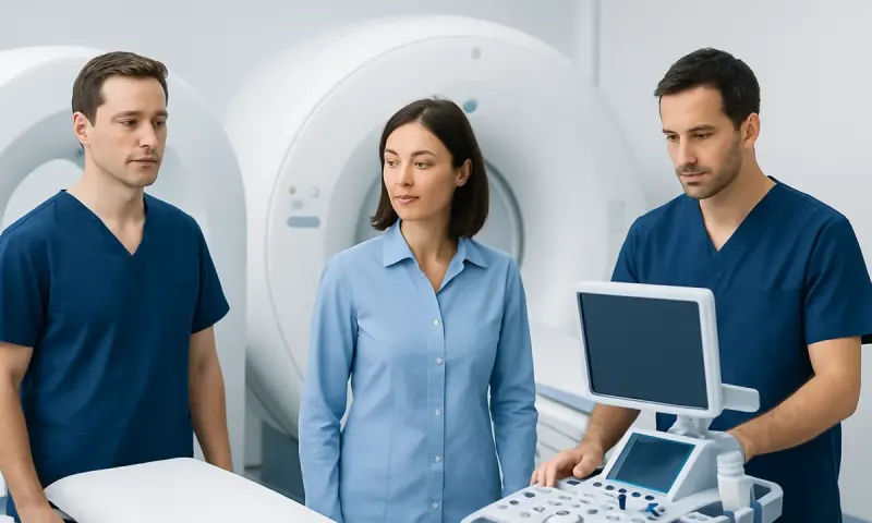
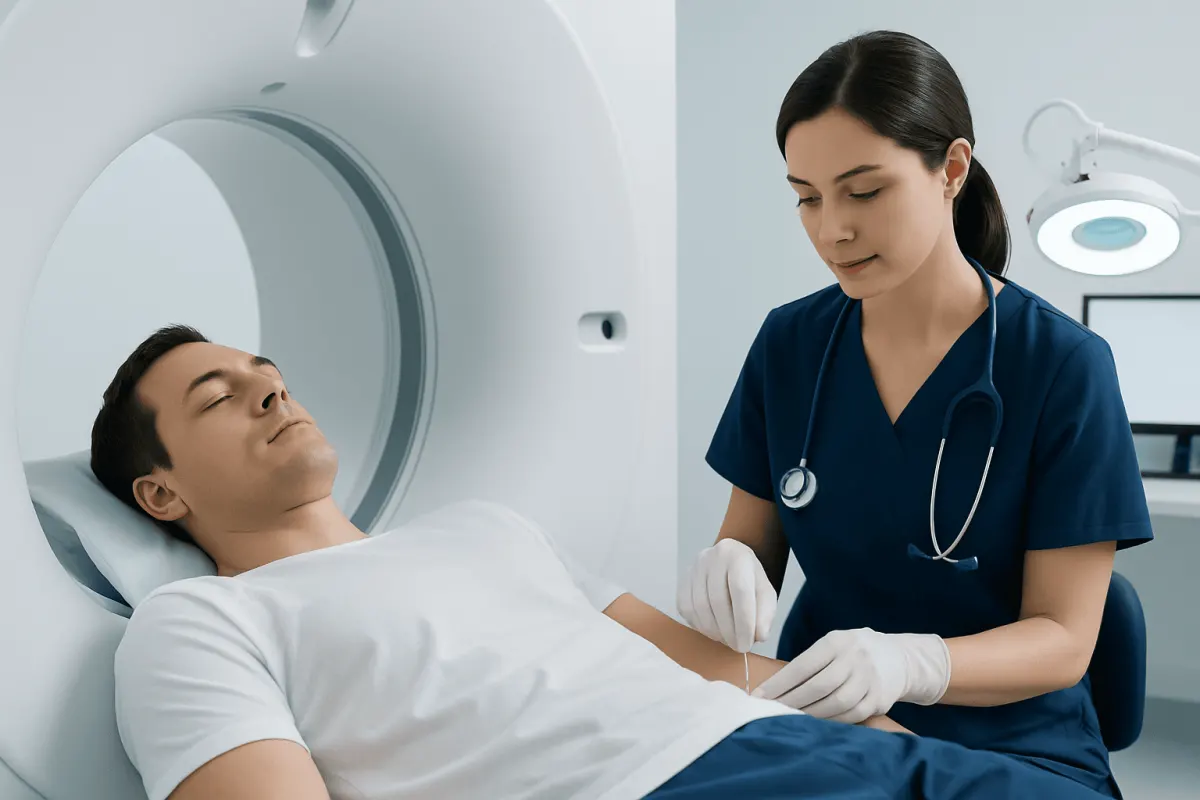
In the question of PET scan versus biopsy, neither test simply replaces the other. A positron emission tomography (PET) scan is an imaging examination that shows how actively tissues are using a small amount of radiotracer, often a glucose-like substance. A biopsy is a procedure in which a clinician removes cells or a small tissue sample so a pathologist can examine it under a microscope and, when needed, perform additional laboratory tests.
PET scans can help locate suspicious areas, assess the extent of a known condition, monitor response to treatment, or look for recurrence. Biopsies can identify whether an abnormality is cancerous, inflammatory, infectious, or benign, and can provide details such as tumor type and molecular features that may guide treatment.
| Feature | PET scan | Biopsy |
|---|---|---|
| Main purpose | Maps abnormal metabolic activity in the body | Examines cells or tissue to establish a diagnosis |
| What it can show | Location, extent, and activity of possible disease | Cell type, tissue changes, and sometimes genetic or molecular features |
| Can it confirm cancer? | Usually no; it can raise or lower suspicion | Often yes, when an adequate sample is obtained |
| Invasiveness | Non-surgical injection and imaging | Ranges from needle sampling to a minor surgical procedure |
| Common limitation | Inflammation or infection may resemble cancer | The sample may be difficult to obtain or may not represent the whole lesion |
Many PET scans are performed as PET/CT scans, which combine metabolic information from PET with detailed anatomical images from CT. This combined approach can help clinicians identify a suitable site for tissue sampling when biopsy is needed.
How Clinicians Tell PET Findings and Biopsy Results Apart
Clinicians interpret a PET scan in context rather than treating a bright or active area as a diagnosis on its own. The scan is reviewed alongside symptoms, medical history, physical examination findings, blood tests, and other imaging. The pattern of uptake, its location, and whether a change is new or stable over time can all matter.
A biopsy provides direct evidence from the sampled tissue. A pathologist may assess how cells look, whether they are malignant, and whether there are signs of inflammation, infection, scarring, or another process. In cancer care, tissue testing may also help determine the tumor’s origin and identify features relevant to treatment planning.
Both tests have limitations. PET can miss some slow-growing or very small cancers, and it can show false-positive activity from recent surgery, healing, infection, inflammation, or some noncancerous conditions. A biopsy can be nondiagnostic if too little tissue is collected, if the needle does not reach the most informative area, or if the abnormality is difficult to access safely.
For this reason, a multidisciplinary team may review images and pathology together. If PET results and biopsy findings do not match the clinical picture, clinicians may recommend repeat imaging, a different biopsy technique, or sampling from another area.
Which Is More Accurate, a Biopsy or a PET Scan?
For confirming whether a specific suspicious lesion is cancer and determining its precise type, a biopsy is generally more definitive than a PET scan. The tissue sample allows direct examination of cells, which is why pathology is commonly considered the diagnostic standard for many cancers.
However, “accuracy” depends on the question being asked. A PET scan may be more useful for evaluating whether there are additional areas of active disease elsewhere in the body, for estimating the extent of some known cancers, or for assessing whether treatment appears to be reducing metabolic activity.
A biopsy is not automatically perfect. Its reliability depends on obtaining enough representative tissue from the right location and having it assessed with appropriate pathology methods. If a result is inconclusive but clinical concern remains, the care team may discuss another biopsy or further evaluation.
In practical terms, PET helps clinicians understand where disease may be active, while biopsy helps establish what the abnormal tissue is. Using both appropriately can provide a clearer and safer diagnostic pathway than relying on either test alone.
Which Should Be Done First, a PET Scan or a Biopsy?
There is no single correct order for every person. Often, a biopsy is performed first when there is an accessible suspicious lump, lesion, or organ abnormality that needs confirmation. Establishing a tissue diagnosis can help clinicians decide which scans are appropriate next and whether staging studies are needed.
In other situations, a PET scan may come first. For example, it can help identify the most metabolically active and safest area to biopsy when there are several abnormalities, when the original finding is difficult to reach, or when clinicians need to assess how widespread a suspected disease may be before planning a procedure.
Timing can also matter because inflammation after surgery or biopsy can sometimes increase uptake on a PET scan and make interpretation more complex. The medical team considers this along with urgency, symptoms, the suspected diagnosis, and whether delaying a procedure could affect care.
People should ask their clinician what question each test is intended to answer and how the result will change the next step. The decision is individualized, particularly in the assessment of suspected cancer, where imaging, pathology, and specialist input are commonly coordinated.
What Is the Downside of a PET Scan?
A PET scan is generally well tolerated, but it has important limitations. It involves exposure to a small amount of radiation from the radiotracer and, when combined with CT, radiation from the CT component as well. The expected benefit is weighed against this exposure, especially for children, younger adults, and people who may need repeated imaging.
The most significant diagnostic downside is that increased uptake does not always mean cancer. Infection, inflammatory conditions, normal healing, and some benign growths may look active on PET. Conversely, some cancers may have low uptake or may be too small to detect, so a normal scan does not rule out every possible disease.
Preparation may be inconvenient. People are commonly asked to fast for a period before the scan and to limit strenuous exercise beforehand. Blood glucose levels can affect some PET studies, so people with diabetes should receive individualized preparation instructions from their imaging team.
Rarely, a person may experience discomfort from the injection, anxiety in the scanner, or practical difficulties remaining still. Pregnancy, breastfeeding, kidney concerns related to CT contrast when contrast is used, and certain medications should be discussed before the appointment. The imaging department can explain the safety measures relevant to the planned examination.
Why Would a Doctor Order a PET Scan Instead of a CT Scan?
A CT scan mainly shows anatomy: the size, shape, and location of organs, lymph nodes, masses, bleeding, or structural changes. A PET scan shows metabolic activity, which may reveal functional changes before an abnormality becomes obvious through size or appearance alone. A doctor may therefore order PET when the key question concerns how biologically active a finding is or whether there may be active disease in more than one location.
PET is often paired with CT because the tests complement one another. The CT portion helps precisely locate an area of PET uptake, while PET adds functional information to the structural detail. This can be helpful in the evaluation and follow-up of selected cancers, certain infections, and some inflammatory disorders.
CT remains the preferred or first test in many situations. It is widely used for urgent symptoms, injuries, lung and abdominal evaluation, and detailed assessment of anatomy. A PET scan is not a general replacement for CT; it is requested when its additional information is likely to influence diagnosis, staging, treatment planning, or follow-up.
If cancer has already been diagnosed, imaging findings may help guide cancer treatment planning. The treating team selects scans based on the type of cancer, the area being assessed, prior test results, and the person’s individual circumstances.
What to Do After a PET Scan or Biopsy
After a PET scan, the radiologist or nuclear medicine physician interprets the images and sends a report to the referring clinician. Results are usually considered with prior scans and other tests. A report describing uptake or a suspicious area does not by itself establish a diagnosis, and a clinician should explain what the finding means in the individual situation.
After a biopsy, minor soreness, bruising, or a small amount of bleeding may occur depending on the procedure. The care team gives specific instructions about wound care, activity, and medicines. Pathology results can take time because some samples need special stains, cultures, or molecular testing in addition to routine microscopic examination.
People should keep follow-up appointments and bring a current list of medicines, allergies, previous imaging, and relevant medical records. Helpful questions include whether the result is definitive, whether further testing is necessary, which specialist should be involved, and what symptoms should prompt earlier contact.
Acıbadem Health Point’s multidisciplinary specialists and JCI-accredited hospitals support international patients who require coordinated imaging, pathology review, and treatment planning for complex diagnostic concerns.
When to Seek Medical Care
Medical assessment is appropriate for a new or persistent lump, unexplained bleeding, ongoing cough, unexplained weight loss, persistent pain, fever that does not improve, or other symptoms that are concerning or worsening. These symptoms have many possible causes, and timely evaluation can help clarify the reason without assuming a diagnosis.
Anyone who has been advised to have a PET scan or biopsy should contact the ordering clinician if they do not understand the reason for the test, preparation instructions, or expected next steps. It is reasonable to ask about benefits, possible risks, alternatives, and how results will influence care.
Following a biopsy, urgent medical advice is needed for heavy or ongoing bleeding, rapidly increasing swelling, severe or worsening pain, fever, pus-like drainage, breathing difficulty, or signs of an allergic reaction. The appropriate emergency service should be used for severe symptoms.
A person should not delay evaluation because imaging or biopsy results feel worrying. Most abnormal findings require careful interpretation, and the next step may range from routine monitoring to further testing or treatment.
Frequently asked questions
01Can a PET scan detect cancer without a biopsy?
A PET scan can identify areas that may be suspicious for cancer, but it usually cannot confirm cancer on its own. Inflammation, infection, and healing tissues can also show increased uptake. A biopsy is often needed to identify the cause and determine the exact cell type.
02Can a biopsy be normal after an abnormal PET scan?
Yes. An active area on PET may be caused by a noncancerous process, or the biopsy may not have sampled the relevant part of the lesion. Clinicians compare the pathology result with imaging and the overall clinical picture to decide whether observation, repeat testing, or another biopsy is appropriate.
03Does a PET scan hurt?
The scan itself is painless, although there is a brief needle stick for the radiotracer injection. People need to lie still on an imaging table, which can be uncomfortable for some. The team can help with concerns such as pain, anxiety, or difficulty remaining still.
04How long do PET scan and biopsy results take?
PET scan reports are commonly available after the images have been reviewed, although the exact timing varies by facility and clinical urgency. Biopsy results may take longer because tissue processing and additional laboratory tests may be needed. The ordering clinician can explain the expected timeline.
05Is a PET scan more accurate than a CT scan?
PET and CT scans are accurate for different purposes. CT provides detailed structural information, while PET identifies areas of altered metabolic activity. Combining them in a PET/CT study can provide more complete information in selected clinical situations.
06Can a PET scan tell whether cancer has spread?
A PET scan can help identify areas elsewhere in the body that may contain active cancer and is commonly used for staging certain cancers. However, not every cancer type is best assessed with PET, and suspicious areas may still need confirmation with biopsy or other tests. The care team interprets PET results alongside other imaging and pathology.
This article is for general information only and is not a substitute for professional medical advice. Please consult a qualified doctor about your individual situation.
Oncology care in Turkey — second opinion and treatment plan
JCI-accredited · board-certified surgeons · reply within 24h
Add us as a Preferred Source to see more of our trusted health content across Google Search, AI Overviews and Discover.