Cat Scan vs CT Scan: Are They the Same Test?
There is no medical difference between a CAT scan and a CT scan. CAT means computed axial tomography, an earlier name for computed tomography (CT), which uses X-rays and computer processing to create detailed images of the body.
Overview: Cat Scan vs CT Scan
Cat scan vs CT scan has no difference. Both terms refer to computed tomography, an imaging examination that combines multiple X-ray images with computer processing to create detailed cross-sectional views of the body. “CAT” stands for computed axial tomography, while “CT” stands for computed tomography; CT is now the term used more often in clinical practice.
A CT scan may examine one area, such as the head, chest or abdomen, or several body regions after injury or when a widespread condition is suspected. The images can be viewed as thin slices and reconstructed into three-dimensional views, helping clinicians assess structures that may not be clear on a standard X-ray.
CT is a diagnostic tool, not a diagnosis by itself. A radiologist interprets the images in the context of a person’s symptoms, examination findings and other tests, then sends a report to the clinician who ordered the scan.
How a CT Scan Works and What It Can Show
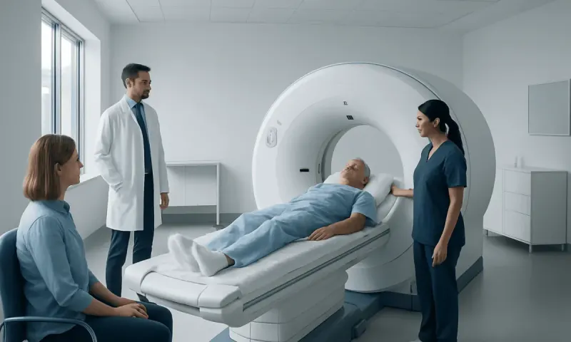
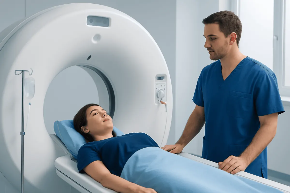
During CT imaging, a patient lies on a motorized table that moves slowly through a large, doughnut-shaped scanner. An X-ray tube rotates around the body while detectors collect information from many angles. A computer converts this information into detailed images of internal structures.
CT is especially useful for evaluating bones, lungs, certain abdominal organs, blood vessels and injuries. It can help identify fractures, internal bleeding, kidney stones, lung abnormalities, appendicitis, infections, blood clots in selected settings and many other conditions. Depending on the clinical question, CT may also help guide procedures such as a biopsy or drainage.
In a cat scan vs ct scan with contrast comparison, the scan itself remains the same. Contrast is an additional substance, commonly iodine-based and given through a vein, that can improve visibility of blood vessels and tissues. Some CT scans use oral contrast for the digestive tract, while others do not need contrast at all.
A CT scan can provide rapid information in urgent situations. This is particularly valuable after significant trauma, with sudden severe symptoms, or when clinicians need to assess internal organs and blood vessels quickly.
Why Would a Doctor Recommend a CAT Scan?

A doctor may recommend a CAT scan when detailed internal images are needed to clarify symptoms, confirm or rule out a suspected condition, assess an injury, plan treatment or monitor a known condition. The choice is individualized: the clinician considers the question that needs answering, the body area involved, urgency, previous imaging and the person’s health history.
For example, a head CT may be used after a head injury or to evaluate certain sudden neurological symptoms. A chest CT can provide more detail than a chest X-ray when assessing the lungs or chest structures. CT of the abdomen and pelvis may help investigate significant abdominal pain, suspected stones, inflammation, obstruction or trauma.
CT may also be recommended in cancer care to assess the extent of a known cancer, guide a biopsy, evaluate treatment response or monitor for recurrence. An abnormal result does not always mean cancer or another serious disease; imaging findings often require correlation with other tests or follow-up imaging.
Before ordering the scan, the care team should explain its purpose and whether contrast is planned. Patients should mention possible pregnancy, kidney disease, diabetes, thyroid conditions, prior contrast reactions and all medicines or supplements they use.
What Can a CT Scan Show That an MRI Cannot?
CT and MRI are complementary tests rather than direct substitutes. CT uses X-rays and is generally faster, while MRI uses a magnetic field and radio waves, without ionizing radiation. The most suitable test depends on the suspected problem and which type of tissue needs assessment.
CT is often better suited to showing fine bone detail, including many fractures, and can be very effective for detecting acute bleeding in the head. It is also frequently preferred for evaluating the lungs because air-filled lung tissue is well assessed with CT. CT can rapidly image the chest, abdomen and pelvis in emergencies and can be used to visualize blood vessels with CT angiography.
MRI usually offers greater soft-tissue detail in areas such as the brain, spinal cord, nerves, muscles, ligaments and certain joints. MRI can be more informative for many soft-tissue injuries and some neurological conditions. However, it typically takes longer and may not be appropriate for everyone with certain implanted or metallic devices.
In the broader comparison of cat scan vs ct scan vs mri, CAT and CT are the same examination. A PET scan is different again: in a cat scan vs ct scan vs pet scan discussion, PET evaluates metabolic activity using a small amount of radioactive tracer, and it may be combined with CT to match metabolic findings to anatomy.
Candidacy and the CT Scan Procedure: Step by Step
Most people can have a CT scan safely when it is clinically appropriate. The imaging team reviews relevant health information beforehand, particularly pregnancy status, known allergies or previous reactions to contrast, kidney function concerns and medicines that may need special consideration. A clinician may select a non-contrast CT, an alternative imaging test, or additional precautions when needed.
On arrival, the patient may be asked to change into a gown and remove jewelry, glasses, hearing aids or metal objects near the area being scanned. Fasting instructions vary. For many non-contrast scans, no preparation is needed; for some contrast studies, the imaging center may ask the patient not to eat for a period beforehand.
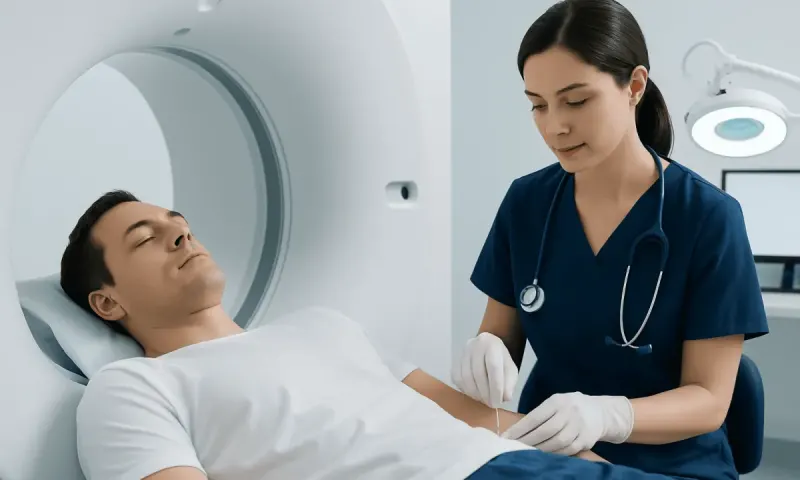
If intravenous contrast is used, a small cannula is placed in a vein. During injection, some people briefly notice warmth spreading through the body, a metallic taste, or a sensation of needing to urinate. These feelings usually pass within seconds. The patient then lies still while the table moves through the scanner, following brief breathing instructions if requested.
Clear communication is available throughout the examination. The technologist operates the scanner from an adjacent room but can see, hear and speak with the patient. Remaining still helps reduce motion blur and improves image quality.
How Long Do You Stay in a CAT Scan? Recovery and Results
The actual image acquisition in a CAT scan is often only a few minutes. The full visit commonly takes around 15 to 30 minutes, though it can take longer if contrast preparation, additional image sequences or observation after a prior contrast issue is needed. The exact timing depends on the body area and scan type.
There is usually no recovery period after a CT scan. People can generally return to normal daily activities immediately. After contrast, drinking fluids may be advised unless a clinician has recommended fluid restriction for another health reason. The body normally removes the contrast through the kidneys.
Radiology images are reviewed by a radiologist, who prepares a report for the ordering clinician. Timing varies by setting and urgency: emergency studies may be interpreted rapidly, while routine outpatient results can take longer. The clinician who requested the scan is best placed to explain what the findings mean for the individual.
If itching, rash, breathing difficulty, facial swelling, severe dizziness or other concerning symptoms develop after contrast, urgent medical advice is needed. Delayed reactions are uncommon, but patients should report them to their healthcare team.
Why Don’t Doctors Like to Do CT Scans? Benefits, Risks and Safer Use
Doctors do use CT scans regularly when they are likely to provide important information, but they avoid unnecessary scans because CT involves ionizing radiation. Radiation exposure from one scan is generally small, yet exposure can add up across repeated studies. The aim is to use the lowest suitable radiation dose and choose another test when it can answer the clinical question equally well.
CT can offer major benefits: it is fast, widely useful, detailed and often available in urgent situations. In many circumstances, the information gained can support prompt treatment decisions and outweigh the potential risks. Modern CT protocols are tailored to the area being scanned and, where possible, to the patient’s size and clinical indication.
Contrast material has a small risk of allergic-like reactions, ranging from mild symptoms to rare serious reactions. It can also require additional consideration in people with significantly reduced kidney function. The care team can assess these risks and plan appropriately before imaging.
People who are pregnant or think they may be pregnant should tell their clinician and imaging team before the scan. Imaging is not automatically ruled out during pregnancy, especially in emergencies, but the team will carefully consider whether CT is necessary and how to minimize exposure.
When to Seek Medical Care
Medical care should be sought urgently for symptoms such as sudden weakness or trouble speaking, a severe sudden headache, chest pain, serious breathing difficulty, major injury, uncontrolled bleeding, fainting, confusion, or severe and worsening abdominal pain. A clinician will decide whether CT or another test is appropriate based on the symptoms and examination.
For non-urgent concerns, an appointment is appropriate when persistent or unexplained symptoms are affecting daily life, such as recurring severe headaches, ongoing abdominal pain, unexplained blood in urine or stool, persistent cough, or pain after an injury that is not improving. Imaging should be guided by a qualified clinician rather than requested solely for reassurance.
Patients can ask why a scan is being recommended, whether contrast is needed, what alternatives may be suitable and how the results could affect care. Acıbadem Health Point’s multidisciplinary specialists and JCI-accredited hospitals evaluate and treat patients requiring diagnostic imaging and coordinated follow-up care.
Frequently asked questions
01Is a CAT scan the same as a CT scan?
Yes. CAT scan and CT scan are two names for the same imaging test, computed tomography. CAT is an older abbreviation for computed axial tomography, while CT is the modern term used more commonly.
02Is a CT scan painful?
A CT scan itself is not painful. The patient needs to lie still on a table, and some positions may be uncomfortable if there is an injury or pain. If intravenous contrast is used, there may be a brief warm sensation or metallic taste during the injection.
03Do all CT scans require contrast?
No. Whether contrast is needed depends on the part of the body being examined and the clinical question. Some scans are clearer without contrast, while others use it to show blood vessels, inflammation, organs or certain abnormalities more clearly.
04Can a CT scan detect cancer?
CT can identify findings that may suggest cancer, such as a mass, enlarged lymph nodes or spread to other organs. However, CT alone does not always confirm a cancer diagnosis. Further imaging, blood tests or a biopsy may be needed depending on the findings.
05How is a CT scan different from a PET scan?
CT primarily shows detailed anatomy, including organs, bones and blood vessels. PET shows areas of altered metabolic activity after a radioactive tracer is administered. PET and CT are often combined so clinicians can compare metabolic activity with precise anatomical images.
06Are CT scans safe?
CT scans are considered safe when medically justified and performed with appropriate protocols. They use radiation, so clinicians balance the expected diagnostic benefit against the small potential risk and avoid unnecessary repeat scans. The imaging team also takes precautions when contrast or pregnancy is relevant.
This article is for general information only and is not a substitute for professional medical advice. Please consult a qualified doctor about your individual situation.
Joint and spine care in Turkey — expert assessment & treatment
JCI-accredited · board-certified surgeons · reply within 24h
Add us as a Preferred Source to see more of our trusted health content across Google Search, AI Overviews and Discover.