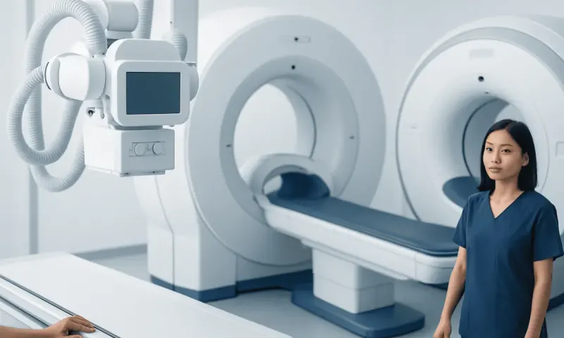
PET Scan vs MRI Cost: What Affects the Price?
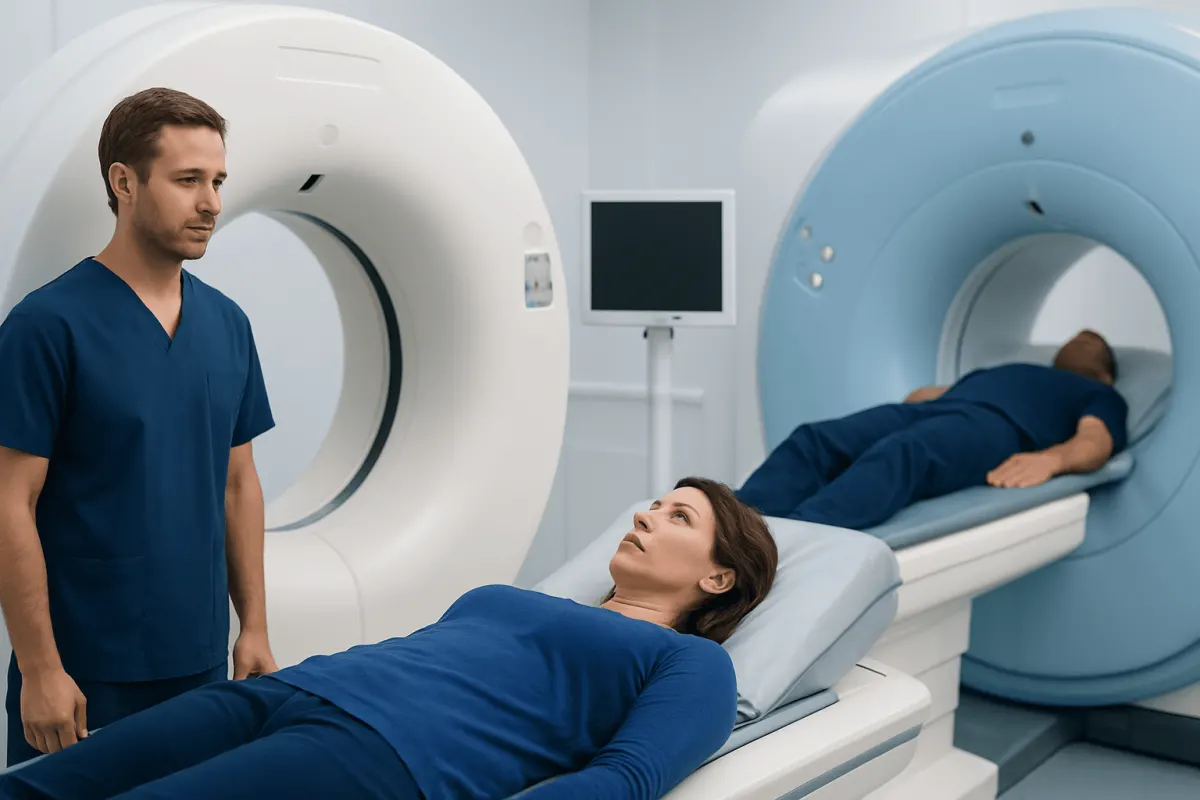
PET scan vs MRI cost cannot be compared fairly with one fixed figure because charges vary by country, scan area, imaging materials, facility and insurance coverage. More importantly, PET and MRI answer different clinical questions: PET shows metabolic activity, while MRI provides highly detailed images of soft tissues and organs.
PET Scan vs MRI Cost: A Side-by-Side Comparison
PET scan vs MRI cost varies widely and should not be used alone to decide which examination to have. A PET scan often involves a specialized radioactive tracer, preparation time and imaging equipment that may combine PET with CT or MRI. MRI costs can vary according to the body area scanned, scan duration, use of contrast material, type of scanner, facility and local payment or insurance arrangements.
These tests are not interchangeable. PET identifies areas where cells are using energy differently from expected, whereas MRI creates detailed structural images. A clinician selects the scan that is most likely to answer a specific question, such as whether a cancer is active, whether a brain symptom may relate to tissue changes, or whether a joint injury involves a ligament.
- PET: Shows metabolic or biochemical activity using an injected tracer; frequently combined with CT.
- MRI: Shows detailed soft tissue anatomy using magnets and radio waves; it does not use ionizing radiation.
- CT: Uses X-rays to provide rapid cross-sectional anatomical images, particularly useful for lungs, bones, bleeding and many urgent conditions.
- Planning costs: Patients can request a written estimate that clarifies the scan, tracer or contrast, radiologist interpretation, facility charge and any separate appointments.
When comparing pet scan cost vs mri cost, it is helpful to ask what information the doctor needs and whether a combined examination is proposed. A lower-cost test is not necessarily useful if it cannot answer the clinical question, while a more complex test may not be necessary when a simpler examination is appropriate.
How PET, MRI and CT Answer Different Questions
In a pet scan vs ct scan vs mri scan comparison, PET is primarily a functional imaging test, MRI is a detailed soft-tissue imaging test, and CT is a fast structural imaging test that uses X-rays. PET images may show areas of increased or reduced tracer uptake, which can help identify biologically active disease. CT and MRI show the location, size, shape and relationship of structures within the body.
PET is commonly performed as PET/CT. The CT component helps locate areas of tracer uptake precisely and provides additional anatomical information. In selected settings, PET/MRI may be used, particularly where excellent soft-tissue definition and metabolic information are both valuable. The best method depends on the organ being assessed, the suspected diagnosis and previous test results.
MRI is often preferred for the brain, spinal cord, nerves, muscles, ligaments, pelvic organs and many liver or soft-tissue findings. CT is often preferred when speed matters or when evaluating lung disease, fractures, internal bleeding or certain abdominal concerns. A clinician may use more than one modality because each scan supplies different information rather than because one result is necessarily uncertain.
People may use the terms PET scan vs MRI vs CAT, with CAT scan being another name for CT scan. In clinical practice, the referral should state the medical reason for imaging, allowing the radiology and nuclear medicine team to tailor the study safely and effectively.
What Can a PET Scan Show That an MRI Cannot?

A PET scan can show patterns of metabolic activity that may not be visible on an MRI image. Many PET examinations use a glucose-like tracer, because some cancer cells and inflamed tissues take up more glucose than surrounding tissue. This can help clinicians identify potentially active disease, assess whether treatment is working, or look for disease elsewhere in the body.
MRI may show a mass or area of altered tissue very clearly, but it cannot directly measure tracer uptake in the way PET does. Conversely, an area of increased PET uptake is not automatically cancer. Infection, inflammation, healing tissue and some normal organs can also take up tracer. PET results must therefore be interpreted alongside symptoms, medical history, laboratory findings and anatomical imaging.
For cancer care, PET may contribute to staging and treatment planning in selected cancers, as well as evaluating suspected recurrence. It can be part of the assessment of conditions discussed in cancer care, but the type of cancer, its location and the clinical setting determine whether PET is useful. Some tumors do not take up the most commonly used tracer strongly, and other imaging may be more informative.
A PET scan does not replace MRI when precise soft-tissue detail is needed. For example, MRI can be particularly valuable for defining the relationship of a lesion to the spinal cord, brain structures, nerves or joints. The two tests may therefore be complementary.
Why Would a Doctor Order a PET Scan Instead of a CT Scan?
A doctor may order a PET scan instead of a CT scan when the key question concerns tissue activity rather than anatomy alone. CT can show that a lymph node is enlarged or that there is a lung nodule, for example. PET may help determine whether a finding has increased tracer uptake and whether there are other active areas that could influence further testing or treatment planning.
PET/CT is often considered when staging certain cancers, investigating possible recurrence, monitoring response to treatment, or evaluating selected inflammatory or infectious conditions. The CT portion provides anatomical detail, while the PET portion adds information about metabolism. This combined approach may reduce uncertainty in appropriate circumstances, but it is not required for every cancer or every abnormal CT finding.
CT may be the better first test for many concerns because it is widely available, fast and excellent for several types of structural assessment. In emergency care, CT can be especially important for suspected trauma, bleeding, stroke evaluation pathways or acute chest and abdominal conditions. A PET scan generally requires advance preparation and is not typically an emergency imaging tool.
The decision is individualized. Doctors consider the suspected diagnosis, urgency, previous imaging, whether a biopsy is being planned, kidney function, pregnancy status and the likely effect of scan findings on management. Patients can ask what decision the scan is expected to help make.
Which Is More Claustrophobic, MRI or PET Scan?
MRI is generally more difficult for people with claustrophobia than PET. During an MRI, the patient usually lies in a relatively narrow tunnel within a powerful magnet, often for a longer period. The machine can also make loud tapping or knocking sounds, although ear protection is provided.
A PET scan may involve lying still on a scanning table, usually after a quiet uptake period while the tracer distributes through the body. PET/CT scanners are commonly more open and shorter than conventional MRI systems. However, some people may still feel uneasy about remaining still, the medical setting, injections or the scanning process.
Patients should tell the referring doctor and imaging team about claustrophobia, anxiety, pain, mobility limitations or previous difficulty with scans before the appointment. The team may explain each step, offer practical comfort measures, consider a more open MRI system where clinically suitable, or discuss whether medication for anxiety is appropriate. Any sedating medicine should only be used under medical guidance, and patients may need someone to accompany them home.
Communication is important. Imaging teams usually provide a call button or a way to contact staff during the examination, and breaks may sometimes be possible depending on the scan type and clinical requirements.
What Is the Downside of a PET Scan?
The main downside of a PET scan is that it uses a radioactive tracer, which involves a small amount of ionizing radiation. When PET is combined with CT, the total radiation exposure includes both the tracer and the CT component. The expected clinical benefit should outweigh this exposure, and clinicians avoid unnecessary imaging.
PET also has limitations in accuracy. Increased tracer uptake can occur with infection, inflammation or normal physiological processes, leading to findings that need further evaluation. In addition, some cancers and very small lesions may not be detected reliably. A PET result is one part of a wider assessment and may occasionally lead to follow-up imaging, blood tests or biopsy.
Preparation can be more involved than for some other scans. Patients may need to fast, follow instructions about physical activity, and keep warm and relaxed before the scan. Blood glucose levels can affect some PET studies, so people with diabetes should receive individualized instructions from their healthcare team.
Pregnant or breastfeeding patients, and those who may be pregnant, should inform their doctor before any nuclear medicine examination. The tracer injection can cause brief discomfort, while serious reactions are uncommon. The imaging team will review safety considerations and give aftercare guidance appropriate to the tracer used.
How to Choose the Right Scan and Prepare
The patient does not usually need to choose between PET and MRI independently. The referring clinician selects an imaging pathway based on the symptom, examination findings, medical history and the question that needs answering. If a scan has been recommended, patients may ask whether the goal is diagnosis, staging, treatment monitoring, follow-up or planning a procedure.
Before MRI, patients should report implants, pacemakers, clips, cochlear devices, metal fragments, medication patches and possible pregnancy. Many implants are MRI-compatible, but they must be checked carefully. Before PET, patients should disclose diabetes, pregnancy or breastfeeding, recent infections, recent surgery, strenuous exercise and medicines, since these can affect preparation or interpretation.
For people receiving cancer treatment, imaging may be coordinated with chemotherapy planning or radiotherapy planning when clinically relevant. Timing matters because treatment-related inflammation can affect imaging appearances. The treating team can advise when a scan is most informative.
Acıbadem Health Point’s multidisciplinary specialists and JCI-accredited hospitals diagnose and treat conditions requiring advanced imaging for international patients. Patients should bring prior reports and image files where possible, as comparison with earlier examinations can improve interpretation.
When to Seek Medical Care
People should seek medical advice for new, persistent or worsening symptoms that concern them, such as an unexplained lump, ongoing unexplained weight change, persistent pain, coughing up blood, a seizure, new weakness, severe headaches with neurological changes, or symptoms that continue despite initial treatment. These symptoms have many possible causes and do not automatically mean a serious condition, but timely assessment is important.
Emergency medical care is needed for symptoms such as sudden weakness or facial drooping, difficulty speaking, sudden severe headache, chest pain, severe breathing difficulty, fainting, major injury or uncontrolled bleeding. Emergency teams may use urgent imaging such as CT or MRI depending on the situation; PET is not generally used for immediate emergency assessment.
A scan should not be requested solely because of a cost comparison or concern about a single symptom. A qualified clinician can determine whether imaging is needed, which test has the best balance of information and safety, and whether another step such as examination, laboratory testing or biopsy should come first.
Frequently asked questions
01Is a PET scan more expensive than an MRI?
A PET scan may involve additional resources, including a radioactive tracer and nuclear medicine staff, while MRI costs depend on factors such as scan duration, body area and contrast use. Actual charges vary significantly by country, facility, insurance coverage and the details of the examination. Patients can request an itemized estimate from the imaging provider.
02Is PET better than MRI for cancer?
Neither test is always better for cancer. PET can provide information about metabolic activity and may help with staging or monitoring selected cancers, while MRI offers detailed images of soft tissues and certain organs. The most useful test depends on the cancer type, location and clinical question.
03Does a PET scan use radiation?
Yes. PET uses a radioactive tracer, and PET/CT also includes radiation from the CT component. The amount and type of exposure depend on the examination, and clinicians use PET when the expected information is likely to be useful for care.
04Does MRI use radiation?
No. MRI uses strong magnetic fields and radio waves rather than ionizing radiation. However, MRI is not suitable for every person or every clinical question, particularly when certain implants or metal fragments are present.
05How long do PET and MRI scans take?
The imaging portion of a PET scan often takes less than an hour, but the total appointment is longer because time is needed after the tracer injection. MRI duration varies by body part and the number of image sequences, and some examinations may take longer. The imaging center can provide specific timing before the appointment.
06Can PET and MRI results be wrong?
All medical tests have limitations. PET uptake can reflect inflammation or infection as well as cancer, and MRI findings can sometimes be nonspecific or incidental. Radiologists and clinicians interpret results together with symptoms, examination findings and other tests, and may recommend follow-up when needed.
This article is for general information only and is not a substitute for professional medical advice. Please consult a qualified doctor about your individual situation.
Joint and spine care in Turkey — expert assessment & treatment
JCI-accredited · board-certified surgeons · reply within 24h
Add us as a Preferred Source to see more of our trusted health content across Google Search, AI Overviews and Discover.