CAT Scan vs Other Imaging Tests: How They Compare
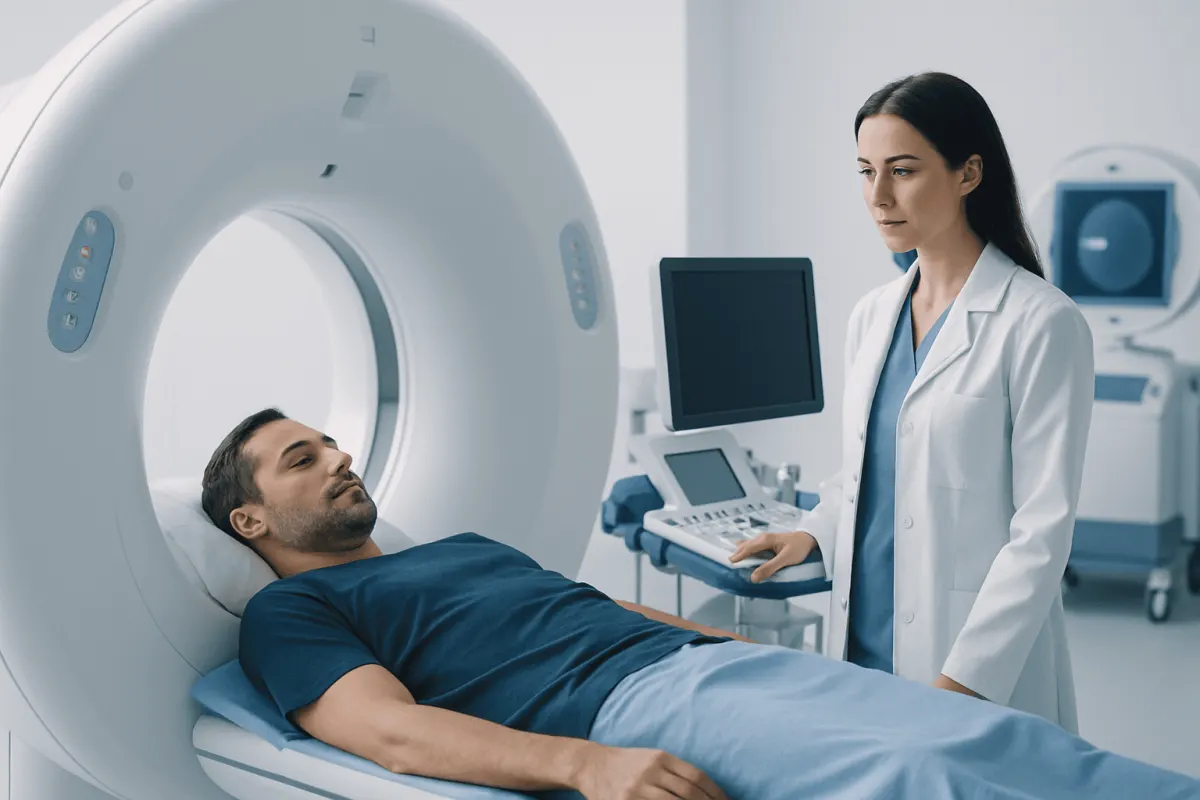
A CAT scan and a CT scan are the same examination: “CAT” is an older term for computed axial tomography, now generally called computed tomography (CT). The best scan depends on the body area, the clinical question, urgency, pregnancy status, prior imaging, and whether contrast material would add useful detail.
CAT Scan vs Other Imaging Tests: A Side-by-Side Comparison
A CAT scan is a CT scan. CAT stands for computed axial tomography, while CT stands for computed tomography; modern healthcare teams usually say CT. Both names describe an imaging test that uses a series of X-rays and computer processing to create detailed cross-sectional images of the body.
In a cat scan vs CT scan comparison, there is therefore no difference in technology, preparation, radiation exposure or typical results. The meaningful comparison is between CT and other tests, such as MRI, PET and ultrasound. Each test answers different clinical questions, so one is not automatically better than another.
- CT (CAT scan): Uses X-rays; fast; excellent for bones, lungs, many abdominal conditions, internal bleeding and emergency assessment.
- MRI: Uses magnetic fields and radio waves; no ionizing radiation; particularly useful for soft tissues, the brain, spinal cord, nerves, joints and selected pelvic conditions.
- PET/CT or PET/MRI: Uses a small amount of radiotracer to show metabolic activity; often used for selected cancer staging, treatment response, heart viability or neurological evaluations.
- Ultrasound: Uses sound waves; no ionizing radiation; commonly used for pregnancy, gallbladder, kidneys, blood flow and many superficial structures.
- X-ray: Uses a small amount of radiation; quick and useful for many bone and chest questions, but less detailed than CT.
A clinician considers the suspected condition, the part of the body being assessed and whether the result may change immediate care. They also consider whether a person can remain still, has an implanted device, may be pregnant, has reduced kidney function, or has had a previous reaction to contrast material.
How a Clinician Chooses Between CT, MRI, PET and Ultrasound
Clinicians do not select imaging based on a general ranking. They start with the question that needs answering. For example, after a significant injury, CT can rapidly identify fractures, internal bleeding and injury to organs. In a person with new neurological symptoms, a CT scan may be used urgently to look for bleeding, while MRI may later provide more detailed information about brain tissue, the spinal cord or nerves.
For cat scan vs CT scan brain searches, the terms still refer to the same test. A head CT is commonly used when speed matters, such as after head trauma or when stroke-like symptoms begin suddenly. MRI may be preferred when the situation is stable and clinicians need greater detail for certain causes of headache, seizures, inflammation, tumors or smaller areas of ischemic stroke.
The cat scan vs CT scan vs MRI decision also depends on practical factors. CT typically takes only minutes and is more widely suitable for people who cannot tolerate a long, enclosed examination. MRI often takes longer and may not be appropriate for some implants or metal fragments, although many modern devices are MRI-compatible under specific conditions. MRI also may require special planning for people with severe claustrophobia.
In cat scan vs CT scan vs PET scan comparisons, PET is not usually a substitute for an initial CT. PET adds functional information: it can highlight tissue with increased or reduced metabolic activity. It is often paired with CT or MRI to help locate and characterize findings, especially in selected oncology care. A PET result is interpreted carefully because inflammation and infection can also cause increased uptake.
How to Decide CT With or Without Contrast?

CT contrast is a medication, usually iodine-based, given into a vein or sometimes by mouth or another route depending on the body area. It helps blood vessels, organs, inflammation, infections and some tumors stand out more clearly. A non-contrast CT can still be the right examination when contrast would not improve the answer or when a specific finding is better seen without it.
For example, non-contrast CT is often used to assess certain types of head injury or suspected bleeding in the brain, and it may be used for some suspected kidney stones. CT with intravenous contrast can be especially helpful when assessing blood vessels, appendicitis and other abdominal concerns, infections, masses, or suspected clots in particular clinical settings. The exact protocol is tailored to the symptoms and the question being investigated.
In a cat scan vs CT scan with contrast comparison, the scan itself is still CT; the difference is whether contrast is used. Before contrast, the imaging team may ask about previous contrast reactions, asthma or allergies, kidney disease, diabetes medicines, thyroid conditions, pregnancy and current medications. Kidney function testing may be appropriate for some people, particularly when there are risk factors for impaired kidney function.
Most people tolerate contrast well. A temporary warm sensation, metallic taste or feeling of needing to urinate can occur shortly after injection and usually passes quickly. Serious reactions are uncommon, but staff are trained to recognize and treat them. Patients should report any past contrast reaction and tell the team promptly if they develop itching, hives, breathing difficulty or swelling after contrast.
Why Don't Doctors Like to Do CT Scans?
Doctors do use CT scans when they are likely to provide clinically useful information. The concern is not that CT is inherently unsafe; rather, it uses ionizing radiation, and clinicians aim to avoid imaging that is unlikely to affect care. This approach is sometimes called justification: the expected benefit should outweigh the potential risk.
CT generally delivers more radiation than a standard X-ray because it collects many images. Radiation exposure from medical imaging is carefully managed, and modern scanners use protocols designed to use the lowest dose that can still answer the clinical question. Avoiding unnecessary repeat scans is particularly important for children, younger adults and people who have already had substantial imaging, while still ensuring needed tests are not delayed.
Other reasons a clinician may select a different test include the need for better soft-tissue detail, a desire to avoid radiation during pregnancy when possible, or the possibility that ultrasound or MRI can provide the needed answer. Contrast-related considerations, such as a previous severe reaction or significantly reduced kidney function, may also influence the plan.
A patient should not decline an urgently recommended CT because of general concerns about radiation. In suspected serious injury, stroke, internal bleeding, pulmonary embolism or other time-sensitive conditions, a CT may be the fastest and most informative test. The clinician can explain why it is recommended and whether a reasonable alternative exists.
How to Understand CT Scan Results?
A CT result is usually written by a radiologist, a doctor trained to interpret medical imaging. The report commonly includes the reason for the scan, the technique used, whether contrast was given, a detailed description of what was seen and an “impression.” The impression is the radiologist’s concise summary of the most important findings.
Words such as “normal,” “no acute abnormality” or “no evidence of” can be reassuring in the appropriate clinical context. “No acute abnormality” generally means the scan did not show an urgent new problem; it does not necessarily rule out every possible explanation for symptoms. Terms such as “incidental finding,” “lesion,” “nodule,” “density” or “follow-up recommended” do not by themselves establish a diagnosis. They describe an observation that may need comparison with prior imaging, additional tests or monitoring.
The requesting clinician interprets the report alongside symptoms, medical history, physical examination and laboratory results. A scan can identify structural changes, but it cannot always explain pain, fatigue, dizziness or other symptoms. Conversely, a normal scan does not mean symptoms are unimportant; it may help guide the next step in evaluation.
Patients can ask for a copy of the report and images and should discuss the impression with the clinician who ordered the scan. Helpful questions include what the finding means in their situation, whether it needs follow-up, whether prior scans were compared, and what symptoms should prompt earlier reassessment.
What Test Is Better Than a CT Scan?
No imaging test is always better than a CT scan. The most appropriate test is the one most likely to answer the clinical question safely and efficiently. CT is often superior for quickly evaluating complex fractures, lung tissue, certain abdominal emergencies and active internal bleeding. It can also be valuable when MRI is unavailable, unsuitable or would take too long in an emergency.
MRI can be better when detailed views of soft tissues are needed. This includes many conditions affecting the brain, spinal cord, nerves, ligaments, tendons, cartilage and bone marrow. Ultrasound may be better as a first examination for gallbladder symptoms, pelvic concerns, pregnancy, testicular symptoms and evaluation of blood flow, because it has no ionizing radiation and can show movement in real time.
PET combined with CT or MRI may be more informative than CT alone for particular cancer-related questions, such as assessing metabolic activity or looking for disease in multiple areas of the body. However, PET is not appropriate for every cancer question and is not generally used for routine emergency evaluation. A biopsy, blood test, endoscopy or clinical follow-up may also be more useful than another scan in some circumstances.
Patients can ask what the test is intended to detect, what alternatives exist and how the result would affect treatment. This shared discussion is especially useful when imaging is elective rather than urgent. When advanced imaging or coordinated specialty input is needed, Acıbadem Health Point’s multidisciplinary specialists and JCI-accredited hospitals support diagnosis and treatment for international patients.
What to Do Before and After a CT Scan
Preparation depends on the type of CT. For many non-contrast scans, no special preparation is required. If contrast is planned, the imaging department may provide instructions about eating and drinking beforehand. Patients should follow the instructions they receive rather than stopping medicines or fasting on their own.
Before the appointment, patients should tell the care team about pregnancy or a possible pregnancy, breastfeeding, kidney problems, thyroid disease, diabetes, previous reactions to contrast, allergies, implanted devices and all medications or supplements. They should also bring details of earlier scans when available, since comparison with prior images can prevent unnecessary tests and clarify whether a finding is new or unchanged.
During CT, the person lies on a table that moves through a short, ring-shaped scanner. The team communicates from a nearby room and can see and hear the patient. Remaining still is important, and short breath-holds may be requested for chest or abdominal imaging. The scan itself is painless.
After a contrast-enhanced scan, most people can return to usual activities unless given different instructions. Drinking fluids may be advised if medically appropriate. Patients should seek urgent help for breathing difficulty, facial or throat swelling, widespread hives, fainting or other signs of a severe allergic reaction after leaving the imaging center.
When to Seek Medical Care
Imaging should be arranged through a qualified clinician except in emergency services, where the team determines what is needed. A person should seek emergency care for sudden weakness or numbness on one side, facial drooping, trouble speaking, sudden severe headache, loss of consciousness, seizures, severe chest pain, major breathing difficulty, coughing blood, serious injury, or severe abdominal pain with fainting or signs of shock.
Prompt medical assessment is also appropriate for persistent or worsening symptoms such as an unexplained new severe headache, ongoing vomiting, significant abdominal pain, blood in urine or stool, unexplained weight loss, a persistent cough, or new neurological changes. The correct next step may be CT, MRI, ultrasound, laboratory testing or another assessment depending on the individual situation.
Patients with non-urgent questions about a planned scan should contact the clinician who ordered it or the radiology department. They can ask why the test is recommended, whether contrast is needed, how to prepare and when results are expected. Clear communication helps ensure that imaging is appropriate and that results are acted on safely.
Frequently asked questions
01Is a CAT scan the same as a CT scan?
Yes. CAT scan is an older term for computed axial tomography, while CT means computed tomography. In current clinical practice, CT is the more commonly used name, but both refer to the same imaging technology.
02Is a CT scan painful?
A CT scan is not painful. The person needs to lie still on a table while it moves through the scanner, and brief breath-holds may be requested. If contrast is injected, some people notice short-lived warmth or a metallic taste.
03Does a CT scan always need contrast?
No. Whether contrast is used depends on what clinicians need to see. Some conditions are best assessed without contrast, while contrast can make blood vessels, organs, inflammation and certain abnormalities easier to evaluate.
04Is MRI safer than CT?
MRI does not use ionizing radiation, which can make it preferable in some situations. However, it is not automatically the better or safer choice for every person, since it takes longer and may be unsuitable with certain implants or metal fragments. The best test depends on the medical question and individual circumstances.
05Can a CT scan detect cancer?
CT can identify abnormalities that may suggest cancer and can help show their size, location and spread. It cannot always confirm whether a finding is cancerous. Further imaging, laboratory testing, follow-up scans or a biopsy may be needed for diagnosis.
06How long does it take to receive CT scan results?
Timing varies by setting and urgency. In emergency care, results may be reviewed quickly to guide immediate decisions, while non-urgent reports may take longer. The ordering clinician or imaging center can explain when and how results will be communicated.
This article is for general information only and is not a substitute for professional medical advice. Please consult a qualified doctor about your individual situation.
Joint and spine care in Turkey — expert assessment & treatment
JCI-accredited · board-certified surgeons · reply within 24h
Add us as a Preferred Source to see more of our trusted health content across Google Search, AI Overviews and Discover.